. 2005 Dec;132(24):5565-75.
doi: 10.1242/dev.02155. Epub 2005 Nov 9.

Normal myoblast fusion requires myoferlin

Affiliations

Normal myoblast fusion requires myoferlin

Katherine R Doherty et al. Development. 2005 Dec.

Abstract

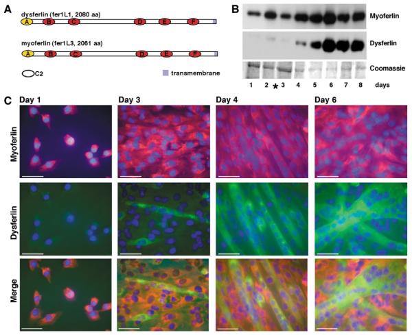
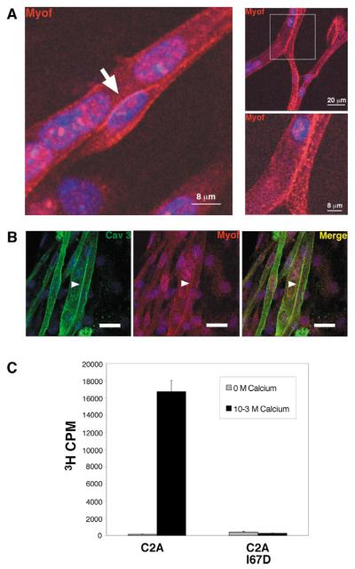
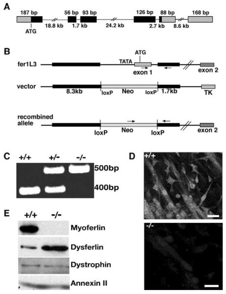
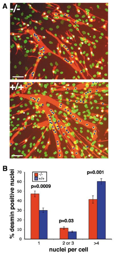
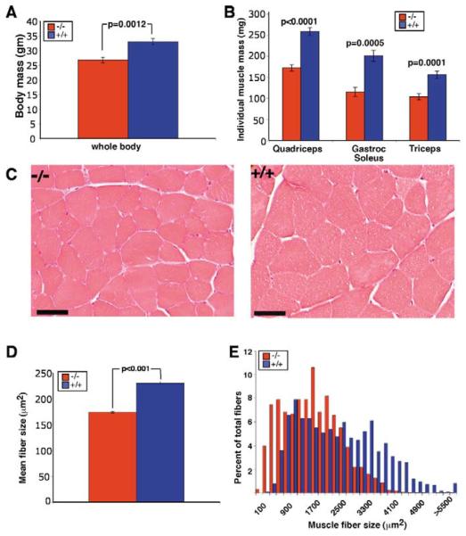
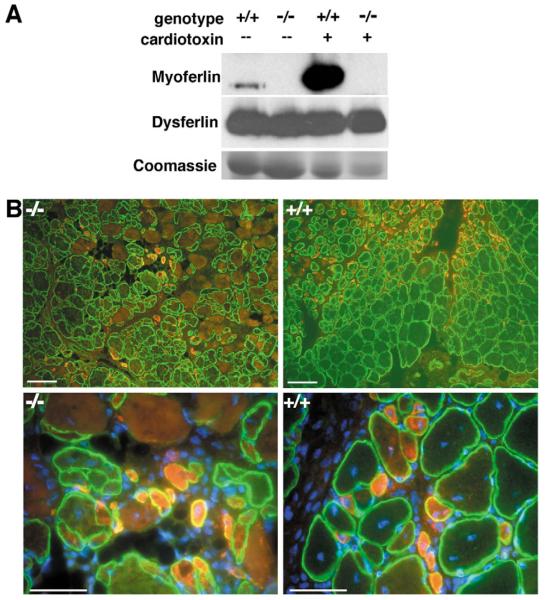
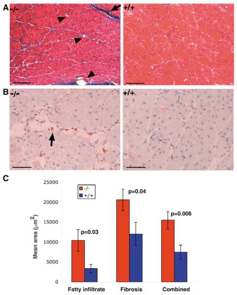
Muscle growth occurs during embryonic development and continues in adult life as regeneration. During embryonic muscle growth and regeneration in mature muscle, singly nucleated myoblasts fuse to each other to form myotubes. In muscle growth, singly nucleated myoblasts can also fuse to existing large, syncytial myofibers as a mechanism of increasing muscle mass without increasing myofiber number. Myoblast fusion requires the alignment and fusion of two apposed lipid bilayers. The repair of muscle plasma membrane disruptions also relies on the fusion of two apposed lipid bilayers. The protein dysferlin, the product of the Limb Girdle Muscular Dystrophy type 2 locus, has been shown to be necessary for efficient, calcium-sensitive, membrane resealing. We now show that the related protein myoferlin is highly expressed in myoblasts undergoing fusion, and is expressed at the site of myoblasts fusing to myotubes. Like dysferlin, we found that myoferlin binds phospholipids in a calcium-sensitive manner that requires the first C2A domain. We generated mice with a null allele of myoferlin. Myoferlin null myoblasts undergo initial fusion events, but they form large myotubes less efficiently in vitro, consistent with a defect in a later stage of myogenesis. In vivo, myoferlin null mice have smaller muscles than controls do, and myoferlin null muscle lacks large diameter myofibers. Additionally, myoferlin null muscle does not regenerate as well as wild-type muscle does, and instead displays a dystrophic phenotype. These data support a role for myoferlin in the maturation of myotubes and the formation of large myotubes that arise from the fusion of myoblasts to multinucleate myotubes.


Figures

Figure 1
Figure 1
Ferlin protein expression during muscle development. (A) Myoferlin and dysferlin are 230 kDa transmembrane proteins in the ferlin family. Their amino acid sequences are 74% similar and contain six cytoplasmic C2 domains. Myoferlin and dysferlin are expressed in skeletal muscle and in the muscle cell line C2C12. (B) During C2C12 cell differentiation, immunoblotting showed that myoferlin was expressed earlier in differentiation than was dysferlin. The asterisk indicates the switch from growth to differentiation media. These timepoints represent cultures containing a mixture of myoblasts and myotubes. (C) Images taken during C2C12 differentiation, corresponding to the timepoints shown in B. Scale bar: 50 μm. The same cell cultures were co-stained for myoferlin (red) and dysferlin (green). Myoferlin was expressed earlier and was expressed highly in singly nucleated myoblasts. Dysferlin expression was detected only in multinucleated myotubes.
Figure 2
Figure 2
Myoferlin is concentrated at sites of membrane fusion. (A) Confocal images of differentiating myoblasts, where myoferlin (red) appeared at the membrane, concentrated at sites of cell-cell contact (arrow and boxed area). (B) Regions of enhanced myoferlin immunoreactivity (red; and arrowhead) corresponded with membrane, as they were also positive for caveolin 3 immunoreactivity (green; arrowhead). Scale bar: 20 μm. (C) The C2A domain of myoferlin was generated as a GST-fusion protein and tested for calcium-sensitive lipid binding to 3H-labeled vesicles containing 50% phosphotidylcholine and 50% phosphotidylserine. Binding to C2A required the presence of calcium. Myoferlin C2A engineered with the mutation I67D showed no phospholipid binding in the presence of calcium. This mutation is predicted to disrupt the hydrophobic packing of the β-strands of the myoferlin C2A domain.
Figure 3
Figure 3
Targeted homologous disruption of the murine myoferlin (fer1L3) locus. (A) Gene structure of the first six exons of myoferlin, showing the start codon (ATG). Those regions that encode the C2A domain are shown in black. (B) A neomycin-containing cassette was used to replace the transcriptional and translational start site of myoferlin. The thick black bars represent the homology arms present in the targeting construct. (C) PCR confirmed the replacement of exon 1 with neomycin. (D) Immunofluorescence microscopy using the MYOF3 antibody reveals that myoferlin null myoblasts do not express myoferlin protein. Scale bar: 20 μm. (E) Partially differentiated cultures of primary myoblasts from myoferlin null mice expressed no myoferlin protein, as detected by immunoblotting with an anti-myoferlin antibody whose epitope does not reside in the deleted region. An increased level of dysferlin expression was noted, while levels of other membrane associated proteins, dystrophin and annexin II, were unchanged.
Figure 4
Figure 4
Myoferlin null myoblasts show impaired fusion in vitro. (A) Myoblasts isolated from littermate control and myoferlin null neonatal mice were plated at equal densities and induced to differentiate. After 4 days of differentiation, cells were fixed and stained with anti-desmin antibodies (red) and Sytox nuclear dye (green). Scale bar: 50 μm. (B) Desmin is expressed only in myogenic cells, so the efficiency of fusion was determined by quantifying the number of singly nucleated desmin-positive cells (white squares), those containing two to three (green squares) nuclei, and those containing four or more nuclei (blue squares). The percentage of nuclei not associated with desmin staining was equivalent in wild-type and myoferlin null cultures, and these nuclei were not included when determining fusion efficiency. Myoferlin null cultures displayed significantly more desmin-positive single nuclei than did littermate control cultures. Myotubes containing four or more nuclei were reduced in myoferlin null cultures. Eight 10× fields from each genotype were used for quantification, comprising 2390 wild-type and 2485 myoferlin null nuclei.
Figure 5
Figure 5
Myoferlin null mice have a decreased body mass and muscle mass, and the muscle fibers of myoferlin null animals are decreased in area and size. Individual muscles were dissected, weighed and preserved for histology. (A) Body mass was significantly less in myoferlin null mice than in littermate control mice. (B) Individual muscle mass was also reduced in myoferlin null mice compared with in littermate controls. (C) Representative cross-sections of myoferlin null and wild-type quadriceps muscle. Scale bar: 50 μm. Multiple images of cross-sections through the belly of the quadriceps were used to quantify fiber size, revealing that the average area of myoferlin null fibers is reduced. (D) The mean wild-type fiber size was 2301 μm2 whereas the myoferlin null mean was 1740 μm2. (E) The distribution of fiber sizes was also examined and showed that myoferlin null muscle is composed of smaller fibers, and specifically lacks the largest fibers. 1740 wildtype and 2240 myoferlin null fibers were measured from 21 images of each genotype.
Figure 6
Figure 6
Myoferlin is highly upregulated after muscle injury. (A) Immunoblot of muscle extracts 5 days after cardiotoxin injection showing marked upregulation of myoferlin after injury. Dysferlin was not upregulated after cardiotoxin-induced injury in either myoferlin null or wild-type mice. (B) Myoferlin null muscle does not regain its normal architecture after cardiotoxin injection. At low magnification (scale bar: 100 μm), dystrophin staining (green) shows smaller, more irregular fibers in myoferlin null muscle than in wild-type muscle. Embryonic myosin heavy chain (red) indicates regenerating myofibers. Higher magnification images (scale bar: 50 μm) show an increase in the number of centrally placed nuclei, which is indicative of recent fusion, in wild type compared with in myoferlin null muscle. Nuclei are stained with DAPI (blue).
Figure 7
Figure 7
Myoferlin null muscle regenerates less effectively and shows more fibrofatty infiltrate than does littermate control muscle after cardiotoxin injection. (A) In Mason Trichrome-stained sections of gastrocnemius muscles 9 days after cardiotoxin injection, fatty infiltrates appear white (arrowheads) and fibrotic tissue appears blue (arrows). Scale bar: 100 μm. (B) In sections stained with Oil Red O and Hematoxylin counterstain 13 days after injection, areas of fatty infiltrate appear as round white cells with orange droplets in the myoferlin null tissue. Scale bar: 50 μm. (C) ImageJ was used to quantify the area covered by fatty infiltrates and fibrotic tissue in four images of Trichrome-stained tissue, such as those shown in A, at each of three time points, days 9, 11 and 13 post-injection. Significantly more muscle was replaced with fibrofatty tissue in myoferlin null mice than in wild type.

References

    1. Antonio J, Gonyea WJ. Skeletal muscle fiber hyperplasia. Med. Sci. Sports Exerc. 1993;25:1333–1345. - PubMed
    1. Bai J, Wang P, Chapman ER. C2A activates a cryptic Ca(2+)-triggered membrane penetration activity within the C2B domain of synaptotagmin I. Proc. Natl. Acad. Sci. USA. 2002;99:1665–1670. - PMC - PubMed
    1. Bansal D, Miyake K, Vogel SS, Groh S, Chen CC, Williamson R, McNeil PL, Campbell KP. Defective membrane repair in dysferlin-deficient muscular dystrophy. Nature. 2003;423:168–172. - PubMed
    1. Bashir R, Britton S, Strachan T, Keers S, Vafiadaki E, Lako M, Richard I, Marchand S, Bourg N, Argov Z, et al. A gene related to Caenorhabditis elegans spermatogenesis factor fer-1 is mutated in limb-girdle muscular dystrophy type 2B. Nat. Genet. 1998;20:37–42. - PubMed
    1. Bornemann A, Schmalbruch H. Immunocytochemistry of M-cadherin in mature and regenerating rat muscle. Anat. Rec. 1994;239:119–125. - PubMed

Publication types

MeSH terms