. 2006 Jan;17(1):327-35.
doi: 10.1091/mbc.e05-06-0523. Epub 2005 Nov 9.

An effector domain mutant of Arf6 implicates phospholipase D in endosomal membrane recycling

Affiliations

An effector domain mutant of Arf6 implicates phospholipase D in endosomal membrane recycling

Olivera A Jovanovic et al. Mol Biol Cell. 2006 Jan.

Abstract

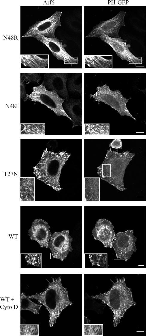
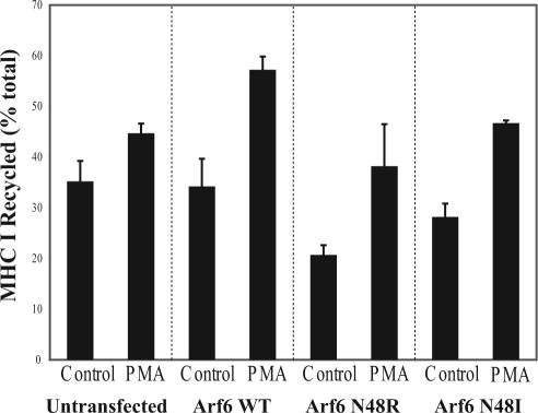
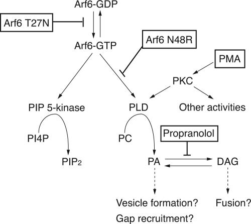
In this study, we investigated the role of phospholipase D (PLD) in mediating Arf6 function in cells. Expression of Arf6 mutants that are defective in activating PLD, Arf6N48R and Arf6N48I, inhibited membrane recycling to the plasma membrane (PM), resulting in an accumulation of tubular endosomal membranes. Additionally, unlike wild-type Arf6, neither Arf6 mutant could generate protrusions or recruit the Arf6 GTPase activating protein (GAP) ACAP1 onto the endosome in the presence of aluminum fluoride. Remarkably, all of these phenotypes, including accumulated tubular endosomes, blocked recycling, and failure to make protrusions and recruit ACAP effectively, could be recreated in either untransfected cells or cells expressing wild-type Arf6 by treatment with 1-butanol to inhibit the formation of phosphatidic acid (PA), the product of PLD. Moreover, most of the defects present in cells expressing Arf6N48R or N48I could be reversed by treatment with agents expected to elevate PA levels in cells. Together, these observations provide compelling evidence that Arf6 stimulation of PLD is required for endosomal membrane recycling and GAP recruitment.


Figures

Figure 1.
Figure 1.
Cells expressing Arf6N48R or N48I or treatment of cells expressing wild-type Arf6 with 1-butanol accumulate tubular endosomes. HeLa cells were transfected with Arf6N48R (A), Arf6N48I (B), or Arf6 (C, D, and F). Some Arf6-expressing cells were incubated with 100 nM cytochalasin D (D), 0.3% 1-butanol (F), or 2-butanol for 30 min. The cells were then fixed and immunolabeled with antibodies to Arf6 and MHCI. The juxtanuclear labeling of MHCI visible in a number of cells represents biosynthetic MHCI in the Golgi complex. Insets show colocalization of MHCI with Arf6. (E) Quantification of cells displaying tubular endosomal phenotype as a percentage of 100 transfected cells counted per experiment. Shown are the mean values with standard deviations from three experiments. Bars, 10 μm.
Figure 2.
Figure 2.
Expression of Arf6N48R or treatment of cells with 1-butanol, but not 2-butanol, inhibits MHCI recycling to the plasma membrane. (A) HeLa cells were transfected with Arf6 or Arf6N48R. Cells were loaded with antibody to MHCI for 30 min according to protocol described in Materials and Methods. Recycling of MHCI was measured by comparing the emptying of the MHCI internal pool within cells after a 30-min chase. Arf6-overexpressing cells were used as the standard against which the mutants were compared and set with an arbitrary value of 100%. (B) The presence of 0.3% 1-butanol during the last 10 min of antibody internalization and subsequent 30-min recycling inhibited MHCI antibody recycling, measured by internal pool emptying. Shown are means and SD from three independent experiments.
Figure 3.
Figure 3.
Tubular endosomes that accumulate in cells expressing Arf6N48R and N48I contain PIP2. HeLa cells were cotransfected with Arf6N48R, Arf6N48I, Arf6T27N, or wild-type Arf6 and PH-GFP for 20 h. Some cells expressing WT Arf6 were treated with cytochalasin D (Cyto D) (200 nM) for 30 min and then fixed, and Arf6 was detected with an antibody. Insets reveal Arf6 tubular endosomes. Bars, 10 μm.
Figure 4.
Figure 4.
Accumulation of tubular endosomes in Arf6N48R- and N48I-expressing cells is rescued by PMA and propranolol treatments. (A) HeLa cells transfected with either Arf6N48R or N48I were treated with 200 nM PMA for 30 min. Some treated cells were then fixed and stained for Arf6, whereas others were washed with media and allowed to recover in media for 90 min before fixation. (B) Quantification of cells displaying a tubular phenotype as a percentage of transfected cells counted. Data collected from 100 cells from at least two experiments. (C) Tubular endosome accumulations of Arf6N48R and N48I are rescued by propranolol treatment. HeLa cells transfected with Arf6N48R or N48I were treated with 0.1 mM propranolol for 30 min. Some cells were fixed, whereas others were allowed to recover by washout with media for 90 min before fixing. (D) Quantification of cells displaying tubular endosomes as a percentage of 100 transfected cells counted from at least two experiments. Mean and SE from two independent experiments. Bars, 10 μm.
Figure 5.
Figure 5.
PMA treatment rescues inhibition of MHCI recycling in Arf6N48R- and N48I-expressing cells. Untransfected cells or cells expressing Arf6, Arf6N48R, or Arf6N48I were loaded with antibody to MHCI for 30 min. Cells were treated with 200 nM PMA during the 30-min recycling phase. The surface MHCI antibody was stripped, and the extent of MHCI recycling was determined as described in Materials and Methods. The fraction of internalized MHCI that was recycled at 30 min is shown for two independent experiments (mean ± SE).
Figure 6.
Figure 6.
GAP recruitment is defective in cells expressing Arf6N48R and N48I, and in cells treated with 1-butanol, and is rescued with propranolol treatment. HeLa cells that were cotransfected with Arf6 (A), Arf6N48R (B), or Arf6N48I (B) and ACAP1 were treated with 30 mM NaF and 50 μM AlCl3 for 30 min and then fixed and probed with antibodies to Arf6 and FLAG epitope to detect ACAP1. Some cells were pretreated with 0.1 mM propranolol for 10 min, 0.3% 1-butanol, or 0.3% 2-butanol before AlF was added for 30 min; the cells were then fixed and stained described in text. (C) Quantification of ACAP1 recruitment to membranes in 100 transfected cells from at least two separate experiments. Bars, 10 μm.
Figure 7.
Figure 7.
Scheme for Arf6 activation of PIP 5-kinase and PLD. Arf6-GTP can activate PIP 5-kinase and PLD leading to the generation of PIP2 and PA, respectively. Arf6T27N inhibits activation of Arf6 and Arf6N48R inhibits activation of PLD but not of PIP 5-kinase. PMA-induced PKC stimulation of PLD, and propranolol inhibition of PA phosphohydrolase lead to alternative means for generation of PA, although PKC may have other activities.

Similar articles

Cited by

References

    1. Aikawa, Y., and Martin, T.F.J. (2003). ARF6 regulates a plasma membrane pool of phosphatidylinositol(4,5)bisphosphate required for regulated exocytosis. J. Cell Biol. 162, 647-659. - PMC - PubMed
    1. Brown, F. D., Rozelle, A. L., Yin, H. L., Balla, T., and Donaldson, J. G. (2001). Phosphatidylinositol 4,5-bisphosphate and Arf6-regulated membrane traffic. J. Cell Biol. 154, 1007-1017. - PMC - PubMed
    1. Brown, F. D., Thompson, N., Saqib, K. M., Clark, J. M., Powner, D., Thompson, N. T., Solari, R., and Wakelam, M. J. (1998). Phospholipase D1 localises to secretory granules and lysosomes and is plasma-membrane translocated on cellular stimulation. Curr. Biol. 8, 835-838. - PubMed
    1. Brown, H. A., Gutowski, S., Moomaw, C. R., Slaughter, C., and Sternweis, P. C. (1993). ADP-ribosylation factor, a small GTP-dependent regulatory protein, stimulates phospholipase D activity. Cell 75, 1137-1144. - PubMed
    1. Caumont, A. S., Galas, M. C., Vitale, N., Aunis, D., and Bader, M. F. (1998). Regulated exocytosis in chromaffin cells. Translocation of ARF6 stimulates a plasma membrane-associated phospholipase D. J. Biol. Chem. 273, 1373-1379. - PubMed

LinkOut - more resources