Comparative Study
. 2005 Nov 9;25(45):10479-93.
doi: 10.1523/JNEUROSCI.2103-05.2005.

Src-family kinases stabilize the neuromuscular synapse in vivo via protein interactions, phosphorylation, and cytoskeletal linkage of acetylcholine receptors

Affiliations
Comparative Study

Src-family kinases stabilize the neuromuscular synapse in vivo via protein interactions, phosphorylation, and cytoskeletal linkage of acetylcholine receptors

Gayathri Sadasivam et al. J Neurosci. .

Abstract

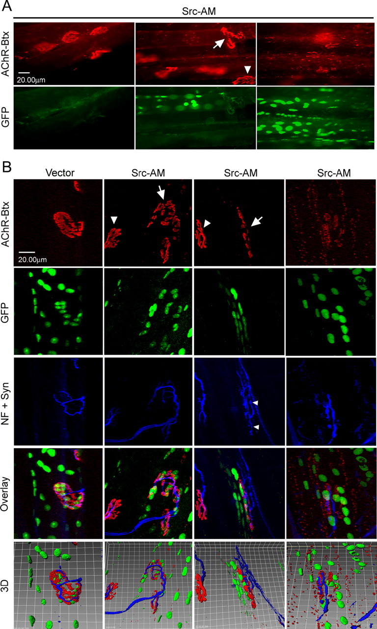
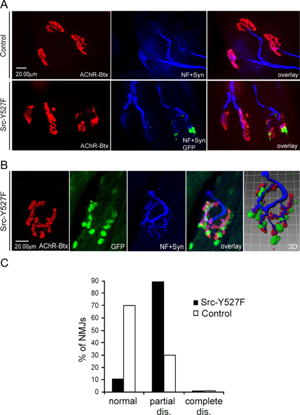
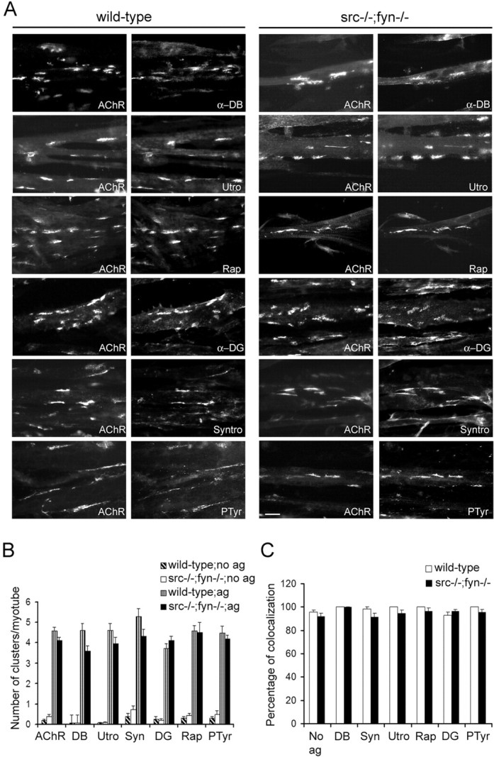
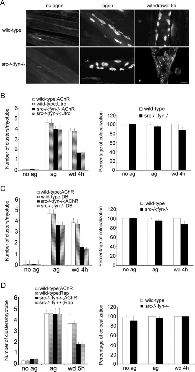
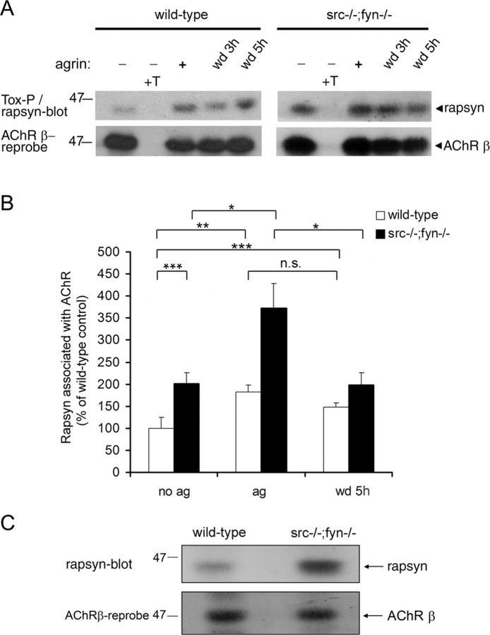
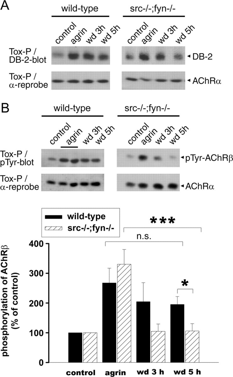
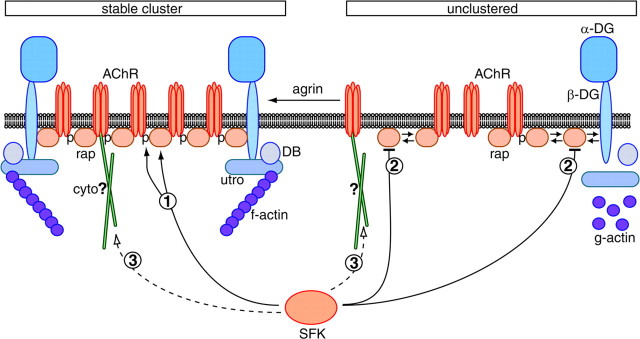
Postnatal stabilization and maturation of the postsynaptic membrane are important for development and function of the neuromuscular junction (NMJ), but the underlying mechanisms remain poorly characterized. We examined the role of Src-family kinases (SFKs) in vivo. Electroporation of kinase-inactive Src constructs into soleus muscles of adult mice caused NMJ disassembly: acetylcholine receptor (AChR)-rich areas became fragmented; the topology of nerve terminal, AChRs, and synaptic nuclei was disturbed; and occasionally nerves started to sprout. Electroporation of kinase-overactive Src produced similar but milder effects. We studied the mechanism of SFK action using cultured src(-/-);fyn(-/-) myotubes, focusing on clustering of postsynaptic proteins, their interaction with AChRs, and AChR phosphorylation. Rapsyn and the utrophin-glycoprotein complex were recruited normally into AChR-containing clusters by agrin in src(-/-);fyn(-/-) myotubes. But after agrin withdrawal, clusters of these proteins disappeared rapidly in parallel with AChRs, revealing that SFKs are of general importance in postsynaptic stability. At the same time, AChR interaction with rapsyn and dystrobrevin and AChR phosphorylation decreased after agrin withdrawal from mutant myotubes. Unexpectedly, levels of rapsyn protein were increased in src(-/-);fyn(-/-) myotubes, whereas rapsyn-cytoskeleton interactions were unaffected. The overall cytoskeletal link of AChRs was weak but still strengthened by agrin in mutant cells, consistent with the normal formation but decreased stability of AChR clusters. These data show that correctly balanced activity of SFKs is critical in maintaining adult NMJs in vivo. SFKs hold the postsynaptic apparatus together through stabilization of AChR-rapsyn interaction and AChR phosphorylation. In addition, SFKs control rapsyn levels and AChR-cytoskeletal linkage.


Figures

Figure 1.
Figure 1.
Electroporation of kinase-inactive Src-AM into soleus muscle leads to disassembly of NMJs. Muscles were electroporated in vivo with a mixture of Src-AM and NLS-GFP or empty control vector and NLS-GFP. Six weeks later, muscles were dissected and whole mounts of fibers were stained with α-BT-rhodamine and a mixture of neurofilament (NF) and synaptophysin (Syn) antibodies (blue). A, Conventional microscopy shows that in GFP-positive fibers, NMJs are partially (middle, arrow) or completely (right) disassembled, whereas GFP-negative fibers show intact NMJs (left, arrowhead in middle). B, Confocal microscopy with 3D reconstruction to visualize the degree of NMJ disassembly and the topology of nerve, AChRs, and synaptic myonuclei. Control vector electroporation leaves NMJs intact (left), whereas Src-AM electroporation disassembles NMJs in GFP-positive fibers (arrows, middle columns) but not GFP-negative fibers (arrowheads, middle columns). Disassembly ranges from partial (middle columns) to complete (right). Occasionally, nerves of disassembled NMJs show sprouting as indicated by the small arrowheads. Clusters of synaptic nuclei are less dense in disassembled NMJs, and the topology of nerve versus AChRs is disturbed.
Figure 2.
Figure 2.
Three-dimensional rotation and quantitation of intact and disassembled endplates of muscle fibers expressing control plasmid or kinase-inactive Src. A-J, 3D reconstructions as shown in Figure 1 B were rotated around the x-axis to illustrate the relative positioning of nerve, AChR clusters, and GFP-stained nuclei. K, L, In experiments as described in Figure 1, the degree of NMJ disassembly was scored as detailed in Materials and Methods. Control fibers score mostly as intact endplates with some partial and no complete disassembly (dis.). Fibers expressing Src-AM (K) or Src-K295M (L) show a high percentage of complete and partial disassembly compared with control. Disassembly is quantified as a percentage of all endplates analyzed within the control, Src-AM, and Src-K295M groups. Fifty endplates were analyzed for each group.
Figure 3.
Figure 3.
Electroporation of constitutively active Src-Y527F leads to partial disassembly of NMJs in vivo. Experiments were performed as for Figures 1 and 2. A, Conventional microscopy shows that after Src-Y527F electroporation, NMJs disassemble partially in GFP-positive fibers but not in GFP-negative fibers or in control muscles that were not electroporated. B, Confocal analysis illustrates the partial disassembly of an endplate, displaying large holes between AChR pretzel fragments. Nerves and synaptic nuclei are arranged accordingly. C, Quantitation of 30 Src-Y527F and 30 control situations shows that Src-Y527F expression increases partial disassembly (dis.) without leading to complete disassembly. NF, Neurofilament; Syn, synaptophysin; Btx, α-bungarotoxin.
Figure 4.
Figure 4.
Electroporation of Src-AM causes specific cytoskeletal changes at the postsynapse and does not induce muscle degeneration/regeneration. A, B, Muscles were electroporated with Src-AM and NLS-GFP, and whole mounts were stained with rhodamine-α-BT and Alexa 350-coupled phalloidin (to visualize actin filaments) (A; blue) or antibodies against α-tubulin (B; pink). The organization of F-actin in costameric structures along myofibers is not affected in Src-AM-expressing, GFP-positive fibers in which NMJs are disassembled (A). Subsynaptic α-tubulin is organized in ring-like structures at intact NMJs (B, arrows), and this arrangement is disturbed at disassembled NMJs in GFP-positive fibers. C, Muscles were electroporated with Src-AM and NLS-GFP or empty control vector and NLS-GFP. Muscles were first processed and fixed as for whole-mount analysis but were then embedded, cryosectioned, and stained with anti-GFP antibodies and DAPI as described in Materials and Methods. Muscle fibers are visible because of low-intensity diffuse GFP staining. In both Src-AM and vector samples, strong GFP signals are always at the fiber periphery and mostly colocalize with DAPI, identifying them as peripheral nuclei (arrows). Thus, Src-AM does not lead to centrally positioned nuclei, excluding the presence of myotubes and degeneration/regeneration. D, Samples as in C were additionally stained with rodamine-α-BT. Fibers lacking nuclear GFP-signal show intact AChR clusters (asterisks), whereas a fiber with disassembled AChR clusters (arrowhead) displays peripheral nuclei (arrows).
Figure 5.
Figure 5.
Agrin induces coclustering of postsynaptic proteins with AChRs in src-/-;fyn-/- myotubes.A, Cells were incubated with 0.5 nm agrin overnight and double labeled with rhodamine-α-BT (AChR) and antibodies recognizing α-dystrobrevin-1 (α-DB), utrophin (Utro), rapsyn (Rap),α-dystroglycan (α-DG), syntrophin isoforms (Syntro), or phosphotyrosine (PTyr), followed by FITC-coupled secondary antibodies. Wild-type and src-/-;fyn-/- myotubes show identical coclustering of AChRs with the respective postsynaptic proteins. Scale bar, 10 μm. B, Clusters of AChRs and postsynaptic proteins were counted in agrin-treated (ag) and untreated (no ag) cells. C, Protein colocalization is expressed as a percentage of AChR clusters that contain the respective postsynaptic protein in agrin-treated cells. “No ag” shows the average of colocalization of each marker with AChRs in non-agrin-treated cells. Values are mean ± SEM, from 20 pictures for each marker and condition. Error bars represent SEM.
Figure 6.
Figure 6.
Src and Fyn are required to stabilize postsynaptic protein clusters along with AChR clusters. Cells were treated overnight with 0.5 nm agrin to induce protein clustering. For studying cluster maintenance, agrin was then withdrawn from cultures for 4-5 h (wd 4 h, wd 5 h). A, Cells were labeled with rhodamine-α-BT (AChR), showing that 5 h of withdrawal causes pronounced AChR cluster disassembly in src-/-;fyn-/- but not wild-type myotubes. Cells were double labeled with rhodamine-α-BT (AChR) and antibodies recognizing utrophin (utro) (B), α-dystrobrevin-1 (DB) (C), or rapsyn (Rap) (D), followed by FITC-coupled secondary antibodies. Illustrative pictures of clusters and their disassembly are shown in supplemental Figure 1 (available at www.jneurosci.org as supplemental material). Quantitation (left) shows that clusters of these postsynaptic proteins disappear in parallel with AChRs after agrin withdrawal. Colocalization (right) indicates the percentage of AChR clusters that contain the respective postsynaptic protein, in the case of agrin withdrawal for remaining AChR clusters. Values are mean ± SEM, from 20 pictures for each marker and condition. Error bars represent SEM. ag, Agrin treated; no ag, untreated.
Figure 7.
Figure 7.
SFKs maintain AChR-rapsyn interaction and negatively regulate rapsyn protein levels. A, Cells were treated overnight with 0.5 nm agrin followed by withdrawal (wd) as indicated. AChRs were precipitated from lysates using α-BT-biotin and streptavidin-agarose (Tox-P), and associated rapsyn was detected by Western blotting. As control, an excess (10 μm) of free soluble toxin (+T) was added to some lysates. In the bottom part, the blots were stripped and reprobed for the AChR β subunits with monoclonal antibody (mAb) 124. B, Experiments were quantitated by densitometric scanning. Rapsyn signals were divided through AChR β signals to ensure equal loading. The graph indicates the percentage of rapsyn associated with AChR, with wild-type cells not treated with agrin set to 100%. Untreated src-/-;fyn-/- myotubes show an increased rapsyn association (approximately twofold) with the AChR. Agrin further increases this interaction approximately twofold, similarly to wild-type cells. In src-/-;fyn-/- myotubes, the agrin-induced increase in rapsyn-AChR interaction decreases after agrin withdrawal but remains more stable in the wild type. Data represent mean ± SEM of at least five experiments. *p < 0.05, **p < 0.01, and ***p < 0.001; unpaired Student's t tests (n.s., not significant). C, Protein-matched aliquots of wild-type and src-/-;fyn-/- myotube lysates were analyzed by immunoblotting using antibodies against rapsyn (Rap-1) and AChR β subunits (mAb 124). The level of rapsyn protein is higher in mutant cells.
Figure 8.
Figure 8.
In src-/-;fyn-/- myotubes, AChR-dystrobrevin association and phosphorylation of AChR β subunits are unstable after agrin withdrawal. Agrin was added overnight, and cells were washed and incubated in withdrawal medium (wd) lacking agrin for 3 or 5 h. AChRs were precipitated with α-BT and subjected to immunoblotting using antibodies against α-dystrobrevin-2 (DB-2) (A) or against phosphotyrosine (B). Blots were stripped and reprobed for AChR α subunits as control. A, Association of AChRs with α-dystrobrevin is increased by agrin in both wild-type and mutant cells. After 5 h of agrin withdrawal, the association is much weaker in the mutant than in the wild type. B, In mutant cells, phosphorylation of AChR β subunits [detected based on molecular weight and comparison with parallel AChR β immunoblots (data not shown)] is normally induced by agrin but rapidly decreases after withdrawal. Quantitation of the phospho-AChR β signal by densitometric scanning, normalized for AChR α, shows a significant decrease in the mutant but not in the wild type after agrin withdrawal (mean ± SEM from at least five experiments; ***p = 0.003; *p = 0.018; n.s., not significant; unpaired Student's t tests).
Figure 9.
Figure 9.
The overall basal cytoskeletal link of AChRs is weakened in src-/-;fyn-/- myotubes but is still strengthened by agrin treatment. Wild-type (A) or src-/-;fyn-/- (B) myotubes were subjected to a first extraction in a buffer containing a low-detergent concentration, ranging from 0.03 to 0.09% Triton X-100 as indicated. Insoluble materials (pellets) were subjected to a second extraction, using 1% Triton X-100. AChRs were precipitated from the soluble low- and high-detergent fractions using α-BT and visualized by anti-AChR α subunit-antibodies in immunoblots. AChR extraction was quantified by densitometric scanning, and AChR signals are shown as a percentage of the sum of both extractions (low plus high detergent). Values are mean ± SEM from at least five experiments. For wild-type cells, 0.06% detergent extracts approximately one-half of the total AChRs, whereas for src-/-;fyn-/- myotubes, only 0.04% of detergent is required to achieve the same, indicating that, in the mutant, the AChR extractability is higher and thus the cytoskeletal link is weaker. C, Mutant cells were incubated overnight with agrin to induce AChR cluster formation. AChRs were first extracted with 0.05% and then with 1% detergent as described above and visualized by α-BT precipitation and AChR α immunoblotting. Agrin causes decreased AChR extractability, indicating stronger cytoskeletal linkage, because less AChRs are found in the first extraction and more in the second. D, Quantitation of experiments as in C shows a significant agrin-induced decrease of AChRs in the first extraction and a significant increase in the second extraction. Values are mean ± SEM from five experiments. *p = 0.03; unpaired Student's t tests. E, Sequential extraction as in C was performed for wild-type and C2C12 myotubes, using 0.06% Triton X-100 in the first extraction and 1% in the second. The percentage of decrease of AChRs in the first extraction, induced by agrin, was quantitated from five experiments and is the same, ∼30 - 40%, as for src-/-;fyn-/- myotubes. Thus, although the overall cytoskeletal link is weaker in src-/-;fyn-/- myotubes, the agrin-induced strengthening of this link is comparable with wild-type and C2C12 cells. Error bars represent SEM.
Figure 10.
Figure 10.
Model of SFK action in stabilization of the postsynaptic apparatus. Unclustered proteins such as rapsyn (rap) are in equilibrium between free and complexed form, indicated by double arrows. Agrin increases AChR-protein interactions in the process of clustering. SFKs act in postsynaptic stabilization by maintaining the AChR-rapsyn interaction (pathway 1). Because rapsyn and AChRs are the most abundant postsynaptic proteins, their interaction plays a core role and holds the postsynaptic apparatus together. This may occur through AChR β phosphorylation (p), which is maintained by SFKs (pathway 1). SFKs also negatively control the overall amount of rapsyn protein (pathway 2). In the absence of Src and Fyn, rapsyn amounts are high and may start saturating binding sites on β-dystroglycan (β-DG) and the AChR, although AChR-dystrobrevin interactions appear normal. SFKs may control another cytoskeletal link (cyto) of the AChR, independent of rapsyn as a linker (hypothetical pathway 3). This would explain the observed weak overall AChR-cytoskeletal linkage in src-/-;fyn-/- myotubes, which is still strengthened after agrin treatment resulting from AChR-UGC association. utro, Utrophin; DB, α-dystrobrevin.

Similar articles

Cited by

References

    1. Apel ED, Merlie JP (1995) Assembly of the postsynaptic apparatus. Curr Opin Neurobiol 5: 62-67. - PubMed
    1. Apel ED, Roberds SL, Campbell KP, Merlie JP (1995) Rapsyn may function as a link between the acetylcholine receptor and the agrin-binding dystrophin-associated glycoprotein complex. Neuron 15: 115-126. - PubMed
    1. Bartoli M, Ramarao MK, Cohen JB (2001) Interactions of the rapsyn RING-H2 domain with dystroglycan. J Biol Chem 276: 24911-24917. - PubMed
    1. Bezakova G, Ruegg MA (2003) New insights into the roles of agrin. Nat Rev Mol Cell Biol 4: 295-308. - PubMed
    1. Borges LS, Ferns M (2001) Agrin-induced phosphorylation of the acetylcholine receptor regulates cytoskeletal anchoring and clustering. J Cell Biol 153: 1-12. - PMC - PubMed

Publication types

MeSH terms

LinkOut - more resources