. 2005 Dec 7;24(23):4007-17.
doi: 10.1038/sj.emboj.7600861. Epub 2005 Nov 10.

Structural basis for UTP specificity of RNA editing TUTases from Trypanosoma brucei

Affiliations

Structural basis for UTP specificity of RNA editing TUTases from Trypanosoma brucei

Junpeng Deng et al. EMBO J. .

Abstract

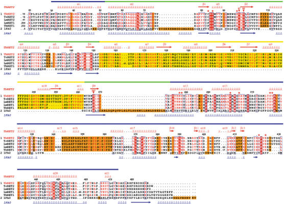
Trypanosomatids are pathogenic protozoa that undergo a unique form of post-transcriptional RNA editing that inserts or deletes uridine nucleotides in many mitochondrial pre-mRNAs. Editing is catalyzed by a large multiprotein complex, the editosome. A key editosome enzyme, RNA editing terminal uridylyl transferase 2 (TUTase 2; RET2) catalyzes the uridylate addition reaction. Here, we report the 1.8 A crystal structure of the Trypanosoma brucei RET2 apoenzyme and its complexes with uridine nucleotides. This structure reveals that the specificity of the TUTase for UTP is determined by a crucial water molecule that is exquisitely positioned by the conserved carboxylates D421 and E424 to sense a hydrogen atom on the N3 position of the uridine base. The three-domain structure also unveils a unique domain arrangement not seen before in the nucleotidyltansferase superfamily, with a large domain insertion between the catalytic aspartates. This insertion is present in all trypanosomatid TUTases. We also show that TbRET2 is essential for survival of the bloodstream form of the parasite and therefore is a potential target for drug therapy.


Figures

Figure 1
Figure 1
Multiple sequence alignment of selected terminal uridylyl transferases. Depicted are RET2 and RET1 from T. brucei, L. major, RET2 from T. cruzi and poly(A) polymerase from Saccharomyces cerevisiae (PDB code: 1fa0). The red secondary structure symbols on top represent those observed in the TbRET2 structure. Those at the bottom, in blue, are from the poly(A) polymerase structure. The NTD, MD and CTD are shown as green, yellow and blue bars above the secondary structure elements, respectively. The unique MD insertion in trypanosomatid TUTases is indicated by a yellow-shaded box. The residues involved in UTP binding at site A are shown with red asterisks. The three conserved aspartates, D97, D99 and D267, are indicated by red inverted triangles and labeled. The figure was generated by the program ESPript (Gouet et al, 1999).
Figure 2
Figure 2
TbRET2 expression is essential for the growth of bloodstream form trypanosomes. (A) TbRET2 conditional knockout cells were generated in which both endogenous alleles were replaced in the presence of a tetracycline-regulated ecotopic RET2 allele (see Materials and methods for details). Duplicate cultures of cells were grown in the presence (RET2 expressed, filled line) or absence (RET2 repressed, dashed line) of tetracycline. The cumulative cell number was calculated by multiplying the number of cells by the dilution. Inset: Western blot of glycerol gradient purified lysates of cells in which TbRET2 was expressed (E) or repressed for 3 days (R) using TbRET2 antiserum. TbRET2 is indicated by the arrow. The upper band is a protein from the whole-cell lysates that crossreacts with the polyclonal antiserum. (B) Recombinant TbRET2 protein activity. Wild-type (WT) and triple mutant (W94R, W1222R and W207R) recombinant RET2 proteins were assayed for nucleotide addition to a 5′-labeled ssRNA (left panel) or a precleaved editing dsRNA substrate (right panel). Diagrams of the RNA substrates are shown and the asterisk denotes the radiolabel. Each assay contained 25 ng protein and 100 μM of the indicated nucleotide. Labeled input RNA is indicated by (I) and input RNA with 1, 2, 3 or 5 added Us are indicated by +1U, +2U, +3U and +5U, respectively.
Figure 3
Figure 3
Overall structure of TbRET2. (A) Ribbon presentation of the molecule. The three domains are shown in green (NTD), yellow (MD) and blue (CTD) (as in Figure 1). The UTP-Mg2+ at site A is shown as ball-and-stick in a cleft between the NTD and the CTD. (BD) Electrostatic potential surface of TbRET2. The positive and negative regions are colored in blue and red, respectively. Nucleotide-binding sites are labeled in yellow. UTP/UMP and Mg2+ are shown as ball-and-stick. Notice the extended blue patch across three domains that suggests an RNA binding region. (B) Front view, UTP-binding site A with a UTP and nucleotide-binding site B with a UMP molecule; (C) side view (90° upward rotation), nucleotide-binding site B with a UMP molecule; (D) back view (180° upward rotation), nucleotide-binding site C with a UMP molecule. Figure generated by PYMOL (DeLano, 2002).
Figure 4
Figure 4
Stereoview of the UTP-binding site A. (A) The UTP-Mg2+ as well as the essential interacting residues are shown as ball-and-stick. The U-specifying Wat1 molecule is shown as a white sphere. The hydrogen bonds are shown as red dashed lines. The secondary structure elements are colored with the same scheme as in Figure 1. (B) 2 mFoDFc electron density map covering the UTP molecule and an Mn2+ ion. The anomalous Fourier map of Mn2+ is shown in red at a 7σ contour level.
Figure 5
Figure 5
UTP-specificity and UMP binding. (A) Interactions of the UTP sugar moiety at site A with the 2′-OH and the side chains of N277 and S278. (B) The uridine base is specifically recognized by Wat1 and D421 and E424. The two hydrogen atoms of Wat1 shown as white spheres makes exquisite hydrogen bonds with Oδδ1 of D421 and Oɛ1 of E424. The latter two residues are conserved among all trypanosomatid TUTases (Figure 1). (C) UMP binding at site B; (D) UMP binding at site C, which is located far from the active site.
Figure 6
Figure 6
TbRET2 in complex with NTPs. (A) 1 mM UTP, 10 mM Mg2+, 5 min soaking time, 2.2 Å resolution. (B) 10 mM CTP, 10 mM Mg2+, 30 min soaking time, 2.5 Å resolution. (C) 10 mM ATP, 10 mM Mg2+, 30 min soaking time, 3.2 Å resolution. (D) 10 mM GTP, 10 mM Mg2+, 30 min soaking time, 3.2 Å resolution. Nonmodel-biased Fourier difference electron density maps are shown in red at a 3.0σ contour level. The positioned NTP models shown, except for UTP, are not refined.
Figure 7
Figure 7
Superimposition of the structures of TbRET2 and poly(A) polymerase (PAP) from Saccharomyces cerevisiae (Bard et al, 2000). (A) Side view. The PAP structure is shown in magenta, the TbRET2 structure is colored according to the same scheme as in Figure 1. Note that, despite a similar topology (see text), the MD of TbRET2 (in orange dashed circle) displays an entirely different position compared to the C-terminal domain of PAP (in magenta dashed circle), with respect to the equivalent catalytic bidomains that are common to both enzymes. (B) Top view of the catalytic bidomain. The UTP at site A in the TbRET2 structure is shown in cyan ball-and-stick, while the ATP in the PAP structure is shown in red. The UTP and ATP occupy similar positions, particularly for the triphosphate moieties.

Similar articles

Cited by

References

    1. Alfano C, Sanfelice D, Babon J, Kelly G, Jacks A, Curry S, Conte M (2004) Structural analysis of cooperative RNA binding by the La motif and central RRM domain of human La protein. Nat Struct Mol Biol 11: 323–329 - PubMed
    1. Aphasizhev R, Aphasizheva I, Nelson RE, Gao G, Simpson AM, Kang X, Falick AM, Sbicego S, Simpson L (2003a) Isolation of a U-insertion/deletion editing complex from Leishmania tarentolae mitochondria. EMBO J 22: 913–924 - PMC - PubMed
    1. Aphasizhev R, Aphasizheva I, Simpson L (2003b) A tale of two TUTases. Proc Natl Acad Sci USA 100: 10617–10622 - PMC - PubMed
    1. Aphasizhev R, Aphasizheva I, Simpson L (2004) Multiple terminal uridylyltransferases of trypanosomes. FEBS Lett 572: 15–18 - PubMed
    1. Aphasizhev R, Sbicego S, Peris M, Jang S, Aphasizheva I, Simpson A, Rivlin A, Simpson L (2002) Trypanosome mitochondrial 3′ terminal uridylyl transferase (TUTase): the key enzyme in U-insertion/deletion RNA editing. Cell 108: 637–648 - PubMed

Publication types

MeSH terms