Vowel sound extraction in anterior superior temporal cortex
- PMID: 16281283
- PMCID: PMC6871493
- DOI: 10.1002/hbm.20201
Vowel sound extraction in anterior superior temporal cortex
Abstract
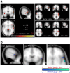
We investigated the functional neuroanatomy of vowel processing. We compared attentive auditory perception of natural German vowels to perception of nonspeech band-passed noise stimuli using functional magnetic resonance imaging (fMRI). More specifically, the mapping in auditory cortex of first and second formants was considered, which spectrally characterize vowels and are linked closely to phonological features. Multiple exemplars of natural German vowels were presented in sequences alternating either mainly along the first formant (e.g., [u]-[o], [i]-[e]) or along the second formant (e.g., [u]-[i], [o]-[e]). In fixed-effects and random-effects analyses, vowel sequences elicited more activation than did nonspeech noise in the anterior superior temporal cortex (aST) bilaterally. Partial segregation of different vowel categories was observed within the activated regions, suggestive of a speech sound mapping across the cortical surface. Our results add to the growing evidence that speech sounds, as one of the behaviorally most relevant classes of auditory objects, are analyzed and categorized in aST. These findings also support the notion of an auditory "what" stream, with highly object-specialized areas anterior to primary auditory cortex.
2005 Wiley-Liss, Inc.
Figures


Similar articles
-
Locating the initial stages of speech-sound processing in human temporal cortex.Neuroimage. 2006 Jul 1;31(3):1284-96. doi: 10.1016/j.neuroimage.2006.01.004. Epub 2006 Feb 28. Neuroimage. 2006. PMID: 16504540
-
Evidence for rapid auditory perception as the foundation of speech processing: a sparse temporal sampling fMRI study.Eur J Neurosci. 2004 Nov;20(9):2447-56. doi: 10.1111/j.1460-9568.2004.03687.x. Eur J Neurosci. 2004. PMID: 15525285
-
Illusory vowels resulting from perceptual continuity: a functional magnetic resonance imaging study.J Cogn Neurosci. 2008 Oct;20(10):1737-52. doi: 10.1162/jocn.2008.20069. J Cogn Neurosci. 2008. PMID: 18211243
-
[Auditory perception and language: functional imaging of speech sensitive auditory cortex].Rev Neurol (Paris). 2001 Sep;157(8-9 Pt 1):837-46. Rev Neurol (Paris). 2001. PMID: 11677406 Review. French.
-
Segmental processing in the human auditory dorsal stream.Brain Res. 2008 Jul 18;1220:179-90. doi: 10.1016/j.brainres.2007.11.013. Epub 2007 Nov 17. Brain Res. 2008. PMID: 18096139 Review.
Cited by
-
Speech-specific tuning of neurons in human superior temporal gyrus.Cereb Cortex. 2014 Oct;24(10):2679-93. doi: 10.1093/cercor/bht127. Epub 2013 May 16. Cereb Cortex. 2014. PMID: 23680841 Free PMC article.
-
Brain Activity Related to Sound Symbolism: Cross-modal Effect of an Aurally Presented Phoneme on Judgment of Size.Sci Rep. 2019 May 7;9(1):7017. doi: 10.1038/s41598-019-43457-3. Sci Rep. 2019. PMID: 31065027 Free PMC article.
-
Age-related effects on word recognition: reliance on cognitive control systems with structural declines in speech-responsive cortex.J Assoc Res Otolaryngol. 2008 Jun;9(2):252-9. doi: 10.1007/s10162-008-0113-3. Epub 2008 Feb 15. J Assoc Res Otolaryngol. 2008. PMID: 18274825 Free PMC article.
-
A review and synthesis of the first 20 years of PET and fMRI studies of heard speech, spoken language and reading.Neuroimage. 2012 Aug 15;62(2):816-47. doi: 10.1016/j.neuroimage.2012.04.062. Epub 2012 May 12. Neuroimage. 2012. PMID: 22584224 Free PMC article. Review.
-
Segregation of vowels and consonants in human auditory cortex: evidence for distributed hierarchical organization.Front Psychol. 2010 Dec 24;1:232. doi: 10.3389/fpsyg.2010.00232. eCollection 2010. Front Psychol. 2010. PMID: 21738513 Free PMC article.
References
-
- Arnott SR, Binns MA, Grady CL, Alain C (2004): Assessing the auditory dual‐pathway model in humans. Neuroimage 22: 401–408. - PubMed
-
- Binder JR, Frost JA, Hammeke TA, Bellgowan PS, Springer JA, Kaufman JN, Possing ET (2000): Human temporal lobe activation by speech and nonspeech sounds. Cereb Cortex 10: 512–528. - PubMed
-
- Binder JR, Liebenthal E, Possing ET, Medler DA, Ward BD (2004): Neural correlates of sensory and decision processes in auditory object identification. Nat Neurosci 7: 295–301. - PubMed
Publication types
MeSH terms
LinkOut - more resources
Full Text Sources

