

Visibility, visual awareness, and visual masking of simple unattended targets are confined to areas in the occipital cortex beyond human V1/V2
- PMID: 16282374
- PMCID: PMC1282175
- DOI: 10.1073/pnas.0508010102
Visibility, visual awareness, and visual masking of simple unattended targets are confined to areas in the occipital cortex beyond human V1/V2
Abstract
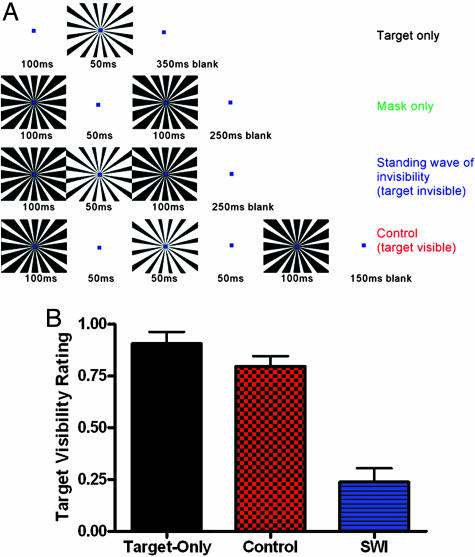
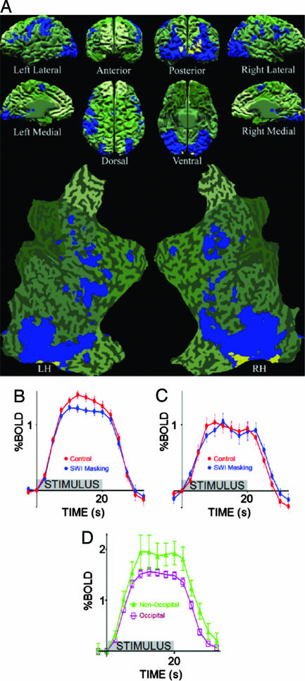
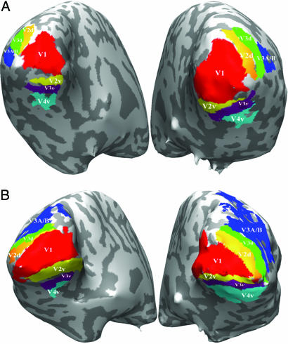
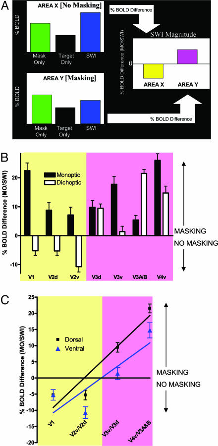
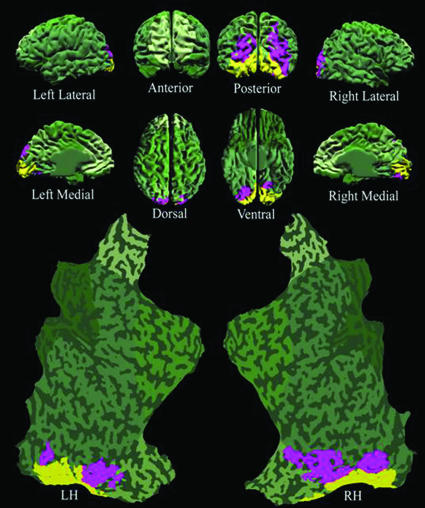
In visual masking, visible targets are rendered invisible by modifying the context in which they are presented, but not by modifying the targets themselves. Here, we localize the neuronal correlates of visual awareness in the human brain by using visual masking illusions. We compare monoptic visual masking activation, which we find within all retinotopic visual areas, with dichoptic masking activation, which we find only in those retinotopic areas downstream of V2. Because monoptic and dichoptic masking are equivalent in magnitude perceptually, the present results establish a lower bound for maintenance of visual awareness of simple unattended targets. Moreover, we find that awareness-correlated circuits for simple targets are restricted to the occipital lobe. This finding provides evidence of an upper boundary in the visual hierarchy for visual awareness of simple unattended targets, thus constraining the location of circuits that maintain the visibility of simple targets to occipital areas beyond V1/V2.
Figures
Similar articles
-
Visual masking approaches to visual awareness.Prog Brain Res. 2006;155:177-215. doi: 10.1016/S0079-6123(06)55011-3. Prog Brain Res. 2006. PMID: 17027388 Review.
-
Dichoptic visual masking reveals that early binocular neurons exhibit weak interocular suppression: implications for binocular vision and visual awareness.J Cogn Neurosci. 2004 Jul-Aug;16(6):1049-59. doi: 10.1162/0898929041502788. J Cogn Neurosci. 2004. PMID: 15298791
-
Regional brain activity associated with visual backward masking.J Cogn Neurosci. 2005 Jan;17(1):13-23. doi: 10.1162/0898929052880011. J Cogn Neurosci. 2005. PMID: 15701236
-
Dichoptic masking and binocular rivalry share common perceptual dynamics.J Vis. 2007 Nov 21;7(14):3.1-11. doi: 10.1167/7.14.3. J Vis. 2007. PMID: 18217798
-
Binocular battles on multiple fronts.Trends Cogn Sci. 2004 Apr;8(4):148-51. doi: 10.1016/j.tics.2004.02.009. Trends Cogn Sci. 2004. PMID: 15551483 Review.
Cited by
-
Does V1 response suppression initiate binocular rivalry?iScience. 2023 Jul 8;26(8):107359. doi: 10.1016/j.isci.2023.107359. eCollection 2023 Aug 18. iScience. 2023. PMID: 37520732 Free PMC article.
-
Subjective decision threshold for accurate visual detection performance in rats.Sci Rep. 2018 Jun 19;8(1):9357. doi: 10.1038/s41598-018-27696-4. Sci Rep. 2018. PMID: 29921866 Free PMC article.
-
Saccades Influence the Visibility of Targets in Rapid Stimulus Sequences: The Roles of Mislocalization, Retinal Distance and Remapping.Front Syst Neurosci. 2016 Jun 28;10:58. doi: 10.3389/fnsys.2016.00058. eCollection 2016. Front Syst Neurosci. 2016. PMID: 27445718 Free PMC article.
-
Ask, and You Shall Receive: A Closer Look on Unsolved Consciousness Issue.Basic Clin Neurosci. 2024 Sep-Oct;15(5):569-582. doi: 10.32598/bcn.2021.2308.1. Epub 2024 Sep 1. Basic Clin Neurosci. 2024. PMID: 40583879 Free PMC article. Review.
-
Blinded by the load: attention, awareness and the role of perceptual load.Philos Trans R Soc Lond B Biol Sci. 2014 Mar 17;369(1641):20130205. doi: 10.1098/rstb.2013.0205. Print 2014 May 5. Philos Trans R Soc Lond B Biol Sci. 2014. PMID: 24639578 Free PMC article.
References
-
- Crick, F. & Koch, C. (1990) Cold Spring Harbor Symp. Quant. Biol. 55, 953-962. - PubMed
-
- Milner, A. D. (1995) Neuropsychologia 33, 1117-1130. - PubMed
-
- He, S., Cavanagh, P. & Intriligator, J. (1996) Nature 383, 334-337. - PubMed
-
- Logothetis, N. K., Leopold, D. A. & Sheinberg, D. L. (1996) Nature 380, 621-624. - PubMed
Publication types
MeSH terms
Grants and funding
LinkOut - more resources
Full Text Sources
