. 2005 Dec;79(23):14668-79.
doi: 10.1128/JVI.79.23.14668-14679.2005.

Murine gammaherpesvirus 68 infection is associated with lymphoproliferative disease and lymphoma in BALB beta2 microglobulin-deficient mice

Affiliations

Murine gammaherpesvirus 68 infection is associated with lymphoproliferative disease and lymphoma in BALB beta2 microglobulin-deficient mice

Vera L Tarakanova et al. J Virol. 2005 Dec.

Abstract

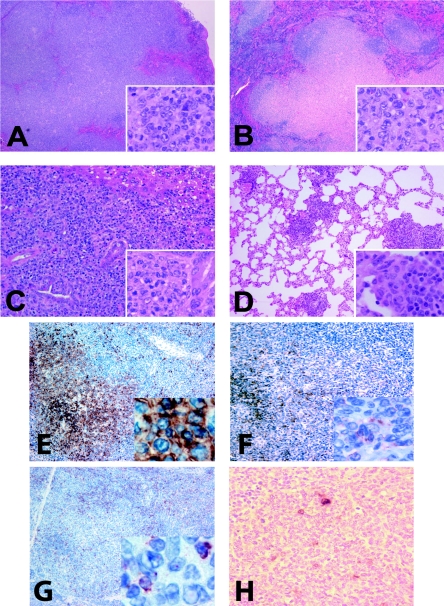
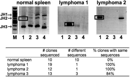
Human gammaherpesvirus infections are associated with development of lymphoproliferative disease. Understanding of the mechanisms of gammaherpesvirus lymphomagenesis during chronic infection in a natural host has been limited by the exquisite species specificity of human gammaherpesviruses and the expense of primates. Murine gammaherpesvirus gammaHV68 is genetically and biologically related to human gammaherpesviruses and herpesvirus saimiri and has been reported to be associated with lymphoproliferative disease in mice (N. P. Sunil-Chandra, J. Arno, J. Fazakerley, and A. A. Nash, Am. J. Pathol. 145:818-826, 1994). We report the development of an animal model of gammaHV68 lymphomagenesis in BALB/c beta2 microglobulin-deficient mice (BALB beta2m-/-). GammaHV68 infection induced two lymphoproliferative lesions: B-cell lymphoma and atypical lymphoid hyperplasia (ALH). ALH lesion histology resembled lesions of Epstein-Barr virus-associated posttransplant lymphoproliferative disease and was characterized by the abnormal infiltration of the white pulp with cells expressing the plasma cell marker CD138. Lymphomas observed in gammaHV68-infected animals were B220+/CD3- large-cell lymphomas. GammaHV68-infected cells were common in ALH lesions as measured by in situ hybridization with a probe specific for viral tRNAs (vtRNAs), but they were scarce in gammaHV68-infected spleens with normal histology. Unlike ALH lesions, gammaHV68 vtRNA-positive cells were rare in lymphomas. GammaHV68 infection of BALB beta2m-/- mice results in lymphoproliferation and lymphoma, providing a valuable tool for identifying viral and host genes involved in gammaherpesvirus-associated malignancies. Our findings suggest that gammaHV68 induces lymphomas via hit-and-run oncogenesis, paracrine effects, or stimulation of chronic inflammation.


Figures

FIG. 1.
FIG. 1.
γHV68 infection increases incidence of lymphoproliferative disease in BALB β2 microglobulin-deficient mice. Age- and sex-matched BALB β2m animals were mock infected or infected with γHV68 and monitored for the development of LPD. Total incidence of LPD (A) was subdivided into incidence of each of the two primary lesions (ALH and lymphoma) in male and female animals. (B and C) Incidence of LPD at indicated times postinfection in γHV68 (B)- and mock (C)-infected groups. n, number of mice.
FIG. 2.
FIG. 2.
Splenic histology of γHV68-infected BALB β2m−/− mice. H&E staining of formalin-fixed paraffin-embedded tissues. (A and B) Normal (×20). (C and D) Follicular hyperplasia (×20). Arrows point to germinal centers abundant throughout the section. (E and F) Atypical lymphoid hyperplasia (×10). (E) A hyperplastic lesion is outlined. (G and H) Lymphoma (×4).
FIG. 3.
FIG. 3.
Histopathology of atypical lymphoid hyperplasia. (A and B) ALH lesions in spleens (×4). The splenic architecture is partially disrupted with expanded white pulp areas. (C) Lung lymphocytic infiltrates seen in γHV68-infected animals with ALH lesions (×4). (Insets in panels A to C) Prominent plasmacytic features of cells in ALH lesions (×100). (D) The majority of the ALH-associated cells stain with the plasma cell marker CD138 (×10). (E) B220 is detected in residual B-cell follicles (×10). Adjacent spleen sections obtained from γHV68-infected BALB β2m−/− mice were stained with hematoxylin and eosin (F and H) or hybridized with antisense digoxigenin-labeled vtRNA probe (G, I, and J) and counterstained with hematoxylin. Sections originated from spleens exhibiting follicular hyperplasia (F and G) or atypical lymphoid hyperplasia (H, I, and J). Magnification: F to I, ×4; J, ×40.
FIG. 4.
FIG. 4.
Histopathology of splenic lymphomas. (A and B) Lymphoma-bearing spleens (×4). The general architecture of the spleen is disrupted by the massive expansion of the white pulp. (Insets in panels A and B) Large lymphoma cells with irregular nuclei with conspicuous nucleoli (×40). Frequent mitotic figures are present. (C) Metastasis to the liver (×10). Lymphoma cells are preferentially localized in the portal areas of the liver surrounding the bile ducts. (Inset) Tumor cells adjacent to a bile duct (×40). (D) Metastasis to the lung (×10). (E to G) HRP immunostaining with hematoxylin counterstain. Magnification at ×10 (insets at ×40). (E) Expression of B-cell marker B220. (F) Expression of the plasma cell marker CD138. (G) Expression of T-cell marker CD3. Peritumoral and tumor-infiltrating small lymphocytes but not lymphoma cells stain positive. (H) Lymphoma-bearing spleen section was hybridized with antisense digoxigenin-labeled vtRNA probe and counterstained with hematoxylin (×40).
FIG. 5.
FIG. 5.
Clonality assessment of γHV68-associated lymphomas. VH-to-DJH gene rearrangement was analyzed by PCR using forward primers specific for members of the Vh558 (lane 1), Vh7183 (lane 2), and VhQ52 (lane 3) families. DH-to-JH gene rearrangements were analyzed with the DhL primer (lane 4). A Jh3 reverse primer was used in all cases. PCR products were analyzed on a 1% agarose gel. M, 100-bp molecular size marker. Bands indicated by boxes were excised from the gel, reamplified using the same primers, cloned, and sequenced. The table shows the sequencing results.

References

    1. Ambinder, R. F. 2000. Gammaherpesviruses and “Hit-and-Run” oncogenesis. Am. J. Pathol. 156:1-3. - PMC - PubMed
    1. Bais, C., B. Santomasso, O. Coso, L. Arvanitakis, E. G. Raaka, J. S. Gutkind, A. S. Asch, E. Cesarman, M. C. Gershengorn, E. A. Mesri, and M. C. Gerhengorn. 1998. G-protein-coupled receptor of Kaposi's sarcoma-associated herpesvirus is a viral oncogene and angiogenesis activator Nature 391:86-89. (Erratum, 392: 6672.) - PubMed
    1. Blaskovic, D., M. Stancekova, J. Svobodova, and J. Mistrikova. 1980. Isolation of five strains of herpesviruses from two species of free living small rodents. Acta Virol. 24:468. - PubMed
    1. Bowden, R. J., J. P. Simas, A. J. Davis, and S. Efstathiou. 1997. Murine gammaherpesvirus 68 encodes tRNA-like sequences which are expressed during latency. J. Gen. Virol. 78:1675-1687. - PubMed
    1. Cardin, R. D., J. W. Brooks, S. R. Sarawar, and P. C. Doherty. 1996. Progressive loss of CD8+ T cell-mediated control of a gamma-herpesvirus in the absence of CD4+ T cells. J. Exp. Med. 184:863-871. - PMC - PubMed

Publication types

MeSH terms