. 2005 Dec;79(23):14708-18.
doi: 10.1128/JVI.79.23.14708-14718.2005.

The neuronal host cell factor-binding protein Zhangfei inhibits herpes simplex virus replication

Affiliations

The neuronal host cell factor-binding protein Zhangfei inhibits herpes simplex virus replication

Oksana Akhova et al. J Virol. 2005 Dec.

Abstract

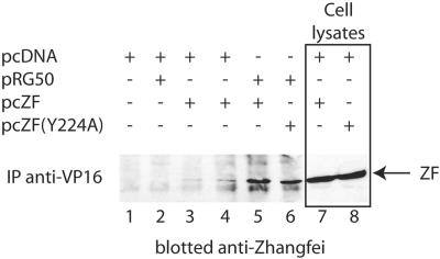
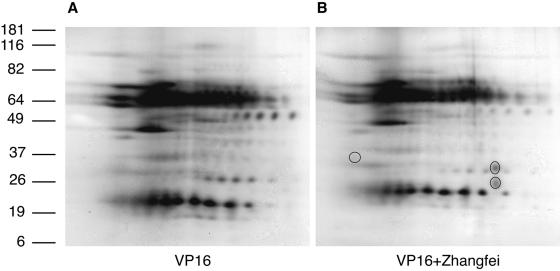
During lytic infection in epithelial cells the expression of herpes simplex virus type 1 (HSV-1) immediate-early (IE) genes is initiated by a multiprotein complex comprising the virion-associated protein VP16 and two cellular proteins, host cellular factor (HCF) and Oct-1. Oct-1 directly recognizes TAATGARAT elements in promoters of IE genes. The role of HCF is not clear. HSV-1 also infects sensory neurons innervating the site of productive infection and establishes a latent infection in these cells. It is likely that some VP16 is retained by the HSV-1 nucleocapsid as it reaches the neuronal nucleus. Its activity must therefore be suppressed for successful establishment of viral latency. Recently, we discovered an HCF-binding cellular protein called Zhangfei. Zhangfei, in an HCF-dependent manner, inhibits Luman/LZIP/CREB3, another cellular HCF-binding transcription factor. Here we show that Zhangfei is selectively expressed in human neurons. When delivered to cultured cells that do not normally express the protein, Zhangfei inhibited the ability of VP16 to activate HSV-1 IE expression. The inhibition was specific for HCF-dependent transcriptional activation by VP16, since a Gal4-VP16 chimeric protein was inhibited only on a TAATGARAT-containing promoter and not a on a Gal4-containing promoter. Zhangfei associated with VP16 and inhibited formation of the VP16-HCF-Oct-1 complex on TAATGARAT motifs. Zhangfei also suppressed HSV-1-induced expression of several cellular genes including topoisomerase IIalpha, suggesting that in addition to suppressing IE expression Zhangfei may have an inhibitory effect on HSV-1 DNA replication and late gene expression.


Figures

FIG. 1.
FIG. 1.
Zhangfei is expressed in neurons. Zhangfei was detected in human tissues using antiserum against Zhangfei (anti-ZF). Serum from which antibodies against Zhangfei had been removed (absorbed) was used as a negative control. (A) Lysates of cells transfected to express Zhangfei (lanes 1) or a negative control (lanes 2) were separated by SDS-polyacrylamide gel electrophoresis and immunoblotted with anti-ZF or absorbed serum. Sections of a parietal lobe (B) and sensory ganglion (C) stained with anti-ZF or absorbed serum are shown. Bar, 50 μm.
FIG. 2.
FIG. 2.
Zhangfei inhibits VP16 activation of the ICP0 promoter. (A) Vero cells were transfected with 100 ng of pAB5 (CAT reporter with ICP0 promoter) and with 50 ng of a plasmid expressing VP16 and increasing amounts (1 to 4 μg) of plasmids expressing Zhangfei (circles) or Zhangfei (Y224A) (squares). The results are expressed as a percentage of CAT activity in cells cotransfected with plasmid expressing VP16 and the CAT reporter pAB5. (B) Expression of Zhangfei was confirmed by immunoblotting using polyclonal serum against Zhangfei.
FIG. 3.
FIG. 3.
Zhangfei inhibits ICP0 transcript and protein expression and reduces the yield of progeny virus in HSV-1-infected cells. Hep2 cells were infected with adenovirus vectors expressing β-galactosidase (lanes and panel 1), Zhangfei (Y224A) (lanes and panel 2), or Zhangfei (lanes and panel 3). Twenty-four hours later the cells were also infected with HSV-1. (A) Northern blot analysis of total RNA, which was harvested from cells probed with radiolabeled ICP0 DNA. The lower panel shows the intensity of ethidium bromide-stained 18S and 28S rRNA bands and indicates that equal amounts of RNA were loaded for each sample. (B) Hep2 cells were stained for immunofluorescence using a monoclonal antibody specific to ICP0. Total cells as well as fluorescent cells in four 400× microscope fields were counted in four independent experiments. (C) Cells expressing ICP0 are shown as percentage of total cells. Bars represent standard deviations of means. (D) VP16, ICP0, and Zhangfei in the samples were detected by immunoblotting. (E) HSV-1 yields by cells expressing β-galactosidase, Zhangfei (Y224A), or Zhangfei 24 h after infection with HSV-1.
FIG. 4.
FIG. 4.
Zhangfei does not deprive VP16 of functional HCF. (A) Vero cells were transfected with 100 ng of pAB5 (CAT reporter with ICP0 promoter) (lanes 1 to 5) or 50 ng of a plasmid expressing VP16 (lanes 2 to 5) and cotransfected with plasmids expressing either LuS221Op (lane 3), LuS221Op (N160G) (lane 4), or Zhangfei (lane 5). Results are expressed as percentages of CAT activity without LuS221Op, LuS221Op (N160G), or Zhangfei. (B) Vero cells were transfected with 100 ng of pAB5 and 50 ng of a plasmid expressing VP16. Increasing amounts of pcZF (0 to 4 μg) were cotransfected along with pcDNA (squares) or a plasmid expressing HCF (circles). Results are expressed as percentages of CAT activity with no Zhangfei.
FIG. 5.
FIG. 5.
Zhangfei suppresses VIC-dependent but not VIC-independent activation by VP16. Vero cells were transfected with pAB5 or pG5EC, a reporter with five Gal4 UASs. Cells were cotransfected with increasing amounts of pcZF (1, 2, and 4 μg) and activator plasmids encoding either VP16 or Gal4-VP16. Squares represent the effect of Zhangfei on pG5EC activated by Gal4-VP16. The effect of Zhangfei on the activation of the ICP0 promoter by VP16 is represented by diamonds, and the effect on activation by Gal4-VP16 is represented as circles.
FIG. 6.
FIG. 6.
Effect of Zhangfei on VP16-induced complex (VIC). Five micrograms of HeLa nuclear extracts was incubated with 5 ng of 32P-labeled DNA probe representing an ICP0 TAATGARAT motif. Samples were separated on a 5% nondenaturing polyacrylamide gel. To some nuclear extracts an HSV-1 lysate containing VP16 was added (lanes 6, 7, and 8).
FIG. 7.
FIG. 7.
Zhangfei forms a complex with VP16. Lysates from Vero cells transfected as indicated in chart above (lanes 1 to 6) were immunoprecipitated with antibodies against VP16. The immunoprecipitates were separated by SDS-polyacrylamide gel electrophoresis and transferred to a nitrocellulose membrane. Zhangfei on the membrane was detected by immunoblotting using anti-Zhangfei serum. Lanes 7 and 8 (boxed) contain lysates of cells expressing Zhangfei or Zhangfei (Y224A) electrophoresed on the same gel as controls.
FIG. 8.
FIG. 8.
The VP16-Zhangfei complex contains other cellular proteins. Results of two-dimensional gel analysis of VP16 immunoprecipitated from cells expressing either VP16 (A) or VP16 as well as Zhangfei (B) are shown. Circles in panel B are spots seen only in immunoprecipitates of cells that expressed both VP16 and Zhangfei (two replicates). Numbers at left are molecular masses in kilodaltons.
FIG. 9.
FIG. 9.
Effect of Zhangfei on the expression of topoisomerase IIα. (A) Hep2 cells were infected with adenovirus vectors expressing β-galactosidase (Adeno-βgal), Zhangfei (Adeno-ZF), or Zhangfei (Y224A) [Adeno-ZF(Y224A)]. The cells were then fixed and stained for topoisomerase IIα. The percentage of total cells expressing topoisomerase IIα is shown, with standard deviations from the means of two experiments indicated. (B) Cells were transfected with plasmids expressing either Zhangfei or Zhangfei (Y224A) and stained for Zhangfei (green) and topoisomerase IIα (red).
FIG. 10.
FIG. 10.
Model for the potential role of Zhangfei in the establishment of HSV-1 latency in sensory neurons. (A) HSV-1 infects an epithelial cell. (B and C) Lytic infection (C) is initiated by the assembly of VIC comprising VP16, HCF, and Oct-1 on TAATGARAT elements on IE promoters. (D) The HSV-1 nucleocapsid along with tegument protein (gray halo around hexagon), possibly including VP16, is transported to the neuronal nucleus by retrograde transport. (E and F). In the neuronal nucleus (E), Zhangfei inhibits VIC formation by interacting with VP16 in an HCF-dependent manner (F). (G and H). Zhangfei also suppresses topoisomerase IIα expression either in an HCF-dependent manner (G) or in an HCF-independent manner (H) by suppressing cellular transcription factors.

Similar articles

Cited by

References

    1. Advani, S. J., R. R. Weichselbaum, and B. Roizman. 2003. Herpes simplex virus 1 activates cdc2 to recruit topoisomerase II alpha for post-DNA synthesis expression of late genes. Proc. Natl. Acad. Sci. USA 100:4825-4830. - PMC - PubMed
    1. Ajuh, P., J. Chusainow, U. Ryder, and A. I. Lamond. 2002. A novel function for human factor C1 (HCF-1), a host protein required for herpes simplex virus infection, in pre-mRNA splicing. EMBO J. 21:6590-6602. - PMC - PubMed
    1. Bearer, E. L., X. O. Breakefield, D. Schuback, T. S. Reese, and J. H. LaVail. 2000. Retrograde axonal transport of herpes simplex virus: evidence for a single mechanism and a role for tegument. Proc. Natl. Acad. Sci. USA 97:8146-8150. - PMC - PubMed
    1. Cai, W., and P. A. Schaffer. 1992. Herpes simplex virus type 1 ICP0 regulates expression of immediate-early, early, and late genes in productively infected cells. J. Virol. 66:2904-2915. - PMC - PubMed
    1. Ebert, S. N., S. S. Shtrom, and M. T. Muller. 1990. Topoisomerase II cleavage of herpes simplex virus type 1 DNA in vivo is replication dependent. J. Virol. 64:4059-4066. - PMC - PubMed

Publication types

Substances

LinkOut - more resources