. 2005 Dec;79(23):14756-68.
doi: 10.1128/JVI.79.23.14756-14768.2005.

Human parainfluenza virus type 4 is incapable of evading the interferon-induced antiviral effect

Affiliations

Human parainfluenza virus type 4 is incapable of evading the interferon-induced antiviral effect

Machiko Nishio et al. J Virol. 2005 Dec.

Abstract

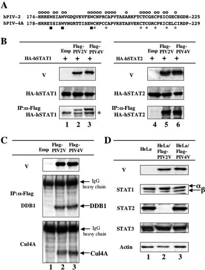
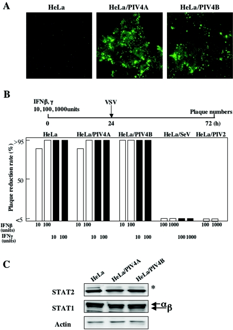
The V proteins of some paramyxoviruses have developed the ability to efficiently inactivate STAT protein function as a countermeasure for evading interferon (IFN) responses. Human parainfluenza virus type 4 (hPIV4) is one of the rubulaviruses, which are members of the family Paramyxoviridae, and has a V protein with a highly conserved cysteine-rich domain that is the hallmark of paramyxovirus V proteins. In order to study the function of the hPIV4 V protein, we established HeLa cells expressing the hPIV4A V protein (HeLa/FlagPIV4V). The hPIV4 V protein had no ability to reduce the level of STAT1 or STAT2, although it associated with STAT1, STAT2, DDB1, and Cul4A. It interfered with neither STAT1 and STAT2 tyrosine phosphorylation nor IFN-induced STAT nuclear accumulation. In addition, HeLa/FlagPIV4V cells are fully sensitive to both beta interferon (IFN-beta) and IFN-gamma, indicating that the hPIV4 V protein has no ability to block IFN-induced signaling. We further established HeLa cells expressing various chimeric proteins between the hPIV2 and hPIV4A V proteins. The lack of IFN-antagonistic activity of the hPIV4 V protein is caused by both the P/V common and V-specific domains. At least two regions (amino acids [aa] 32 to 45 and aa 143 to 164) of hPIV4 V in the P/V common domain and one region (aa 200 to 212) of the C terminus are involved in the inability to evade the IFN-induced signaling. Moreover, we established HeLa cells persistently infected with hPIV4 to make sure of the inability to escape IFN and confirmed that hPIV4 is the only paramyxovirus analyzed to date that can't evade the IFN-induced antiviral responses.


Figures

FIG. 1.
FIG. 1.
hPIV4 V protein complex formation with STAT1, STAT2, DDB1, and Cul4A without STAT degradation. (A) Amino acid sequences of the V-specific regions of hPIV2 and hPIV4A V proteins. The symbols above and below the sequences indicate the positions of conserved cysteine (asterisks), tryptophan (or tyrosine) (squares), and common (circles) residues. (B) Interaction of V proteins with STAT1 or STAT2. BSR T7/5 cells were transfected with either pTM-HA-hSTAT1 (lanes 1 to 3) or pTM-HA-hSTAT2 (lanes 4 to 6) plus pCI carrying the Flag epitope-tagged hPIV2 V gene (lanes 2 and 5), the hPIV4A V gene (lanes 3 and 6), or an empty pCI (lanes 1 and 4). Whole-cell extracts were prepared at 48 h posttransfection, and samples containing equal amounts of total protein were assayed by Western blotting for their levels of HA-hSTAT1 or HA-hSTAT2 and Flag-V proteins using α-HA and α-Flag antibodies, respectively. Other samples were first immunoprecipitated (IP) with Flag affinity gel (Sigma), and the selected material was then assayed by Western blotting for detection of hSTAT proteins. The asterisk on the right indicates the immunoglobulin heavy chain. (C) Interaction of V proteins with DDB1 or Cul4A. HeLa cells were transfected with pCI-Flag-PIV2V (lane 2), pCI-Flag-PIV4V (lane 3), or an empty pCI (lane 1). Whole-cell extracts were prepared at 48 h posttransfection, and samples containing equal amounts of total protein were assayed by Western blotting for detection of Flag-V proteins (α-Flag). Other samples were first immunoprecipitated (IP) with Flag affinity gel (Sigma), and the selected material was then assayed by Western blotting for detection of DDB1 and Cul4A proteins. (D) STAT levels in HeLa cells constitutively expressing V proteins. Samples of cytoplasmic extracts containing equal amounts of total protein of HeLa cells constitutively expressing Flag-PIV2 V (lane 2) or Flag-PIV4 V proteins (lane 3) or nonexpressing HeLa cells (lane 1) were assayed by Western blotting for detection of endogenous STAT1, STAT2, and STAT3 as well as V proteins using anti-Flag, anti-STAT1, anti-STAT2, and anti-STAT3 antibodies, as indicated at the left of each panel.
FIG. 2.
FIG. 2.
Levels of phosphorylated STATs or STAT subcellular localization in HeLa/FlagPIV4V cells treated with IFN. (A) HeLa or HeLa/FlagPIV4V cells were treated with 10, 100, or 1,000 U of IFN-α for the indicated times and assayed by Western blotting for detection of phosphorylated STAT1 or STAT2. The asterisk on the right indicates a cross-reacting host band. (B) After treatment with IFN-α (1,000 U) for 30 min or no treatment, the localization of STAT1 and STAT2 proteins was detected by indirect immunofluorescence staining with anti-STAT1 or anti-STAT2 MAb.
FIG. 3.
FIG. 3.
Interferon susceptibility in HeLa cells expressing Flag-tagged V proteins. (A) GFP expression in HeLa cells following VSV-GFP infection. The cells untreated or pretreated with 1,000 U of IFN-α for 24 h were infected with VSV-GFP. (B) Confluent monolayers of cells were inoculated with 10 or 100 U of IFN-β or IFN-γ, and after incubation for 24 h, these cells were infected with Sindbis virus. After 2 days of further incubation, plaque numbers were counted. All bars express percentages of values for plaque reduction rate.
FIG. 4.
FIG. 4.
Both the P/V common domain and the V-specific domain of hPIV4 V are involved in the inability to interdict IFN-induced signaling. (A) Schematic diagram of the chimera V proteins. Open boxes marked with T's, open boxes, and hatched boxes indicate the Flag epitope tag, amino acids of the hPIV4A V protein, and amino acids of the hPIV2 V protein, respectively. Inhibition of IFN-induced signaling and degradation of STAT2 are summarized in the right panel. (B) Effects of V proteins on IFN-α-stimulated gene activation. 2fTGH cells were transfected with a reporter plasmid [pISRE(f)-luc] and a V expression vector (pCI) or relevant control (see the text), along with pTK-r-luc as an internal reference for transfection efficiency. After incubation for 24 h, the cells were treated with IFN-α (1,000 U/ml) for 14 h or not treated and then lysed. The levels of both firefly (f) and Renilla (r) luciferase activities were determined. Data represent the mean values of the normalized luciferase activities from triplicate samples. GFP alone was used as negative control. (C) STAT1 and STAT2 levels in HeLa cells constitutively expressing V proteins. Samples of cytoplasmic extracts containing equal amounts of total protein of HeLa cells constitutively expressing V proteins (listed above each lane) or nonexpressing HeLa cells (lane 1) were assayed by Western blotting for detection of endogenous STAT1 and STAT2 as well as V proteins using anti-Flag, anti-STAT1, and anti-STAT2 antibodies, as indicated on the left of each panel. The asterisk on the right indicates a cross-reacting host band.
FIG. 5.
FIG. 5.
Subcellular distribution and the ability to interdict IFN signaling of chimera V proteins. (A) Schematic diagram of the chimera V proteins. Shown is an illustration of the chimera protein described in the legend to Fig. 4A. The chimera proteins are named using amino acid numbers of the N terminus of hPIV4A/hPIV2 (PIV4-aa/2) or the amino acid numbers of substitution [PIV2+4(substitution number) or PIV4+2(substitution number)]. Subcellular distribution, inhibition of IFN signaling, and degradation of STAT2 are summarized on the right panel. (B) Subcellular distribution of chimera V proteins as detected by indirect immunofluorescence staining with a MAb against the Flag epitope. (C) Effects of V proteins on IFN-α-stimulated gene activation. A reporter gene assay with luciferase was performed as described in the legend to Fig. 4B. Chimera 1, PIV2; chimera 2, PIV4; chimera 3, PIV4-17/2; chimera 4, PIV4-31/2; chimera 5, PIV4-46/2; chimera 6, PIV4-114/2; chimera 7, PIV2+4(66-72); chimera 8, PIV4+2(46-88)/2; and chimera 9, PIV2+4(143-164). (D) STAT1 and STAT2 levels in HeLa cells constitutively expressing V proteins. Western blotting was performed as described in the legend to Fig. 4C. The asterisk on the right indicates a cross-reacting host band. Numbers under the figure correspond to chimera numbers.
FIG. 6.
FIG. 6.
The amino acids of the C-terminal region important for IFN-induced signaling and STAT degradation. (A) V-specific regions of hPIV2 and hPIV4A V proteins. The symbols below the sequence indicate the positions of conserved cysteine (asterisks) residues, tryptophan (or tyrosine) (squares) residues, and insertion of point mutations. (B) Schematic diagram of the chimera V proteins described in the legend to Fig. 4A. The chimera proteins are named for amino acid numbers of the N terminus of hPIV2/hPIV4A (PIV2-aa/4) or the amino acid number of the substitution [PIV2+4(substitution number)] or point mutation (+mutation). The asterisk indicates the position of the mutated residue. Inhibition of IFN signaling and degradation of STAT are summarized on the right panel. (C) Effects of V proteins on IFN-α-stimulated gene activation. A reporter gene assay with luciferase was performed as described in the legend to Fig. 4B. Chimera 1, PIV2; chimera 2, PIV4; chimera 3, PIV2-177/4; chimera 4, PIV2-195/4; chimera 5, PIV2-209/4; chimera 6, PIV2-209/4+K220S; chimera 7, PIV2+4(178-195); chimera 8, PIV2+4(196-208); and chimera 9, PIV2+4(196-208)+E211F. (D) STAT1 and STAT2 levels in HeLa cells constitutively expressing V proteins. Western blotting was performed as described in the legend to Fig. 4C. The asterisk on the right indicates a cross-reacting host band. Numbers under the figure correspond to chimera numbers.
FIG. 7.
FIG. 7.
Interferon susceptibility and STAT levels in HeLa cells persistently infected with hPIV4. (A) Detection of NP proteins in cloned cells persistently infected with hPIV4A or hPIV4B. Cells were immunostained with a MAb against the NP protein. (B) Interferon susceptibility was performed as described in the legend to Fig. 3B except that VSV was used as a challenge virus. HeLa/SeV or HeLa/PIV2 cells (HeLa cells persistently infected with Sendai virus or hPIV2 virus) were used as positive controls. (C) STAT1 and STAT2 levels. Western blotting was performed as described in the legend to Fig. 4C. The asterisk on the right indicates a cross-reacting host band.

Similar articles

Cited by

References

    1. Andrejeva, J., E. Poole, D. F. Young, S. Goodbourn, and R. E. Randall. 2002. The p127 subunit (DDB1) of the UV-DNA damage repair binding protein is essential for the targeted degradation of STAT1 by the V protein of the paramyxovirus simian virus 5. J. Virol. 76:11379-11386. - PMC - PubMed
    1. Bando, H., K. Kondo, M. Kawano, H. Komada, M. Tsurudome, M. Nishio, and Y. Ito. 1990. Molecular cloning and sequence analysis of human parainfluenza type 4A virus HN gene: its irregularities on structure and activities. Virology 175:307-312. - PubMed
    1. Basler, C. F., X. Wang, E. Muhlberger, V. Volchkov, J. Paragas, H. D. Klenk, A. Garcia-Sastre, and P. Palese. 2000. The Ebola virus VP35 protein functions as a type I IFN antagonist. Proc. Natl. Acad. Sci. USA 97:12289-12294. - PMC - PubMed
    1. Bossert, B., and K. K. Conzelmann. 2002. Respiratory syncytial virus (RSV) nonstructural (NS) proteins as host range determinants: a chimeric bovine RSV with NS genes from human RSV is attenuated in interferon-competent bovine cells. J. Virol. 76:4287-4293. - PMC - PubMed
    1. Buchholz, U. J., S. Finke, and K. K. Conzelmann. 1999. Generation of bovine respiratory syncytial virus (BRSV) from cDNA: BRSV NS2 is not essential for virus replication in tissue culture, and the human RSV leader region acts as a functional BRSV genome promoter. J. Virol. 73:251-259. - PMC - PubMed

Publication types

MeSH terms