. 2005 Dec;79(23):14769-80.
doi: 10.1128/JVI.79.23.14769-14780.2005.

Interactions with pocket proteins contribute to the role of human papillomavirus type 16 E7 in the papillomavirus life cycle

Affiliations

Interactions with pocket proteins contribute to the role of human papillomavirus type 16 E7 in the papillomavirus life cycle

Asha S Collins et al. J Virol. 2005 Dec.

Abstract

Human papillomaviruses (HPVs), most commonly the HPV16 genotype, are the principle etiological determinant for cervical cancer, a common cancer worldwide resulting in over 200,000 deaths annually. The oncogenic properties of HPVs are attributable in part to the virally encoded protein E7, best known for its ability to bind to and induce the degradation of the retinoblastoma tumor suppressor, pRb, and related "pocket proteins" p107 and p130. Previously, we defined a role for E7 in the productive stage of the HPV16 life cycle, which takes place in stratified squamous epithelia. HPV perturbs the normal processes of cell growth and differentiation of stratified squamous epithelia. HPVs reprogram cells to support continued DNA synthesis and inhibit their differentiation in the suprabasal compartment of the epithelia, where cells normally have withdrawn from the cell cycle and initiated a well-defined pattern of terminal differentiation. These virus-induced perturbations, which contribute to the production of progeny HPVs, are dependent on E7. In this study, we define the mechanism of action by which E7 contributes to the productive stage of the HPV16 life cycle. We found that the ability of HPV16 to reprogram suprabasal cells to support DNA synthesis correlates with E7's ability to bind pocket proteins but not its ability to induce their degradation. In contrast, the ability of HPV16 to perturb differentiation correlated with both E7's binding to and degradation of pocket proteins. These data indicate that different hallmarks of the productive stage of the HPV16 life cycle rely upon different sets of requirements for E7.


Figures

FIG. 1.
FIG. 1.
Establishment of wild-type and E7 mutant HPV16 populations that stably harbor the HPV16 genome as an extrachromosomal plasmid (replicon). NIKS cells were cotransfected with a neomycin-resistant plasmid and wild-type or E7 mutant HPV16 genomes. After G418 selection, total genomic DNA was extracted from the transfected populations to determine if the HPV16 genomes were maintained as an extrachromosomal nuclear plasmid (replicon) within NIKS cells. (A) Total genomic DNA extracted from NIKS cells and NIKS cells transfected with wild-type or E7 mutant HPV16 genomes was sheared and subsequently analyzed by Southern blotting. Blots were probed with a full-length HPV16 probe labeled with 32P. The presence of OC and SC forms of the HPV16 genome confirms that the populations harbor HPV16 as a replicon. (B) Total genomic extracts of populations were sheared, digested with BamHI, which cuts the HPV16 genome only once, and then analyzed by Southern analysis as in panel A. As evidenced by the singular band representing the linear form of the HPV16 genome, the NIKS populations transfected with wild-type or E7 mutant genomes contained wild-type genomes with no gross aberrations and/or integrations.
FIG. 2.
FIG. 2.
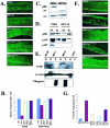
HPV16 significantly degrades pRb in the basal compartment versus the suprabasal compartment yet still can inactivate pRb in the suprabasal compartment, as judged by the induction of an E2F-responsive gene. (A) pRb levels in raft cultures of NIKS cells harboring no (NIKS), wild-type (HPV16), or E7 mutant (HPV16ΔDLYC, HPV16ΔPTLHE, and HPV16E7−Null) HPV16 replicons. Formalin-fixed, paraffin-embedded sections of raft cultures were subjected to pRb-specific immunofluorescence. The white dotted lines indicate the bottom of the basal compartment of the stratified squamous epithelium. (B) Quantification of pRb-positive cells in the basal and suprabasal compartments. For quantification, pRb-positive cells in the basal and the suprabasal compartments of raft sections were counted in 10 random fields (magnification, ×40) of sections from raft cultures of at least three different cell populations for each HPV replicon, as described in Materials and Methods. For each compartment, the average number of pRb-positive cells in the HPV-negative populations was set at a value of 1.0, and the number of pRb-positive cells in the HPV-positive populations is shown relative to that of the HPV-negative populations. A significant decrease in pRb-positive cells in rafts harboring wild-type HPV16 replicons versus NIKS cells was observed with the basal (P = 0.02092) and the suprabasal (P = 0.04331) compartments. (C) Immunoblot analyses of pRb levels in monolayer cultures of keratinocytes. Western blot analyses of cell lysates of monolayer cultures of NIKS cells and NIKS cells harboring wild-type HPV16 replicons were performed. Loading control (LC) levels of a nonrelevant protein were used to confirm that equal amounts of cellular protein were loaded on the gel. (D) Comparison of levels of pRb, p130, and p107 in raft (R) cultures versus monolayer (M) cultures. Western blot analyses were performed with 20 μg (each) of protein-isolated monolayer (M) and raft (R) cultures of NIKS cells and wild-type HPV16 replicons. (E) Comparison of levels of E7 protein in raft cultures versus monolayer cultures. E7-specific Western analysis was performed on 100 μg of protein lysate from monolayer and raft cultures as described in Materials and Methods. The intensity of the E7-specific signal in the two raft cultures was one-third that in the monolayer cultures for the same HPV16-positive cell cultures. The same blot was probed with antibodies either to β-actin to confirm that equivalent amount of cellular protein were loaded or to filaggrin to assess the differentiation state of the cells being analyzed. (F) Expression of the E2F-responsive gene MCM-7 in raft cultures of NIKS cells harboring no HPV16 replicons (NIKS), wild-type HPV16 (HPV16) replicons, or E7 mutant HPV16 (HPV16ΔDLYC, HPV16ΔPTLHE, and HPV16E7−Null) HPV16 replicons. MCM7-specific immunofluorescence was performed on sections of raft cultures. The white dotted lines indicate the bottom of the basal compartments of the stratified squamous epithelium. (G) The number of MCM7-positive cells in the suprabasal compartment versus the total amount of MCM7-positive cells (percentage of MCM7 suprabasal cells) was graphed. To calculate the averages, 10 random fields (magnification, ×40) of three different populations of each replicon were counted.
FIG. 3.
FIG. 3.
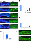
Degradation of the pocket proteins by HPV16E7 does not correlate with E7's ability to induce suprabasal DNA synthesis. (A, left) Eight hours prior to harvesting rafts, rafts of NIKS cells harboring no HPV replicons (NIKS; top row) or NIKS harboring wild-type or E7 mutant HPV16 replicons (second through fifth rows) were incubated with BrdU. BrdU was detected by BrdU-specific immunodetection and visualized by Texas red-conjugated secondary antibody. Raft sections were also costained with DAPI (4′,6′-diamidino-2-phenylindole) nuclear counterstain; DAPI stains nuclei blue. Shown are representative images of the populations studied. The white dotted lines indicate the bottom of the basal compartment of the stratified squamous epithelium in each panel. (Right) Ki67 expression in the same populations was determined by Ki67-specific immunohistochemistry of rafts. Ki67-positive cells are brown. Hematoxylin was used as a counterstain to detect nuclei; hematoxylin-positive cells are blue. Shown are representative images. (B) Relative suprabasal DNA synthesis was determined as described in Materials and Methods for at least three populations of each mutant and wild-type HPV16 replicon. (C) Ki67 expression was quantified as the percentage of suprabasal Ki67 expression as described in Materials and Methods. Ten random fields (magnification, ×40) of three different populations of each replicon were counted to calculate the percentage of suprabasal Ki67 expression. (D) BrdU incorporation in raft cultures that harbor E7ΔPTLHE mutant HPV16 replicons is spatially restricted to the lower levels of the suprabasal compartment. Supraparabasal BrdU incorporation in sections of rafts of NIKS cells harboring wild-type and E7ΔPTLHE mutant HPV16 replicons was determined. The number of BrdU-positive supraparabasal cells was divided by the total number of BrdU-positive suprabasal cells for each set of rafts. To calculate the averages, 10 random fields (magnification, ×40) of three different populations of each replicon were counted. (E) pRb degradation is not required for HPV to reprogram the suprabasal compartment. Sections of raft cultures of NIKS cells harboring no viral DNA (top) or harboring wild-type HPV16 replicons (bottom) were analyzed for pRb expression and BrdU incorporation by coimmunofluorescence. pRb-specific antibodies were used in conjunction with a fluorescein-conjugated secondary antibody to visualize pRb-positive cells. BrdU was detected using a biotin-labeled BrdU-specific primary antibody, followed by incubation with a Texas red-conjugated anti-streptavidin secondary. Cells that contain both pRb and BrdU are yellow.
FIG. 4.
FIG. 4.
The ability of E7 to bind and degrade the pocket proteins correlates with the ability of HPV16 to disrupt differentiation in raft cultures. Immunofluorescence was performed on raft sections from raft cultures of NIKS (first row), HIKS cells harboring wild-type HPV16 replicons (second row), NIKS cells harboring E7ΔDLYC mutant HPV16 replicons (third row), and NIKS cells harboring E7ΔPTLHE mutant HPV16 replicons (fourth row). (A) Early differentiation is not perturbed in the absence of binding or degradation of the pocket proteins by E7. K10 expression was detected by K10-specific primary monoclonal antibody, followed by anti-mouse fluorescein-conjugated secondary antibody. DAPI nuclear counterstaining was also used. (B) Later steps in the differentiation program are not disrupted when E7 cannot disrupt pocket proteins. Raft sections were incubated with monoclonal antibody raised against involucrin, followed by detection with a fluorescein-conjugated anti-mouse secondary antibody. (C) Suprabasal DNA synthesis is not precluded in cells undergoing a normal differentiation program. K10- and BrdU-specific coimmunofluorescence was performed on raft sections; K10 is detected with a fluorescein-conjugated secondary antibody; BrdU is detected with a Texas red secondary antibody. DAPI counterstaining was also applied to sections. The white arrow highlights a cell undergoing suprabasal DNA synthesis and expressing the early differentiation marker K10. (D) Late viral gene expression does not require E7-pocket protein interactions. To determine late viral gene expression in E7 mutant HPV16 replicons, E4-specific immunofluorescence was performed using a fluorescein-conjugated anti-E4 antibody in conjunction with DAPI nuclear counterstaining. (E) L1-specific immunofluorescence was performed on raft sections using the L1-specific monoclonal antibody H16D9.

Similar articles

Cited by

References

    1. Balsitis, S. J., J. Sage, S. Duensing, K. Munger, T. Jacks, and P. F. Lambert. 2003. Recapitulation of the effects of the human papillomavirus type 16 E7 oncogene on mouse epithelium by somatic Rb deletion and detection of pRb-independent effects of E7 in vivo. Mol. Cell. Biol. 23:9094-9103. - PMC - PubMed
    1. Brown, D. C., and K. C. Gatter. 2002. Ki67 protein: the immaculate deception? Histopathology 40:2-11. - PubMed
    1. Cam, H., and B. D. Dynlacht. 2003. Emerging roles for E2F: beyond the G1/S transition and DNA replication. Cancer Cell 3:311-316. - PubMed
    1. Chen, P. L., D. J. Riley, Y. Chen, and W. H. Lee. 1996. Retinoblastoma protein positively regulates terminal adipocyte differentiation through direct interaction with C/EBPs. Genes Dev. 10:2794-2804. - PubMed
    1. Chien, W. M., J. N. Parker, D. C. Schmidt-Grimminger, T. R. Broker, and L. T. Chow. 2000. Casein kinase II phosphorylation of the human papillomavirus-18 E7 protein is critical for promoting S-phase entry. Cell Growth Differ. 11:425-435. - PubMed

Publication types

LinkOut - more resources