. 2005 Dec;79(23):14793-803.
doi: 10.1128/JVI.79.23.14793-14803.2005.

Characterization of adeno-associated virus genomes isolated from human tissues

Affiliations

Characterization of adeno-associated virus genomes isolated from human tissues

Bruce C Schnepp et al. J Virol. 2005 Dec.

Abstract

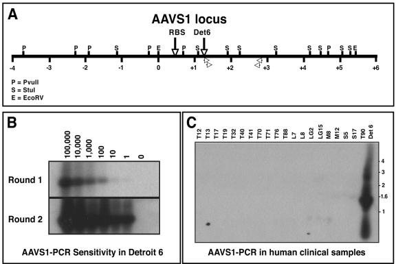
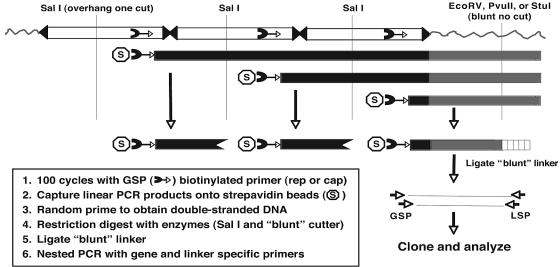
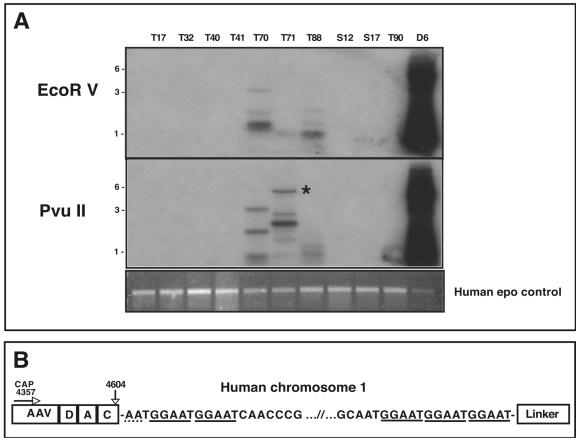
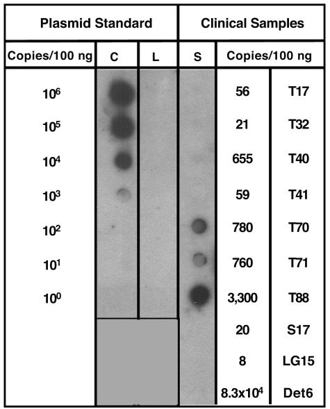
Infection with wild-type adeno-associated virus (AAV) is common in humans, but very little is known about the in vivo biology of AAV. On a molecular level, it has been shown in cultured cells that AAV integrates in a site-specific manner on human chromosome 19, but this has never been demonstrated directly in infected human tissues. To that end, we tested 175 tissue samples for the presence of AAV DNA, and when present, examined the specific form of the viral DNA. AAV was detected in 7 of 101 tonsil-adenoid samples and in 2 of 74 other tissue samples (spleen and lung). In these nine samples, we were unable to detect AAV integration in the AAVS1 locus using a sensitive PCR assay designed to amplify specific viral-cellular DNA junctions. Additionally, we used a second complementary assay, linear amplification-mediated-PCR (LAM-PCR) to widen our search for integration events. Analysis of individual LAM-PCR products revealed that the AAV genomes were arranged predominantly in a head-to-tail array, with deletions and extensive rearrangements in the inverted terminal repeat sequences. A single AAV-cellular junction was identified from a tonsil sample and it mapped to a highly repetitive satellite DNA element on chromosome 1. Given these data, we entertained the possibility that instead of integrated forms, AAV genomes were present as extrachromosomal forms. We used a novel amplification assay (linear rolling-circle amplification) to show that the majority of wild-type AAV DNA existed as circular double-stranded episomes in our tissues. Thus, following naturally acquired infection, AAV DNA can persist mainly as circular episomes in human tissues. These findings are consistent with the circular episomal forms of recombinant AAV vectors that have been isolated and characterized from in vivo transduced tissues.


Figures

FIG. 1.
FIG. 1.
AAVS1 PCR on human samples. (A) Restriction map schematic of the human AAVS1 locus. The Rep binding site (RBS) and Detroit 6 (Det6) integration site are designated by vertical arrows. The location of the AAVS1-specific primers used in the nested PCRs are also shown (open arrowheads). AAVS1 locus numbering follows the convention of Kotin et al. (21), whereby the EcoRI site defining base pair 0 is denoted (positive integers to the right, negative integers to the left). (B) Southern blot hybridization analysis of AAVS1-PCR products using Detroit 6 cellular DNA. Decreasing copies of Detroit 6 genomes (100,000 to 1) were spiked into 1 μg of naïve human genomic DNA and subjected to nested AAVS1 PCR. The PCR products were fractionated on an agarose gel and Southern hybridization was performed using an AAVS1-specific probe. The expected 1.5-kb AAV-AAVS1 junction fragment is readily detected by nested PCR using a single Detroit 6 cell junction equivalent. (C) Southern blot AAVS1-PCR on human clinical samples. AAVS1-PCR was performed on 1 μg of genomic DNA isolated from various human clinical samples. Southern blot hybridization using an AAVS1-specific probe failed to detect any signal (except in Detroit 6 control DNA).
FIG. 2.
FIG. 2.
Schematic of linear amplification mediated PCR (LAM-PCR) to detect random AAV integrants. To isolate DNA sequences flanking integrated wild-type AAV genomes, linear PCR (100 cycles) was performed on total cellular DNA (1 μg) using an AAV-specific biotinylated primer homologous to a conserved region in the cap or rep gene (cap is shown). PCR products were captured on streptavidin beads and converted to double-stranded DNA by random hexanucleotide priming. The double-stranded DNA was then digested with a blunt-cutting restriction enzyme (EcoRV, PvuII, or StuI) to generate a substrate for ligation of a blunt linker (vertical box) to the end of the digested DNA. To reduce the level of competing internal AAV-AAV junctions, double stranded fragments were also digested with SalI, which created an incompatible end for ligation of the blunt adaptor. The resulting DNA fragments were subjected to two rounds of PCR using AAV and linker-specific primers. PCR products were analyzed by Southern hybridization and cloned into an appropriate PCR vector for subsequent analysis.
FIG. 3.
FIG. 3.
LAM-PCR validation and sensitivity using Detroit 6 cells. Southern blot hybridization was performed on LAM-PCR products to determine assay sensitivity. Naïve human total cellular DNA (1 μg), containing various spiked copies of Detroit 6 cell DNA, was used as the template for LAM-PCR using an AAV cap-specific primer and a ligated blunt linker. Positive Southern hybridization (using an AAVS1 probe) detected amplification of as few as 160 Detroit 6 viral-cellular junctions. Shown in the bottom panel is an ethidium bromide stained gel of a human erythropoietin (cap) LAM-PCR genomic fragment that was generated using the same DNA templates used for the AAV LAM-PCR analysis. All samples amplified the expected 500-bp cap genomic fragment following LAM-PCR with an cap gene-specific primer (see Materials and Methods), confirming the DNA integrity of the sample and the reproducibility of the LAM-PCR assay.
FIG. 4.
FIG. 4.
LAM-PCR of positive human clinical samples. (A) Southern blot of LAM-PCR using AAV-cap primers. LAM-PCR was performed using 1 μg of total cellular DNA with either AAV rep or cap primer (cap is shown). The top panel shows LAM-PCR using EcoRV as the blunt-cutting restriction enzyme, while the middle panel shows LAM-PCR using PvuII. The DNA was hybridized with an AAV-specific probe. Detroit 6 DNA (D6) was used as a positive control and gave the predicted size based on the restriction analysis of the AAVS1 locus (see Fig. 1A). Positive hybridization of various sizes can clearly be seen in samples T70, T71, and T88. Analysis of the LAM-PCR products revealed that an approximate 5.5-kb band in sample T71 contained an AAV-cellular junction (starred). The bottom panel shows an ethidium bromide stained gel of the LAM-PCR products using a human epo gene-specific primer as an internal control. All sample amplified the expected 500-bp genomic fragment, confirming the DNA integrity of the samples and the reproducibility of the LAM-PCR assay. (B) Schematic of AAV-cellular junction from tissue T71. The 3′ AAV sequence is represented on the left ranging from bp 4357 (the position of the cap LAM primer) to the junction at bp 4604 in the AAV ITR. The AAV sequence in the LAM-PCR product contained the 3′ cap sequence as well as a complete D, A, and C ITR region. There appears to be only 2 bp of homology between the breakpoint of the AAV ITR and the chromosomal sequence (dotted underline). The LAM-PCR product contained approximately 5 kb of human chromosomal DNA that mapped to position 1q31.1, composed primarily of a (GGAAT)n repeat sequence (solid underline).
FIG. 5.
FIG. 5.
Schematic of AAV-AAV junctions isolated following LAM-PCR. A complete head-to-tail AAV ITR junction is shown at the top of the figure, along with the positions of the cap and rep primers used. Shown below are various AAV-AAV junctions isolated from tissue T70. The majority of junctions analyzed were in a head-to-tail orientation; however, tail-to-tail (T70-14) and head-to-head (T70-E9) orientations were observed. The breakpoints for each junction are given, with deleted sequence designated using a solid line. For several clones (T70-C1, T70-11, and T70-12), we were unable to obtain readable sequence after the D region from either side (designated as dotted lines), which was presumably due to strong secondary structure associated with the ITR structure.
FIG. 6.
FIG. 6.
Linear rolling-circle amplification for the detection of AAV episomes. Total cellular DNA was digested with a restriction enzyme that does not cut within the AAV genome. The DNA was then treated with Plasmid-Safe DNase, which degrades linear fragments but leaves circular, double-stranded DNA intact. The digestion reaction served as a template for linear rolling-circle amplification using AAV-specific primers and φ29 phage DNA polymerase. Large, linear concatameric arrays (U, uncut linear rolling-circle amplification DNA from tissue T88) were produced following amplification of circular AAV episomes. The linear arrays were subsequently digested into unit-length monomers by restriction enzyme digestion with an enzyme that cleaves the AAV genome once (labeled 1 in the figure). The unit-length fragment was then cloned into an appropriate vector for further sequence analysis.
FIG. 7.
FIG. 7.
Wild-type AAV genomes are present predominantly as episomes. To determine the sensitivity of the linear rolling-circle amplification assay, 10-fold dilutions of circular (C) or linear (L) DNA plasmids containing the AAV2 rep and cap genes were spiked into 100ng of genomic DNA from a naïve human tonsil sample. Linear rolling-circle amplification was performed using cap-specific primers and the products were analyzed by dot blot Southern hybridization using an AAV-specific probe. Positive hybridization could be detected at 1,000 circular plasmid copy spike (representing episomal DNA), whereas linear plasmid DNA (representing integrated AAV) could not be amplified to any detectable level. Total cellular DNA (100 ng) from AAV-positive clinical samples (S) was then analyzed by linear rolling-circle amplification, and tissues T70, T71, and T88 showed positive amplification. The AAV genome copy numbers in these particular tissues were at or above the level of sensitivity of linear rolling-circle amplification as determined from the plasmid spike experiments. Detroit 6 (Det6) cells, which have 83,000 integrated AAV genomes in 100ng, failed to amplify the integrated AAV, further confirming linear rolling-circle amplification assay specificity.

Similar articles

Cited by

References

    1. Afione, S. A., C. K. Conrad, W. G. Kearns, S. Chunduru, R. Adams, T. C. Reynolds, W. B. Guggino, G. R. Cutting, B. J. Carter, and T. R. Flotte. 1996. In vivo model of adeno-associated virus vector persistence and rescue. J. Virol. 70:3235-3241. - PMC - PubMed
    1. Atchison, R. W., B. C. Casto, and W. M. Hammon. 1965. Adenovirus-associated defective virus particles. Science 149:754-756. - PubMed
    1. Atchison, R. W., B. C. Casto, and W. M. Hammon. 1966. Electron microscopy of adenovirus-associated virus (AAV) in cell cultures. Virology 29:353-357. - PubMed
    1. Berns, K. I., T. C. Pinkerton, G. F. Thomas, and M. D. Hoggan. 1975. Detection of adeno-associated virus (AAV)-specific nucleotide sequences in DNA isolated from latently infected Detroit 6 cells. Virology 68:556-560. - PubMed
    1. Blacklow, N. R., M. D. Hoggan, and W. P. Rowe. 1967. Isolation of adenovirus-associated viruses from man. Proc. Natl. Acad. Sci. USA 58:1410-1415. - PMC - PubMed

Publication types

LinkOut - more resources