. 2005 Dec;79(23):14945-55.
doi: 10.1128/JVI.79.23.14945-14955.2005.

A hydrophobic domain in the large envelope protein is essential for fusion of duck hepatitis B virus at the late endosome

Affiliations

A hydrophobic domain in the large envelope protein is essential for fusion of duck hepatitis B virus at the late endosome

J Chojnacki et al. J Virol. 2005 Dec.

Abstract

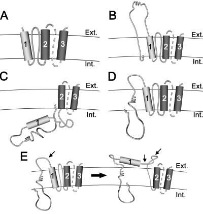
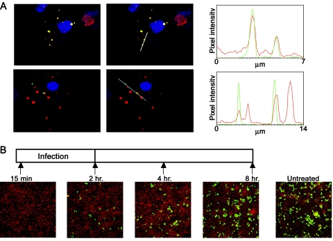
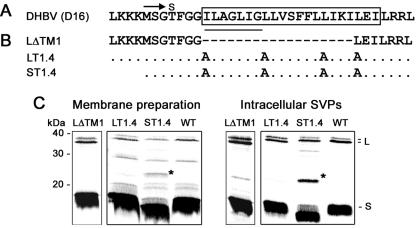
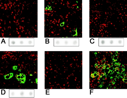
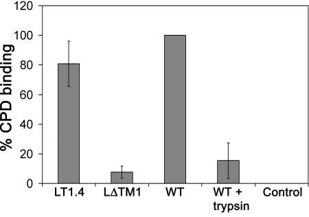
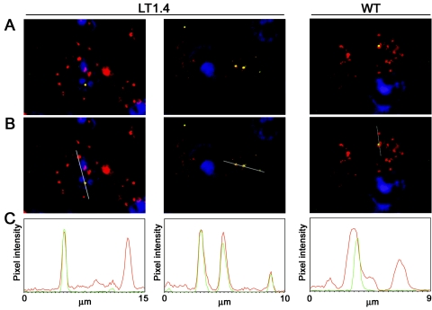
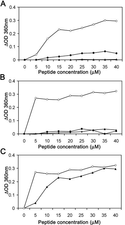
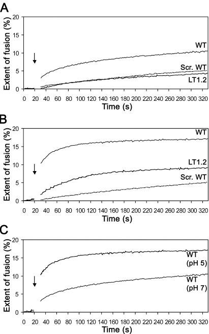
The duck hepatitis B virus (DHBV) envelope is comprised of two transmembrane (TM) proteins, the large (L) and the small (S), that assemble into virions and subviral particles. Secondary-structure predictions indicate that L and S have three alpha-helical, membrane-spanning domains, with TM1 predicted to act as the fusion peptide following endocytosis of DHBV into the hepatocyte. We used bafilomycin A1 during infection of primary duck hepatocytes to show that DHBV must be trafficked from the early to the late endosome for fusion to occur. Alanine substitution mutations in TM1 of L and S, which lowered TM1 hydrophobicity, were used to examine the role of TM1 in infectivity. The high hydrophobicity of the TM1 domain of L, but not of S, was shown to be essential for virus infection at a step downstream of receptor binding and virus internalization. Using wild-type and mutant synthetic peptides, we demonstrate that the hydrophobicity of this domain is required for the aggregation and the lipid mixing of phospholipid vesicles, supporting the role of TM1 as the fusion peptide. While lipid mixing occurred at pH 7, the kinetics of insertion of the fusion peptide was increased at pH 5, consistent with the location of DHBV in the late-endosome compartment and previous studies of the nonessential role of low pH for infectivity. Exchange of the TM1 of DHBV with that of hepatitis B virus yielded functional, infectious DHBV particles, suggesting that TM1 of all of the hepadnaviruses act similarly in the fusion mechanism.


Figures

FIG. 1.
FIG. 1.
Predicted topologies of DHBV envelope proteins. TM1, TM2, and TM3 are indicated by numbered cylinders 1, 2, and 3, respectively. The C-terminal regions of the DHBV envelope proteins, as indicated by the broken lines, remain uncharacterized and may span the membrane more then once. (A) Model of the S protein topology. (B) External L topology with an exposed translocated pre-S domain. (C) Internal L topology, with the pre-S domain and TM1 disposed internally. (D) Intermediate L topology with partially translocated pre-S domain. (E) Proposed model of the conformational change in L. Trypsin digestion sites available before and after the conformational change are denoted by small arrows (12). Ext., external; Int., internal.
FIG. 2.
FIG. 2.
DHBV is targeted to the late endosome. (A) Colocalization of serum-derived DHBV with the endosomal marker, transferrin (red). Cells were incubated with DHBV-positive serum (MOI of ∼100) and human transferrin-Alexa 568 (50 μg/ml) for 1 h at 4°C, followed by 2 h at 37°C. Cells were fixed, immunostained for DHBV (green), and visualized with a confocal microscope. Cell nuclei were stained with TOTO-3 (blue). Panels show representative images with or without the indicated line profiles of pixel intensity (gray line). Graphs represent the line profile output showing pixel intensities of transferrin (red) and DHBV (green). (B) Effects of bafilomycin A1 on DHBV entry. Cells were infected with DHBV serum for 2 h. The time course outline is shown schematically. The arrows and times indicate the addition of 500 nM of bafilomycin A1, which was maintained for the duration of the experiment. Each panel below represents a corresponding immunofluorescence image taken 5 days postinfection by using a monoclonal antibody to pre-S.
FIG. 3.
FIG. 3.
Analysis of the expression and assembly of TM1 mutants. (A) Sequence of DHBV (D16), isolate from the United States, with the boxed region indicating the TM1. The underlined sequence represents a potential fusion peptide site identified previously in the corresponding region of HBV (19). The start of the S domain (Met162) is indicated by an arrow. (B) TM1 mutants. LΔTM1 is a deletion from aa 169 to 186 of L TM1. LT1.4 contains alanine substitutions at positions 169, 176, 183, and 187 in L, whereas ST1.4 contains alanine substitutions at positions 8, 15, 22, and 26 in S. (C) Expression and assembly of DHBV TM1 mutants. LMH cells were cotransfected with WT S and either mutant L (LΔTM1 or LT1.4) or WT L or cotransfected with ST1.4- and WT L-encoding plasmids, as indicated above each panel. Membrane preparations and intracellular SVPs were assessed by Western blotting with anti-S monoclonal antibody (7C12). The L protein, seen as a doublet, represents phosphorylated and unphosphorylated forms. The asterisk indicates a glycosylated form of mutant S (9).
FIG. 4.
FIG. 4.
Infectivity of DHBV TM1 mutants. PDH cells were infected for 24 h with equal amounts of virus (MOI of ∼100) derived from transfected LMH cells, as determined by DNA dot blot (see Materials and Methods). (A) LT1.4, (B) ST1.4, (C) LΔTM1, (D) wild-type DHBV, (E) mock, and (F) 15 μl of positive duck serum. Cells were fixed 7 days postinfection, and DHBV-infected cells were visualized by immunofluorescence using anti-pre-S/S monoclonal antibodies (green) and propidium iodide nuclear stain (red). Images are those of a representative experiment. Viral DNA dot blot peak fractions after sucrose step gradient sedimentation of enveloped virus from LMH cells are indicated in blots below panels A to D.
FIG. 5.
FIG. 5.
Determination of receptor binding of TM1 mutants. Purified intracellular SVPs were incubated with soluble recombinant CPD, followed by sedimentation of receptor-bound SVPs through 20% sucrose and analysis by Western blotting using a rabbit anti-CPD antibody and an anti-pre-S monoclonal antibody. The amount of SVP-bound CPD was quantified and normalized for the amount of L protein in each sample. The percentage of CPD binding was calculated as a percentage relative to that of WT SVPs, which was taken as 100%. “WT + trypsin” represents WT SVPs cleaved with trypsin prior to CPD binding. “Control” represents CPD without SVPs. The means and standard deviations are derived from three independent experiments.
FIG. 6.
FIG. 6.
Cellular uptake of the LT1.4 mutant. PDHs were incubated with either LT1.4 or WT virus (MOI of ∼100) and human transferrin-Alexa 568 (red) (50 μg/ml) for 1 h at 4°C, followed by 2 h at 37°C. Cells were fixed, immunostained for DHBV (green) by using an anti-pre-S monoclonal antibody, and visualized with a confocal microscope. Cell nuclei were stained with TOTO-3 (blue). (A) Acquired images for LT1.4 and WT infection, as indicated above each panel. (B) Acquired images with indicated line profiles of pixel intensity (gray line). (C) Line profile output showing pixel intensities of transferrin (red) and DHBV (green).
FIG. 7.
FIG. 7.
Liposome aggregation induced by synthetic peptides. SUVs (0.14 mM) were mixed with WT (○), LT1.2 (▴), and scrambled WT (•) peptides at either pH 7 (A) or pH 5 (B). Changes in OD360 were measured after a 10-min incubation. (C) Comparison of WT peptide activities at pH 7 (▴) and pH 5 (○). The results are those of a representative experiment.
FIG. 8.
FIG. 8.
Lipid mixing induced by synthetic peptides. Fluorescence of a mix of unlabeled and R18 labeled liposomes (0.14 mM) at a 1:1 ratio was measured prior to peptide addition (50 μM), indicated by the arrow. The R18 dequenching percentage was calculated, as described in Materials and Methods. Percentages of liposome fusion induced by peptides at pH 7 (A) and pH 5 (B). Scr., scrambled. (C) Comparison of the percentages of fusion for WT peptide at pH 7 and pH 5. The 10-s gap in the measurement is due to the manual addition of the peptide, indicated by the arrow. The results are those of a representative experiment.
FIG. 9.
FIG. 9.
Characterization of HBV/DHBV TM1 chimeras. (A) DHBV, HBV, and chimera sequence alignments showing the start of the S domain, indicated by an arrow. The predicted TM1 domains are represented by the boxed sequences. ayw, HBV subtype; D16, DHBV isolate from the United States. (B) Expression, assembly, and export of HBV/DHBV TM1 chimeras from LMH cells cotransfected with the chimeric L plasmid and pCI-S. Membrane preparations, intracellular SVPs, and exported SVPs of L: DHTM1 (lanes 1, 3, and 5, respectively) and of L: DHTM1 Q177L (lanes 2, 4, and 6, respectively) were assessed by Western blotting with anti-S monoclonal antibody. (C) Infectivity of HBV/DHBV TM1 chimeras. PDH cells were infected for 24 h with equal amounts of virus derived from LMH transfected cells, as discussed in Materials and Methods. Cells were fixed 7 days postinfection, and DHBV-infected cells were visualized by immunofluorescence using anti-pre-S/S monoclonal antibodies (green) and propidium iodide nuclear stain (red). Images are those of a representative experiment. Viral DNA dot blot peak fractions are indicated below panels.

Similar articles

Cited by

References

    1. Berting, A., C. Fischer, S. Schaefer, W. Garten, H. D. Klenk, and W. H. Gerlich. 2000. Hemifusion activity of a chimeric influenza virus hemagglutinin with a putative fusion peptide from hepatitis B virus. Virus Res. 68:35-49. - PubMed
    1. Breiner, K. M., and H. Schaller. 2000. Cellular receptor traffic is essential for productive duck hepatitis B virus infection. J. Virol. 74:2203-2209. - PMC - PubMed
    1. Breiner, K. M., S. Urban, and H. Schaller. 1998. Carboxypeptidase D (gp180), a Golgi-resident protein, functions in the attachment and entry of avian hepatitis B viruses. J. Virol. 72:8098-8104. - PMC - PubMed
    1. Bruss, V. 1997. A short linear sequence in the pre-S domain of the large hepatitis B virus envelope protein required for virion formation. J. Virol. 71:9350-9357. - PMC - PubMed
    1. Chandran, K., N. J. Sullivan, U. Felbor, S. P. Whelan, and J. M. Cunningham. 2005. Endosomal proteolysis of the Ebola virus glycoprotein is necessary for infection. Science 308:1643-1645. - PMC - PubMed

Substances

LinkOut - more resources