

doi: 10.1128/JVI.79.23.14971-14975.2005.
Breaking an absolute species barrier: transgenic mice expressing the mink PrP gene are susceptible to transmissible mink encephalopathy
Affiliations
- PMID: 16282497
- PMCID: PMC1287601
- DOI: 10.1128/JVI.79.23.14971-14975.2005
Item in Clipboard
Breaking an absolute species barrier: transgenic mice expressing the mink PrP gene are susceptible to transmissible mink encephalopathy
J Virol.
2005 Dec.
Abstract
Transmissible mink encephalopathy (TME) is a rare disease of the North American mink, which has never been successfully transmitted to laboratory mice. We generated transgenic mice expressing the mink prion protein (PrP) and inoculated them with TME or the mouse-adapted scrapie strain 79A. TME infected mink PrP-transgenic mice on a murine PrP knockout background. The absolute species barrier between the infectious agent of TME and mice was therefore broken. Following TME and 79A infection of mice carrying both mink and murine PrP(C), only proteinase-resistant PrP homologous to the incoming agent was detectable. The presence of the murine PrP(C) prolonged the incubation time of TME substantially.
Figures

(A) Sequence comparison between murine and mink PrP. The sequence of the murine Prnpa allele (36) is shown and differences in the mink PrP sequence (17) are indicated underneath. The numbering is according to the murine PrP. The single letter code for amino acids is used and only the sequences of the mature PrP devoid of the N- and C-terminal signal peptides are shown. Amino acids proposed to be involved in protein X-binding (15) are underlined and amino acids proposed to be involved in the PrPSc/PrPC interface (28) are highlighted in bold and italics. (B) Schematic representation of cosMink. Exonic sequences of the hamster PrP gene are represented by white boxes and the coding region of the mink PrP gene is highlighted in gray.
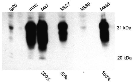
Western blot analysis of transgenic mouse lines carrying the mink PrP gene. Brain homogenates of F1 animals of the four different transgenic lines carrying the mink PrP gene, MK7, MK27, MK39, and MK45 were analyzed alongside of North American mink and tg20, a transgenic mouse line overexpressing the murine Prnp (9). Antibody L42 was used as primary antibody in the Western blot analysis following SDS-PAGE. Molecular size markers are indicated to the right of the blot and estimates of the amount of mink PrP in the different transgenic lines compared to the mink are indicated underneath.
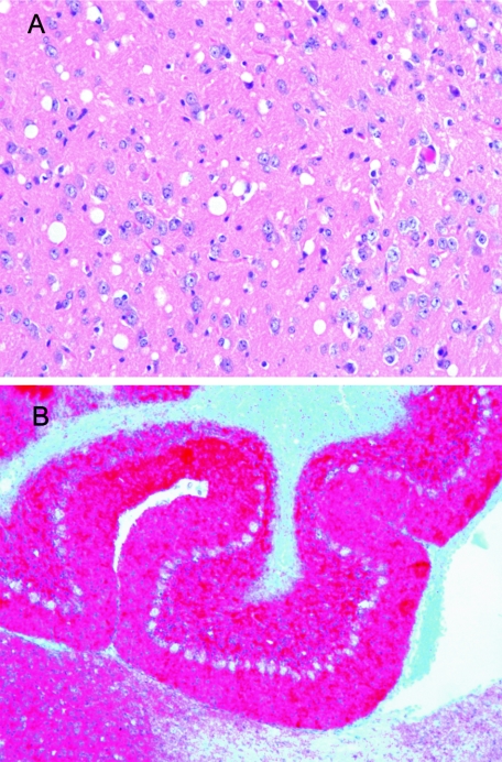
Histopathological analysis of infected MK7/Prnp0/0 mice. (A) Cerebral cortex showing numerous delicate vacuoles (spongiform change) stained with hematoxylin and eosin Original magnification, ×20. (B) Cerebellum showing strong immunostaining for PrPSc in red in the molecular and internal granule cell layer using L42 as primary antibody. Original magnification, ×10.
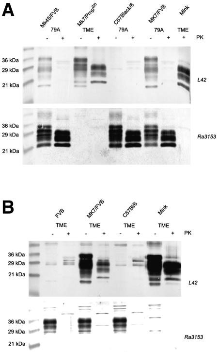
Western blot analysis of PK-resistant PrP in brain homogenates of infected animals. (A) Brain homogenates of transgenic mice (MK45/FVB, MK7/FVB) infected with 79A were compared to normal mice (C57/Black6) infected with the same agent as well as to Mk7/Prnp0/0 mice and mink infected with TME. Homogenates were either treated with PK (+) or left untreated (−). The upper panel was immunostained using L42 as primary antibody, while the lower panel was stained using Ra3153. A molecular marker was loaded in the left lane, and the molecular sizes are indicated. (B) Brain homogenates of normal mice (FVB and C57/Black6), transgenic MK7/FVB and mink, all infected with TME, were compared using either L42 (upper panel) or Ra3153 (lower panel) as the primary antibody.
Similar articles
-
Sc237 hamster PrPSc and Sc237-derived mouse PrPSc generated by interspecies in vitro amplification exhibit distinct pathological and biochemical properties in tga20 transgenic mice.Microbiol Immunol. 2011 May;55(5):331-40. doi: 10.1111/j.1348-0421.2011.00328.x. Microbiol Immunol. 2011. PMID: 21362027
-
Raccoons accumulate PrPSc after intracranial inoculation of the agents of chronic wasting disease or transmissible mink encephalopathy but not atypical scrapie.J Vet Diagn Invest. 2019 Mar;31(2):200-209. doi: 10.1177/1040638718825290. Epub 2019 Jan 29. J Vet Diagn Invest. 2019. PMID: 30694116 Free PMC article.
-
Disease-associated prion protein in neural and lymphoid tissues of mink (Mustela vison) inoculated with transmissible mink encephalopathy.J Comp Pathol. 2012 Nov;147(4):508-21. doi: 10.1016/j.jcpa.2012.03.008. Epub 2012 May 16. J Comp Pathol. 2012. PMID: 22595634 Free PMC article.
-
Prion encephalopathies of animals and humans.Dev Biol Stand. 1993;80:31-44. Dev Biol Stand. 1993. PMID: 8270114 Review.
-
[Mechanisms of neuroinvasion by prions: molecular principles and present state of research].Schweiz Med Wochenschr. 2000 Mar 25;130(12):435-42. Schweiz Med Wochenschr. 2000. PMID: 10780058 Review. German.
Cited by
-
Cross currents in protein misfolding disorders: interactions and therapy.CNS Neurol Disord Drug Targets. 2009 Nov;8(5):363-71. doi: 10.2174/187152709789541998. CNS Neurol Disord Drug Targets. 2009. PMID: 19702573 Free PMC article. Review.
-
Prion replication without host adaptation during interspecies transmissions.Proc Natl Acad Sci U S A. 2017 Jan 31;114(5):1141-1146. doi: 10.1073/pnas.1611891114. Epub 2017 Jan 17. Proc Natl Acad Sci U S A. 2017. PMID: 28096357 Free PMC article.
-
Comparing Prion Proteins Across Species: Is Zebrafish a Useful Model?Mol Neurobiol. 2025 Jan;62(1):832-845. doi: 10.1007/s12035-024-04324-z. Epub 2024 Jun 25. Mol Neurobiol. 2025. PMID: 38918277 Free PMC article. Review.
-
Transgenic Rabbits Expressing Ovine PrP Are Susceptible to Scrapie.PLoS Pathog. 2015 Aug 6;11(8):e1005077. doi: 10.1371/journal.ppat.1005077. eCollection 2015 Aug. PLoS Pathog. 2015. PMID: 26248157 Free PMC article.
-
The prion hypothesis: from biological anomaly to basic regulatory mechanism.Nat Rev Mol Cell Biol. 2010 Dec;11(12):823-33. doi: 10.1038/nrm3007. Epub 2010 Nov 17. Nat Rev Mol Cell Biol. 2010. PMID: 21081963 Free PMC article. Review.
References
-
- Bessen, R. A., and R. F. Marsh. 1992. Identification of two biologically distinct strains of transmissible mink encephalopathy in hamsters. J. Gen. Virol. 73:329-334. - PubMed
-
- Bruce, M. E., P. A. McBride, M. Jeffrey, and J. R. Scott. 1994. PrP in pathology and pathogenesis in scrapie-infected mice. Mol. Neurobiol. 8:105-112. - PubMed
-
- Büeler, H., M. Fischer, Y. Lang, H. Bluethmann, H. P. Lipp, S. J. DeArmond, S. B. Prusiner, M. Aguet, and C. Weissmann. 1992. Normal development and behaviour of mice lacking the neuronal cell-surface PrP protein. Nature 356:577-582. - PubMed
-
- Buschmann, A., E. Pfaff, K. Reifenberg, H. M. Muller, and M. H. Groschup. 2000. Detection of cattle-derived BSE prions using transgenic mice-overexpressing bovine PrP(c). Arch. Virol. Suppl. 16:75-86. - PubMed
Publication types
MeSH terms
Substances
LinkOut - more resources
Full Text Sources
Molecular Biology Databases
Research Materials
