

Tumour-derived microvesicles carry several surface determinants and mRNA of tumour cells and transfer some of these determinants to monocytes
- PMID: 16283305
- PMCID: PMC11030663
- DOI: 10.1007/s00262-005-0075-9
Tumour-derived microvesicles carry several surface determinants and mRNA of tumour cells and transfer some of these determinants to monocytes
Abstract
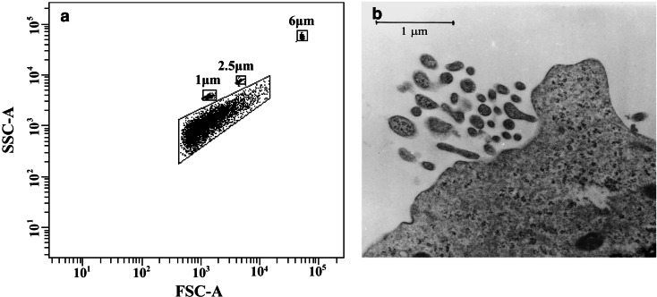
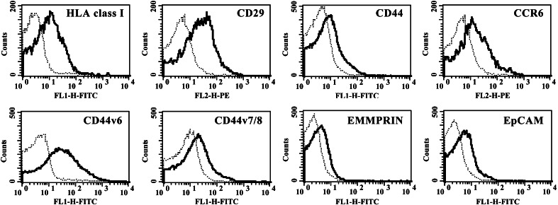
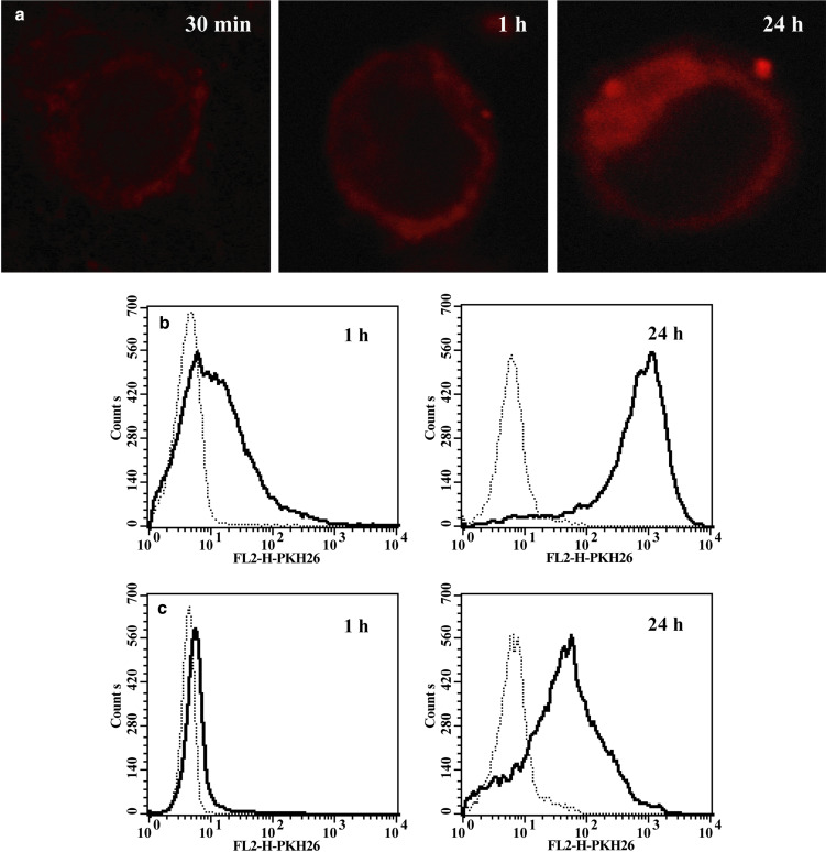
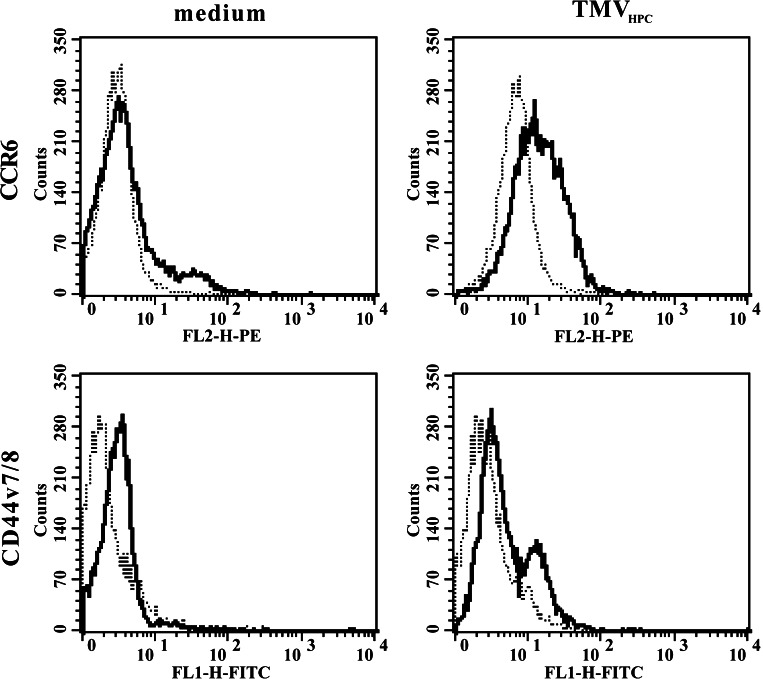
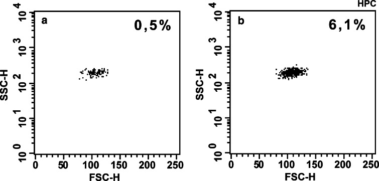
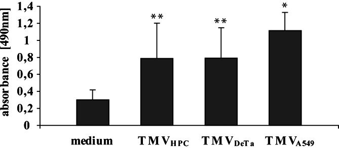
This study was designed to determine the characteristics of tumour cell-derived microvesicles (TMV) and their interactions with human monocytes. TMV were shed spontaneously by three different human cancer cell lines but their release was significantly increased upon activation of the cells with phorbol 12-myristate 13-acetate (PMA). TMV showed the presence of several surface determinants of tumour cells, e.g. HLA class I, CD29, CD44v7/8, CD51, chemokine receptors (CCR6, CX3CR1), extracellular matrix metalloproteinase inducer (EMMPRIN), epithelial cell adhesion molecule (EpCAM), but their level of expression differed from that on cells they originated from. TMV also carried mRNA for growth factors: vascular endothelial growth factor (VEGF), hepatocyte growth factor (HGF), interleukin-8 (IL-8) and surface determinants (CD44H). TMV were localized at the monocytes surface following their short exposure to TMV, while at later times intracellularly. TMV transferred CCR6 and CD44v7/8 to monocytes, exerted antiapoptotic effect on monocytes and activated AKT kinase (Protein Kinase B). Thus, TMV interact with monocytes, alter their immunophenotype and biological activity. This implicates the novel mechanism by which tumour infiltrating macrophages may be affected by tumour cells not only by a direct cell to cell contact, soluble factors but also by TMV.
Figures


References
-
- Albanese J, Meterissian S, Kontogiannea M, Dubreuil C, Hand A, Sorba S, Dainiak N. Biologically active Fas antigen and its cognate ligand are expressed on plasma membrane-derived extracellular vesicles. Blood. 1998;91:3862–3874. - PubMed
-
- Andreola G, Rivoltini L, Castelli C, Huber V, Perego P, Deho P, Squarcina P, Accornero P, Lozupone F, Lugini L, Stringaro A, Molinari A, Arancia G, Gentile M, Parmiani G, Fais S. Induction of lymphocyte apoptosis by tumor cell secretion of FasL-bearing microvesicles. J Exp Med. 2002;195:1303–1316. doi: 10.1084/jem.20011624. - DOI - PMC - PubMed
-
- Baj-Krzyworzeka M, Majka M, Pratico D, Ratajczak J, Vilaire G, Kijowski J, Reca R, Janowska-Wieczorek A, Ratajczak MZ. Platelet-derived microparticles stimulate proliferation, survival, adhesion, and chemotaxis of hematopoietic cells. Exp Hematol. 2002;30:450–459. doi: 10.1016/S0301-472X(02)00791-9. - DOI - PubMed
-
- Baran J, Weglarczyk K, Mysiak M, Guzik K, Ernst M, Flad HD, Pryjma J. Fas (CD95)-Fas ligand interactions are responsible for monocyte apoptosis occurring as a result of phagocytosis and killing of Staphylococcus aureus. Infect Immun. 2001;69:1287–1297. doi: 10.1128/IAI.69.3.1287-1297.2001. - DOI - PMC - PubMed
Publication types
MeSH terms
Substances
LinkOut - more resources
Full Text Sources
Other Literature Sources
Research Materials
Miscellaneous
