

Review
doi: 10.1113/jphysiol.2005.097873.
Epub 2005 Nov 10.
Cardiac memory ... new insights into molecular mechanisms
Affiliations
- PMID: 16284076
- PMCID: PMC1464312
- DOI: 10.1113/jphysiol.2005.097873
Item in Clipboard
Review
Cardiac memory ... new insights into molecular mechanisms
J Physiol.
.
Abstract
'Cardiac memory' describes an electrocardiographic T wave vector change, recorded during normal sinus rhythm that reflects the QRS complex vector during prior periods of ventricular pacing or arrhythmia. In this brief review we consider the mechanisms responsible for cardiac memory, which offer a unique window for relating molecular determinants of repolarization to their expression in the function of ion channels and in the electrophysiology of the heart. Understanding the steps that translate the molecular mechanisms for memory into clinical expression in this relatively straightforward model facilitates our comprehension of the complex pathways that order normal cardiac repolarization and repolarization changes.
Figures

Upper panels show canine ECG during control sinus rhythm, during ventricular pacing at about 5% faster than sinus rate, and – a few minutes after returning to sinus rhythm – on days 7, 14 and 21 of pacing. Note that the QRS complex is inverted during ventricular pacing and that the T wave in the subsequent panels in sinus rhythm becomes progressively inverted, following the direction of the paced QRS complex. The two left bottom panels show the vectorcardiogram of the same dog in control (sinus rhythm) and during ventricular pacing. Note the change in vector shape, angle and amplitude of the QRS complex. The right lower panel shows an enlargement of the T wave vector during control sinus rhythm and in sinus rhythm on days 14 and 21. Note that the T wave vector has moved in the direction of the paced QRS, and also shows an increased amplitude. Modified from Shvilkin et al. (1998).

A and B, representative records of a control epicardial myocyte (Epi) and an angiotensin II-exposed myocyte (Epi + A-II), respectively: note the marked diminution of current in the latter. C shows a summary of current–voltage data for the entire series. There is a significant reduction in current throughout the voltage range studied. D and E show action potentials from comparable cells. In D the phase 1 notch attributable to Ito is indicated by an arrow. The notch disappears on exposure to angiotensin II (E). Modified from Yu et al. (2000).

HEK 293 cells were transiently transfected with Kv4.3–V5–KChIP2-myc and the HA-AT1 receptor. The distribution of AT1 receptors and Kv4.3 was visualized after cell fixation with 3.7% formaldehyde using anti-HA-TRITC and anti-V5-FITC antibody before and after treatment with 1 μm angiotensin for 1 h. The distribution of Kv4.3 and AT1 receptors is shown before (A) and after treatment with angiotensin II (B). Nuclei (blue colour) were counterstained with 4,6-diamidino-2-phenylindole (DAPI). Note that the AT1 receptor is located predominantly on the cell surface where it colocalizes with Kv4.3 in the absence of angiotensin II (A). Angiotensin II induces internalization of the AT1 receptor (B), and with this Kv4.3 is also removed from the cell surface. The majority of the internalized AT1 receptors colocalize with Kv4.3 in intracellular vesicles. Reproduced with permission from Doronin et al. (2004).
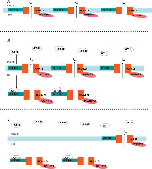
A, coassembled AT-1 receptor–Kv4.3–KChIP2 macromolecular complexes inserted in the cell membrane. Channel opening results in outward K+ current. In B, angiotensin II binds to a subset of receptors, resulting in the internalization of the macromolecular complex. The net result is a loss of functioning channels in the cell membrane and a reduction in current (C).
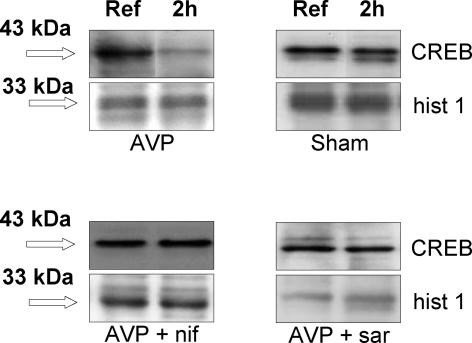
In these experiments, anaesthetized dogs were subjected to atrioventricular pacing with a short PR interval to ensure 100% capture of the ventricles during the 2 h period of pacing. Individual panels show results from an atrioventricular paced dog (AVP), a sham control (instrumented but not paced), an AV paced dog that received the L-type Ca2+ channel blocker nifedipine (AVP + nif), and an AV paced dog that received the AT-I and AT-II receptor blocker saralasin (AVP + sar). Ref indicates reference biopsy; 2 h, biopsy taken after 2 h of AVP. Loading conditions were controlled using an antibody against histone 1 (hist 1). Note the marked reduction in CREB levels at 2 h in the AV paced dog. There was no change in CREB in the sham animal or in those treated with nifedipine or saralasin. Reproduced with permission from Patberg et al. (2003).

Upper panel: epicardial action potentials recorded from slabs of left ventricular tissue removed from a dog paced for 3 weeks to induce cardiac memory, and from a sham control. Note that in the setting of memory, the phase 1 notch diminishes, the plateau is increased in height and action potential duration is prolonged. Lower left panel shows Kv4.3 mRNA for a control dog and one in cardiac memory. Note that message is reduced in the memory animal (Cyc = cyclophyllin). Lower right panel shows Ito conductance for a group of controls and a group of memory animals. There is a significant decrease in channel conductance in the setting of memory. Modified from Yu et al. (1999).

Pacing alters activation and stretch, resulting in angiotensin II synthesis/release and the trafficking and internalization of the AT-1 receptor–Kv4.3–KChIP2 complex from its membrane site and a reduction in current. Angiotensin II also induces an increase in L-type Ca2+ current. Other potential sources for increased Cai2+ would be the Na+–Ca2+ exchanger and stretch-activated channels. It appears that Cai2+ may be a second messenger activating changes in transcriptional factors in the nucleus. The transcriptional factor thus far studied, CREB, is reduced. An association with KChIP2 reduction has been demonstrated here. The other factors and linkages that may be involved have not been identified. However, long-term changes in Ito, IKr and ICa,L have been demonstrated as well, all of which would be expected to contribute to the altered action potentials and ECG changes of cardiac memory.
Similar articles
-
Cardiac memory: a work in progress.Heart Rhythm. 2009 Apr;6(4):564-70. doi: 10.1016/j.hrthm.2009.01.008. Epub 2009 Jan 16. Heart Rhythm. 2009. PMID: 19324320 Review.
-
Cardiac memory: The slippery slope twixt normalcy and pathology.Trends Cardiovasc Med. 2015 Nov;25(8):687-96. doi: 10.1016/j.tcm.2015.02.011. Epub 2015 Feb 25. Trends Cardiovasc Med. 2015. PMID: 25842262 Review.
-
Determinants of CREB degradation and KChIP2 gene transcription in cardiac memory.Heart Rhythm. 2010 Jul;7(7):964-70. doi: 10.1016/j.hrthm.2010.03.024. Epub 2010 Mar 24. Heart Rhythm. 2010. PMID: 20346417 Free PMC article.
-
Altered ventricular stretch contributes to initiation of cardiac memory.Heart Rhythm. 2008 Jan;5(1):106-13. doi: 10.1016/j.hrthm.2007.09.008. Epub 2007 Sep 19. Heart Rhythm. 2008. PMID: 18055271
-
Persistent T-wave changes after alteration of the ventricular activation sequence. New insights into cellular mechanisms of 'cardiac memory'.Circulation. 1993 Oct;88(4 Pt 1):1811-9. doi: 10.1161/01.cir.88.4.1811. Circulation. 1993. PMID: 8403326
Cited by
-
Greater accuracy and broadened applicability of phase reduction using isostable coordinates.J Math Biol. 2018 Jan;76(1-2):37-66. doi: 10.1007/s00285-017-1141-6. Epub 2017 May 25. J Math Biol. 2018. PMID: 28547210
-
Ventricular stimulus site influences dynamic dispersion of repolarization in the intact human heart.Am J Physiol Heart Circ Physiol. 2016 Sep 1;311(3):H545-54. doi: 10.1152/ajpheart.00159.2016. Epub 2016 Jul 1. Am J Physiol Heart Circ Physiol. 2016. PMID: 27371682 Free PMC article.
-
Emergent activity, heterogeneity, and robustness in a calcium feedback model of the sinoatrial node.Biophys J. 2023 May 2;122(9):1613-1632. doi: 10.1016/j.bpj.2023.03.024. Epub 2023 Mar 21. Biophys J. 2023. PMID: 36945778 Free PMC article.
-
The multiple electrocardiographic manifestations of ventricular repolarization memory.Curr Cardiol Rev. 2014 Aug;10(3):190-201. doi: 10.2174/1573403x10666140514102021. Curr Cardiol Rev. 2014. PMID: 24827802 Free PMC article. Review.
-
Precordial T-wave inversion of "cardiac memory" pattern after high-dose methylprednisolone pulse therapy.Intern Emerg Med. 2008 Dec;3(4):375-8. doi: 10.1007/s11739-008-0121-7. Epub 2008 Feb 15. Intern Emerg Med. 2008. PMID: 18274710 No abstract available.
References
-
- Alessandrini RS, McPherson DD, Kadish AH, Kane BJ, Goldberger JJ. Cardiac memory: a mechanical and electrical phenomenon. Am J Physiol. 1997;272:H1952–H1959. - PubMed
-
- Anyukhovsky EP, Sosunov EA, Gainullin RZ, Rosen MR. The controversial M cell. J Cardiovasc Electrophysiol. 1999;10:244–260. - PubMed
-
- Chandra P, Rosen TS, Herweg B, Danilo P, Rosen MR. Left atrial pacing induces memory and is associated with atrial tachyarrhythmias. Cardiovasc Res. 2003;60:307–314. - PubMed
-
- Chandra P, Rosen TS, Herweg B, Plotnikov AN, Danilo P, Rosen MR. Atrial gradient as a potential predictor of atrial fibrillation. Heart Rhythm. 2005;2:404–410. - PubMed
-
- Chatterjee K, Harris AM, Davies JG, Leatham A. T-wave changes after artificial pacing. Lancet. 1969;1:759–760. - PubMed
Publication types
MeSH terms
Substances
Grants and funding
LinkOut - more resources
Full Text Sources
Medical