

doi: 10.1126/science.1118398.
Structure of a V3-containing HIV-1 gp120 core
Affiliations
- PMID: 16284180
- PMCID: PMC2408531
- DOI: 10.1126/science.1118398
Item in Clipboard
Structure of a V3-containing HIV-1 gp120 core
Science.
.
Abstract
The third variable region (V3) of the HIV-1 gp120 envelope glycoprotein is immunodominant and contains features essential for coreceptor binding. We determined the structure of V3 in the context of an HIV-1 gp120 core complexed to the CD4 receptor and to the X5 antibody at 3.5 angstrom resolution. Binding of gp120 to cell-surface CD4 would position V3 so that its coreceptor-binding tip protrudes 30 angstroms from the core toward the target cell membrane. The extended nature and antibody accessibility of V3 explain its immunodominance. Together, the results provide a structural rationale for the role of V3 in HIV entry and neutralization.
Figures
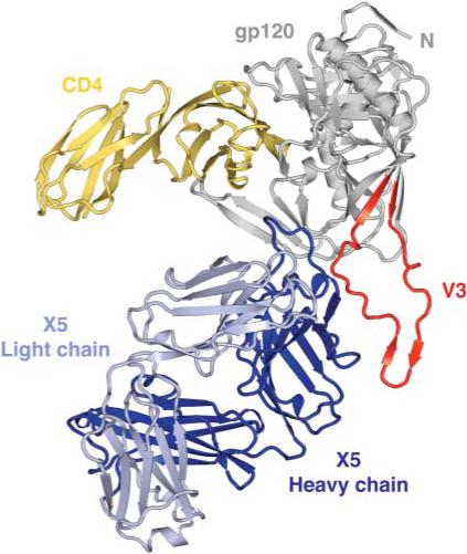
Structure of an HIV-1 gp120 core with V3. The crystal structure of core gp120
(gray) with an intact V3 (red) is shown bound to the membrane-distal two
domains of the CD4 receptor (yellow) and the Fab portion of the ×5
antibody (dark and light blue). In this orientation, the viral membrane
would be positioned toward the top of the page and the target cell toward
the bottom.
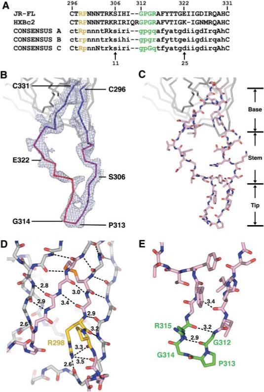
V3 sequence and structure. (A) V3 sequence. The sequences of
JR-FL (17) and HXBc2 are shown along
with the consensus sequence of clades A, B, and C. For the consensus
sequences, absolutely conserved residues are shown in uppercase, with
variable residues in lowercase (37).
Single-letter aminoacid abbreviations: A, Ala; C, Cys; D, Asp; E, Glu; F,
Phe; G, Gly; H, His; I, Ile; K, Lys; M, Met; N, Asn; P, Pro; Q, Gln; R, Arg;
S, Ser; T, Thr; V, Val; Y, Tyr. The conserved (Arg-Pro) and
(Gly-Pro-Gly-Arg) motifs are colored yellow and green, respectively, and are
highlighted with the same colors in (D) and (E). (B) V3
electron density and B values.
2Fobs –
Fcalc density is shown for the entire V3
region and contoured at 1σ. V3 is color-coded by B
value from blue (lower atomic mobility) to red (higher mobility).
(C) V3 structure. The entire V3 is shown (color code:
salmon, carbon atoms; red, oxygen atoms; dark blue, nitrogen atoms; orange,
disulfide bond). Regions corresponding to the fixed base, accordion-like
stem, and β-hairpin tip are labeled. (D) Close-up view
of the V3 base. From its N terminus (Cys296), V3 extends the
antiparallel sheet on the outer domain of gp120. After hydrogen bonding for
three residues, additional sheet contacts are interrupted by two conserved
residues: Arg298, whose side-chain hydrogen bonds to three
carbonyl oxygens, including two on the neighboring outer domain strand; and
Pro299, which initiates the separation of outgoing and
returning V3 strands. In the returning strand, antiparallel β-sheet
interactions with core gp120 recommence with the carbonyl of residue 297 and
continue to the disulfide at Cys331. Main-chain atoms are shown
for the core and V3 base, colored the same as in (C). Hydrogen bonds are
depicted with dashed lines, with select distances in Å. All atoms of
the highly conserved Arg298, Pro299, and
Cys296-Cys331 disulfide are shown, with Arg and
Pro carbons highlighted in yellow and disulfide in orange. (E)
Conformation of the V3 tip. From Ser306 to Gly312, the
main chain assumes a standard β-conformation, which terminates in a
Gly-Pro-Gly-Arg β-turn (residues 312 to 315) (29, 38). After
the turn, the returning density is less well defined, indicative of some
disorder. All atoms of the tip are colored as in (C), with carbon atoms of
the conserved tip highlighted in green. Hydrogen bonds that stabilize the
β hairpin are shown as in (D).
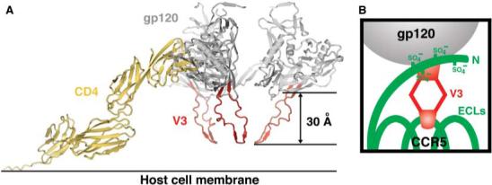
Modeled trimer and co-receptor schematic. (A) V3 in the context
of a trimer at the target cell surface. The structure of the CD4-triggered
gp120 with V3 was superimposed onto the structure of four-domain CD4 (39) and the trimer model obtained by
quantification of surface parameters (32). In this orientation, the target cell membrane and
coreceptor are expected to be positioned toward the bottom of the page.
(B) Schematic of coreceptor interaction. CCR5 (green) is
shown with its tyrosine-sulfated N terminus (at residues 3, 10, 14, and 15)
and three extracellular loops (ECLs). V3 (red) is shown with its conserved
base interacting with the sulfated CCR5 N terminus and its flexible legs
allowing its conserved V3 tip to reach the second ECL of CCR5.
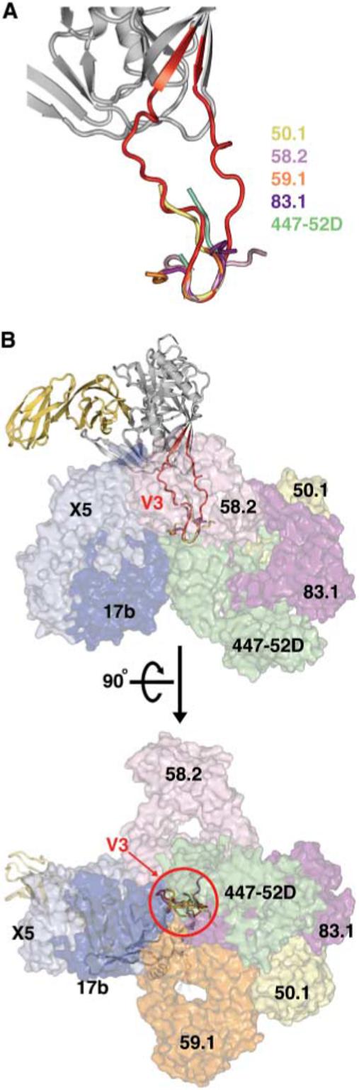
Accessibility of V3 to neutralizing antibodies. The molecular surfaces of
neutralizing antibodies that block coreceptor binding are shown superimposed
onto gp120 in the context of V3; antibodies 17b and ×5 bind to the
conserved coreceptor binding site on the core, whereas monoclonal antibodies
50.1, 58.2, 59.1, 83.1, and 447−52D bind to V3. (A)
Superposition of V3 structures. Core with V3 is shown with V3 peptides as
extracted from peptide–anti-V3 neutralizing antibody complexes after
superposition of the conserved V3 tip. (B) Antibody
accessibility of V3. Core gp120 with V3 (ribbon representation) is shown in
two perpendicular views with Fab fragments (molecular surface
representation) of antibodies that bind at the coreceptor binding site on
either core or V3. V3 is completely surrounded by neutralizing antibodies,
suggesting a high degree of accessibility for generating an immune
response.
References
Publication types
MeSH terms
Substances
Associated data
- Actions
Grants and funding
LinkOut - more resources
Full Text Sources
Other Literature Sources
Molecular Biology Databases
Research Materials
