. 2005 Dec;17(12):3409-21.
doi: 10.1105/tpc.105.035774. Epub 2005 Nov 11.

NAB1 is an RNA binding protein involved in the light-regulated differential expression of the light-harvesting antenna of Chlamydomonas reinhardtii

Affiliations
Free PMC article

NAB1 is an RNA binding protein involved in the light-regulated differential expression of the light-harvesting antenna of Chlamydomonas reinhardtii

Jan H Mussgnug et al. Plant Cell. 2005 Dec.
Free PMC article

Abstract

Photosynthetic organisms respond to changes in ambient light by modulating the size and composition of their light-harvesting complexes, which in the case of the green alga Chlamydomonas reinhardtii consists of >15 members of a large extended family of chlorophyll binding subunits. How their expression is coordinated is unclear. Here, we describe the analysis of an insertion mutant, state transitions mutant3 (stm3), which we show has increased levels of LHCBM subunits associated with the light-harvesting antenna of photosystem II. The mutated nuclear gene in stm3 encodes the RNA binding protein NAB1 (for putative nucleic acid binding protein). In vitro and in vivo RNA binding and protein expression studies have confirmed that NAB1 differentially binds to LHCBM mRNA in a subpolysomal high molecular weight RNA-protein complex. Binding of NAB1 stabilizes LHCBM mRNA at the preinitiation level via sequestration and thereby represses translation. The specificity and affinity of binding are determined by an RNA sequence motif similar to that used by the Xenopus laevis translation repressor FRGY2, which is conserved to varying degrees in the LHCBM gene family. We conclude from our results that NAB1 plays an important role in controlling the expression of the light-harvesting antenna of photosystem II at the posttranscriptional level. The similarity of NAB1 and FRGY2 of Xenopus implies the existence of similar RNA-masking systems in animals and plants.


Figures

Figure 1.
Figure 1.
Identification of stm3 by Fluorescence Spectroscopy and of the Nucleus-Encoded NAB1 Gene in C. reinhardtii. (A) PSII chlorophyll a fluorescence snapshot (55 μmol·m−2·s−1, 620 nm of actinic light) of wild-type and stm3 colonies on Tris-acetate-phosphate (TAP) agar plates in a fluorescence video imager. Colonies were preilluminated for 20 min with 480 nm of blue light to fully gain state 2 conditions. (B) DNA gel blot hybridizations with wild-type and stm3 genomic DNA. Hybridizations were performed with a pSP124S-specific probe. Gray arrows indicate unspecific hybridization, and the black arrow indicates specific hybridization. M, marker. (C) Model of the NAB1 gene. DNA sequences were identified in the stm3 genome by LMS-PCR (Strauss et al., 2001) and subsequent sequence analysis. Black boxes represent exons, and the arrow marks the plasmid pSP124S insertion site. (D) Model of the NAB1 protein with an N-terminal CSD and a C-terminal RRM, and comparison with the Xenopus protein FRGY2. B/A, basic/aromatic region. (E) DNA gel blot hybridizations with wild-type and stm3 genomic DNA. Hybridizations were performed with a radioactively labeled 738-bp NAB1-specific probe. (F) Amino acid sequence of the CSD of NAB1, and alignment with the Xenopus protein FRGY2. Boxes represent β-helix structures, and arrows represent functionally important Phe residues.
Figure 2.
Figure 2.
Evidence for the Disruption of NAB1 Transcription in stm3, and Identification of the NAB1 Complemented Strain nc1 with a Reversed stm3 Phenotype. (A) RNA gel blot hybridizations with wild-type and stm3 mRNA. Hybridizations were performed with a NAB1-specific probe. (B) DNA analysis of the isolated strain nc1 by PCR with primers amplifying either the 5′ (open arrow) or the 3′ (black arrow) region of the NAB1 gene. (C) RT-PCR analysis to detect NAB1 expression using the same primers as in (A) amplifying the 3′ region. The black arrow marks specific NAB1 amplification, the gray arrow marks unspecific amplification, and the open arrow marks degraded RNA. Arrows on the NAB1 gene model mark primer positions used in (A) and (B). (D) Fluorescence video image taken from wild-type, stm3, and nc1 colonies on TAP agar plates preincubated with PSII light to gain maximal state 2. (E) Room-temperature (RT) fluorescence induction curves (Kautsky curves) of the wild type, stm3, and nc1 over a period of 2 min. Dark-precultivated colonies were illuminated with 55 μmol·m−2·s−1 actinic light on TAP agar plates, and fluorescence was recorded in a fluorescence video imager. (F) Coomassie blue–stained SDS-PAGE gel and immunoblot with anti-NAB1. Wild-type, stm3, and nc1 soluble proteins were derived from cell cultures. rNAB1, purified recombinant NAB1 protein.
Figure 3.
Figure 3.
Localization of NAB1 in the Cytosol by Immunogold Labeling. Electron micrographs of wild-type and stm3 cell sections showing anti-NAB1 immunogold-labeled NAB1 proteins as black dots located in the cytosol of the wild type (zoomed sections b, c, and d) but not in the nucleus (zoomed section a), mitochondria, or chloroplast (zoomed section e). Cp, chloroplast; Cyt, cytosol; M, mitochondrion; N, nucleus.
Figure 4.
Figure 4.
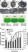
Characteristic Phenotypes of stm3. (A) Electron micrographs of wild-type, stm3, and nc1 cell sections (×13,000 and ×50,000) showing super-stacked thylakoid membranes and higher starch incorporation in stm3 cells compared with wild-type and nc1 cells. Bars = 1 μm. At bottom is an image of the dark-green phenotype of stm3 after growth in TAP medium and the corresponding chlorophyll (Chl) values. All cultures were set up to equal cell densities (OD750 = 0.7); cells were counted in a cell counter as a control. (B) Growth rates and cellular chlorophyll concentrations during cultivation of wild-type and stm3 cell cultures in 400 μmol·m−2·s−1 moderate high light. Standard errors given for cell density and chlorophyll values are based on 10 independent measurements.
Figure 5.
Figure 5.
Effect of NAB1 Deletion on LHC Antenna Protein Expression. (A) One-dimensional electrophoresis of wild-type and stm3 cell extracts based on equal cell density (OD750, controlled by silver staining) and immunoblotting with anti-LHCII. The black arrow marks the LHCBM4/6 protein band, and the gray arrow marks the LHCBM2/8 protein band, as identified by MALDI-TOF analysis. (B) Immunoblots probed with anti-LHCBM4/6 peptide antibodies (Hippler et al., 2001) after protein separation with pH-dependent two-dimensional gel electrophoresis of wild-type, stm3, and nc1 thylakoid membranes. Spots 22, 23, and 24 were assigned according to Hippler et al. (2001). (C) Native gel electrophoresis of wild-type and stm3 thylakoid membranes based on equal chlorophyll concentrations. FP, free pigments; LM, LHCII monomers; LT, LHCII trimers; RC, reaction centers. (D) RNA gel blot analysis of LHCBM4 and LHCBM6 mRNA concentrations in standard light-cultivated wild-type and stm3 cultures. Gene-specific probes were derived from the 3′ UTR, and actin was used as a control for equal loading.
Figure 6.
Figure 6.
Function of NAB1 as an RNA Binding Translation Regulator. (A) Sequence alignment of LHCBM1 to LHCBM9 [(1) to (9)] cDNAs from C. reinhardtii with the consensus motif (CSDCS) recognized by FRGY2 from Xenopus. Identical positions are indicated by a black background, and sequence identity levels are given at right. (B) In vitro RNA binding competition studies. Autoradiogram after UV cross-linking of recombinant NAB1 protein, radiolabeled LHCBM6-CSDCS RNA probe, and a 5-, 10-, and 50-fold molar excess of the indicated nonlabeled competitor RNAs. The exposure time was identical for all lanes. Competition with PSBD 5′ UTR RNA was performed independently. Quantification of RNA binding intensities in relation to the NAB1 signal without competitor (0× value) from one representative experiment was estimated by signal densitometry and plotted as a diagram. (C) In vitro RNA binding studies. Autoradiogram after UV cross-linking of recombinant N-terminal 83–amino acid NAB1 peptide fragment (carrying the complete CSD) and radiolabeled LHCBM6-CSDCS RNA probe (for details, see [B]). Indicated competitor RNAs were added in 5-, 10-, and 50-fold molar excess to the reactions. (D) Analysis of high molecular weight subpolysomal and polysomal RNA–protein fractions derived from wild-type and stm3 cell extracts after 15 to 45% sucrose gradient centrifugation. Isolated RNA from each fraction separated by gel electrophoresis (top panel), and LHCBM6 mRNA levels determined by RNA slot blotting (middle panel; standard errors are based on five independent measurements). Identification of fractions containing NAB1 proteins by anti-NAB1 immunoblotting of nontreated samples (− RNase) and samples pretreated with RNase before centrifugation and separation (+ RNase) (bottom panel). (E) In vivo mRNA binding studies. Anti-NAB1 immunoblot showing purification of immunoprecipitated (IP) native NAB1 from cell extracts, and analysis of bound mRNA by slot-blot analysis and real-time RT-quantitative (Q)-PCR using probes and primers specific for LHCBM6, LHCBM4, and ACTIN. The agarose gel shows transcript abundance after 25 cycles. mRNAs from input cell extracts were used as a positive control. ACTIN, LHCBM4, and LHCBM6 transcripts became detectable after 14 to 16 cycles. Samples without reverse transcriptase (−RT) or template were used as negative controls. LHCBM6 transcripts in RT-Q-PCR studies, using immunoprecipitated NAB1-derived mRNA as a template, became detectable after 16 cycles, whereas LHCBM4 and ACTIN transcripts did not appear before cycles 28 and 39, respectively. Rates for relative transcript abundance are calculated from five independent measurements as 2−[C(T) transcript − C(T) actin], where C(T) = cycle values. The LHCBM6 values were set to 100%.
Figure 7.
Figure 7.
Wild-Type and stm3 Protein Expression Studies of HA-Tagged LHCBM Proteins Carrying Either the LHCBM4-Type CSDCS Motif (HA-Tagged LHCBM4) or the LHCBM6-Type CSDCS Motif (HA-Tagged LHCBM6) under Different Light Regimes. (A) Coomassie blue–stained SDS protein gels as controls for equal loading. (B) Anti-HA tag immunoblots to detect protein levels of HA-tagged LHCBM4 and LHCBM6 in the wild type and stm3 grown in standard light (40 μmol·m−2·s−1) or after treatment for 48 h with moderate high light (180 μmol·m−2·s−1). (C) Quantification of LHCBM4/6 protein expression levels using GelScan 2.0. (D) Quantification control experiment. Dilution series (5, 2.5, and 1.25 μL) of wild-type HA-LHCBM6 samples grown in standard light and compared with a 5-μL sample grown in moderate high light. (E) Real-time RT-quantitative-PCR studies to evaluate LHCBM6 mRNA levels derived from wild-type and stm3 HA-LHCBM6 cell extracts. Standard errors in (C) and (E) are based on three independent measurements.

Similar articles

Cited by

References

    1. Allen, J.F. (1992). Protein phosphorylation in regulation of photosynthesis. Biochim. Biophys. Acta 1098, 275–335. - PubMed
    1. Allen, J.F., and Forsberg, J. (2001). Molecular recognition in thylakoid structure and function. Trends Plant Sci. 6, 317–326. - PubMed
    1. Allen, K.D., and Staehelin, L.A. (1991). Resolution of 16 to 20 chlorophyll protein complexes using a low ionic-strength native green gel system. Anal. Biochem. 194, 214–222. - PubMed
    1. Anderson, S.L., and Kay, S.A. (1995). Functional dissection of circadian clock-regulated and phytochrome-regulated transcription of the Arabidopsis Cab2 gene. Proc. Natl. Acad. Sci. USA 92, 1500–1504. - PMC - PubMed
    1. Barkan, A. (1988). Proteins encoded by a complex chloroplast transcription unit are each translated from both monocistronic and polycistronic mRNAs. EMBO J. 7, 2637–2644. - PMC - PubMed

Publication types

MeSH terms

Associated data

LinkOut - more resources