. 2005 Nov 22;44(46):15203-15.
doi: 10.1021/bi051633z.

Multiple roles of a conserved GAF domain tyrosine residue in cyanobacterial and plant phytochromes

Affiliations

Multiple roles of a conserved GAF domain tyrosine residue in cyanobacterial and plant phytochromes

Amanda J Fischer et al. Biochemistry. .

Abstract

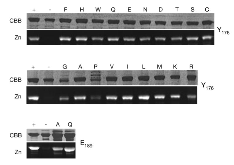
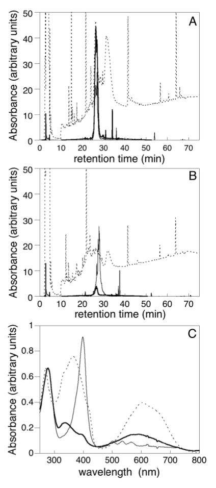
The phytochrome family of red/far-red photoreceptors has been optimized to support photochemical isomerization of a bound bilin chromophore, a process that triggers a conformational change and modulates biochemical output from the surrounding protein scaffold. Recent studies have established that the efficiency of this photochemical process is profoundly altered by mutation of a conserved tyrosine residue (Tyr176) within the bilin-binding GAF domain of the cyanobacterial phytochrome Cph1 [Fischer, A. J., and Lagarias, J. C. (2004) Harnessing phytochrome's glowing potential, Proc. Natl. Acad. Sci. U.S.A. 101, 17334-17339]. Here, we show that the equivalent mutation in plant phytochromes behaves similarly, indicating that the function of this tyrosine in the primary photochemical mechanism is conserved. Saturation mutagenesis of Tyr176 in Cph1 establishes that no other residue can support comparably efficient photoisomerization. The spectroscopic consequences of Tyr176 mutations also reveal that Tyr176 regulates the conversion of the porphyrin-like conformation of the bilin precursor to a more extended conformation. The porphyrin-binding ability of the Tyr176Arg mutant protein indicates that Tyr176 also regulates the ligand-binding specificity of apophytochrome. On the basis of the hydrogen-bonding ability of Tyr176 substitutions that support the nonphotochemical C15-Z,syn to C15-Z,anti interconversion, we propose that Tyr176 orients the carboxyl side chain of a conserved acidic residue to stabilize protonation of the bilin chromophore. A homology model of the GAF domain of Cph1 predicts a C5-Z,syn, C10-Z,syn, C15-Z,anti configuration for the chromophore and implicates Glu189 as the proposed acidic residue stabilizing the extended conformation, an interpretation consistent with site-directed mutagenesis of this conserved acidic residue.


Figures

Figure 1
Figure 1
Domain structure, chromophore configuration, and assembly of phytochromes. (A) Domain organization of the Cph1, BphP, and Phy subfamilies. The N-terminal photosensory region contains the P2 PAS domain, the P3 GAF bilin lyase domain, and the P4 PHY domain. Plant phytochromes (Phy) also possess a Ser/Thr-rich N-terminal P1 domain. The C-terminal regulatory region of these phytochromes contains a domain homologous to histidine kinase transmitter modules. Plant phytochrome C termini possess an additional inserted region that contains two additional PAS domains (labeled PAS1 and PAS2). Cys sites of covalent attachment to the bilin chromophore are indicated. (B) Conformations of the free chromophore precursor phycocyanobilin (PCB) and its protonated form during assembly. Rings are labeled in bold, and the carbons of the ring system are numbered. (Left) A cyclic, deprotonated C5 Z,syn, C10 Z,syn, C15 Z,syn configuration of the initially bound PCB is shown. (Center) Upon protonation of the ring system, PCB adopts a more extended conformation, here shown as C5 Z,syn, C10 Z,syn, C15 Z,anti. (Right) The covalent attachment of PCB to Cph1 results in a covalent thioether linkage to Cys259 with R stereochemistry (). (C) Assembly reaction of PCB with wild-type phytochrome Cph1 is shown (). After initially binding in a cyclic conformation, PCB becomes protonated and adopts a more extended conformation (“ext”). The thioether linkage is then formed (“ext, cov”). KD, initial binding constant for PCB and apoCph1; KPT, equilibrium constant for proton transfer and concomitant chromophore rearrangement; kchem, rate constant for covalent attachment.
Figure 2
Figure 2
Spectroscopic and fluorescent properties of PCB adducts of recombinant Arabidopsis PhyB(N450) wild-type (- - -) and Y276H (—) proteins. (A) Absorbance difference spectra were normalized for equal absorbance at the blue band, demonstrating that the amount of photoconversion is highly reduced for the Y276H mutant compared to the wild-type. (B) Absorbance spectra normalized to the blue band, showing that there is little difference in the bilin configuration or λmax between the wild-type and Tyr276 mutant. (C) Fluorescence excitation (thin line) and emission (thick line) spectra of the wild type and mutant (samples were adjusted to have equal absorbance at 648 nm).
Figure 3
Figure 3
Substitution mutants at Tyr176 and Glu189 alter chromophore configuration. (A) Absorbance spectra were normalized to the blue band. Tyr176 substitutions exhibiting various ranges of R/B ratios are shown with representative absorbance spectra. The spectra shown are wild type, Y176H, Y176A, and Y176I. (B) A plot of the R/B ratio versus the maximal absorbance wavelength for the long wavelength band is shown for the wild-type and mutant proteins with substitutions at Tyr176 (○) or Glu189 (□). Proteins whose spectra are shown in A are indicated in one-letter code. Substitutions at Tyr176 which resulted in fluorescence, are shown in •, while the wild type and other substitutions are shown as ○ and were fitted to a smooth hyperbolic function (—).
Figure 4
Figure 4
Y176R mutant protein affects chromophore-binding specificity. (A) Absorbance (thick solid line), fluorescence excitation (thin solid line), and fluorescence emission (dashed line) spectra of the Y176R mutant Cph1Δ protein. The multiple fluorescence excitation peaks and sharp fluorescence emission peak at approximately 620 nm are characteristic of a porphyrin (), while the broader absorbance around 620 nm is indicative of a second population with bound bilin. (B) Absorbance and fluorescence excitation and emission spectra of the Y176H mutant Cph1Δ protein for comparison. Spectra are the same as in A.
Figure 5
Figure 5
Stereoviews of the Cph1 bilin lyase domain homology model. (A) Proposed Cph1 bilin lyase fold shows a central twisted β sheet, with Tyr176 and Glu189 (blue) on adjacent strands (β1 and β2, respectively). Both side chains point up into the proposed chromophore-binding pocket, with the nucleophilic Cys259 (blue) above Tyr176 and Glu189. β strands are numbered on the basis of their position in the sequence starting from the N terminus. (B) PCB is shown attached to Cys259, with Tyr176 and Glu189 lying below the chromophore (colors: C, gray; H, white; N, blue; O, red; and S, yellow). For Tyr176 and Glu189, atoms within 3.4 Å of PCB are also shown as solvent-accessible surfaces (Tyr176, yellow; and Glu189, orange). PCB atoms within 3.4 Å of Tyr176 are shown as a solvent-accessible surface (light blue). (C) Interactions between Glu189 and PCB. The Glu189 side chain is hydrogen-bonded to the B/C-ring system of the protonated chromophore, stabilizing the positive charge. The rings of PCB are labeled for clarity.
Supplemental Figure 1
The Cph1 GAF domain and chromophore-binding pocket. (A) Sequence alignment of the Cph1 GAF domain and the two GAF domains of mouse phosphodiesterase 2A (PDB code 1MC0: (1)) with Tyr176 and Glu189 in red. (B) Residues within 3 Å of PCB (red) are continually colored by identity ranging from dark blue, absolutely conserved, to bright red, variable using an alignment of Cph1, the five phytochrome proteins in the A. thaliana genome, and BphP proteins from P. aeruginosa and Deinococcus radiodurans (2, 3).
Supplemental Figure 2
SDS/PAGE (CBB) and zinc-blot (Zn) analysis of Cph1Δ mutant proteins as PCB adducts. 3 μg of total protein was run per lane except for Y176P (30 μg). Wildtype holoCph1Δ (+) and apoCph1Δ (−) are shown for each gel. The remaining lanes are labeled with the amino acid substituted for Tyr at position 176 (top two panels) or for Glu at position 189 (bottom panel). Y176P is largely insoluble; therefore a ten-fold excess of total protein was loaded in order to demonstrate its presence. The Cph1Δ proteins all migrate with a relative molecular mass of 60 kDa.
Supplemental Figure 3
Analysis of chromopeptides. Peptides derived from wildtype (A) and Y176R (B) Cph1 were analyzed by HPLC: 220 nm (dashed), 400 nm (porphyrin, thin line), and 650 nm (bilin, thick line). (C) Absorbance spectra for wildtype (dashed) and Y176R (peak 1, thick line; peak 2, thin line) chromopeptides.

References

    1. Smith H. Phytochromes and light signal perception by plants—An emerging synthesis. Nature. 2000;407:585–591. - PubMed
    1. Schäfer, E., and Nagy, F., Eds. (2005) Photomorphogenesis in Plants and Bacteria: Function and Signal Tranduction Mechanisms, 3rd ed., pp 667, Springer Publishers, Dordrecht, The Netherlands.
    1. Nagy F, Schäfer E. Phytochromes control photomorphogenesis by differentially regulated, interacting signaling pathways in higher plants. Annu Rev Plant Biol. 2002;53:329–355. - PubMed
    1. Quail PH. Phytochrome photosensory signalling networks. Nat Rev Mol Cell Biol. 2002;3:85–93. - PubMed
    1. Chen M, Chory J, Fankhauser C. Light signal transduction in higher plants. Annu Rev Gen. 2004;38:87–117. - PubMed

Publication types