In vitro selection, characterization, and application of deoxyribozymes that cleave RNA
- PMID: 16286368
- PMCID: PMC1283523
- DOI: 10.1093/nar/gki930
In vitro selection, characterization, and application of deoxyribozymes that cleave RNA
Abstract
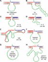
Over the last decade, many catalytically active DNA molecules (deoxyribozymes; DNA enzymes) have been identified by in vitro selection from random-sequence DNA pools. This article focuses on deoxyribozymes that cleave RNA substrates. The first DNA enzyme was reported in 1994 and cleaves an RNA linkage. Since that time, many other RNA-cleaving deoxyribozymes have been identified. Most but not all of these deoxyribozymes require a divalent metal ion cofactor such as Mg2+ to catalyze attack by a specific RNA 2'-hydroxyl group on the adjacent phosphodiester linkage, forming a 2',3'-cyclic phosphate and a 5'-hydroxyl group. Several deoxyribozymes that cleave RNA have utility for in vitro RNA biochemistry. Some DNA enzymes have been applied in vivo to degrade mRNAs, and others have been engineered into sensors. The practical impact of RNA-cleaving deoxyribozymes should continue to increase as additional applications are developed.
Figures


Similar articles
-
In vitro evolution of an RNA-cleaving DNA enzyme into an RNA ligase switches the selectivity from 3'-5' to 2'-5'.J Am Chem Soc. 2003 May 7;125(18):5346-50. doi: 10.1021/ja0340331. J Am Chem Soc. 2003. PMID: 12720447
-
Deoxyribozymes: DNA catalysts for bioorganic chemistry.Org Biomol Chem. 2004 Oct 7;2(19):2701-6. doi: 10.1039/B411910J. Epub 2004 Sep 3. Org Biomol Chem. 2004. PMID: 15455136 Review.
-
Catalytic DNA (deoxyribozymes) for synthetic applications-current abilities and future prospects.Chem Commun (Camb). 2008 Aug 14;(30):3467-85. doi: 10.1039/b807292m. Epub 2008 Jul 1. Chem Commun (Camb). 2008. PMID: 18654692 Review.
-
Catalytic DNA: Scope, Applications, and Biochemistry of Deoxyribozymes.Trends Biochem Sci. 2016 Jul;41(7):595-609. doi: 10.1016/j.tibs.2016.04.010. Epub 2016 May 25. Trends Biochem Sci. 2016. PMID: 27236301 Free PMC article. Review.
-
Zn2+-dependent deoxyribozymes that form natural and unnatural RNA linkages.Biochemistry. 2005 Jun 28;44(25):9217-31. doi: 10.1021/bi050146g. Biochemistry. 2005. PMID: 15966746 Free PMC article.
Cited by
-
DNAzyme as a rising gene-silencing agent in theranostic settings.Neural Regen Res. 2022 Sep;17(9):1989-1990. doi: 10.4103/1673-5374.335157. Neural Regen Res. 2022. PMID: 35142687 Free PMC article. No abstract available.
-
Splitting aptamers and nucleic acid enzymes for the development of advanced biosensors.Nucleic Acids Res. 2020 Apr 17;48(7):3400-3422. doi: 10.1093/nar/gkaa132. Nucleic Acids Res. 2020. PMID: 32112111 Free PMC article. Review.
-
DNAzyme-Catalyzed Site-Specific N-Acylation of DNA Oligonucleotide Nucleobases.Angew Chem Int Ed Engl. 2024 Feb 12;63(7):e202317565. doi: 10.1002/anie.202317565. Epub 2024 Jan 11. Angew Chem Int Ed Engl. 2024. PMID: 38157448 Free PMC article.
-
DNA-catalyzed covalent modification of amino acid side chains in tethered and free peptide substrates.Biochemistry. 2011 May 31;50(21):4741-9. doi: 10.1021/bi200585n. Epub 2011 May 3. Biochemistry. 2011. PMID: 21510668 Free PMC article.
-
Nanovehicular intracellular delivery systems.J Pharm Sci. 2008 Sep;97(9):3518-90. doi: 10.1002/jps.21270. J Pharm Sci. 2008. PMID: 18200527 Free PMC article. Review.
References
-
- Doudna J.A., Cech T.R. The chemical repertoire of natural ribozymes. Nature. 2002;418:222–228. - PubMed
-
- Collins C.A., Guthrie C. The question remains: is the spliceosome a ribozyme? Nat. Struct. Biol. 2000;7:850–854. - PubMed
-
- Villa T., Pleiss J.A., Guthrie C. Spliceosomal snRNAs: Mg2+-dependent chemistry at the catalytic core? Cell. 2002;109:149–152. - PubMed
-
- Ban N., Nissen P., Hansen J., Moore P.B., Steitz T.A. The complete atomic structure of the large ribosomal subunit at 2.4 Å resolution. Science. 2000;289:905–920. - PubMed
-
- Cech T.R. The ribosome is a ribozyme. Science. 2000;289:878–879. - PubMed
Publication types
MeSH terms
Substances
LinkOut - more resources
Full Text Sources
Other Literature Sources

