. 2005 Nov 21;171(4):603-14.
doi: 10.1083/jcb.200507002. Epub 2005 Nov 14.

p62/SQSTM1 forms protein aggregates degraded by autophagy and has a protective effect on huntingtin-induced cell death

Affiliations

p62/SQSTM1 forms protein aggregates degraded by autophagy and has a protective effect on huntingtin-induced cell death

Geir Bjørkøy et al. J Cell Biol. .

Abstract

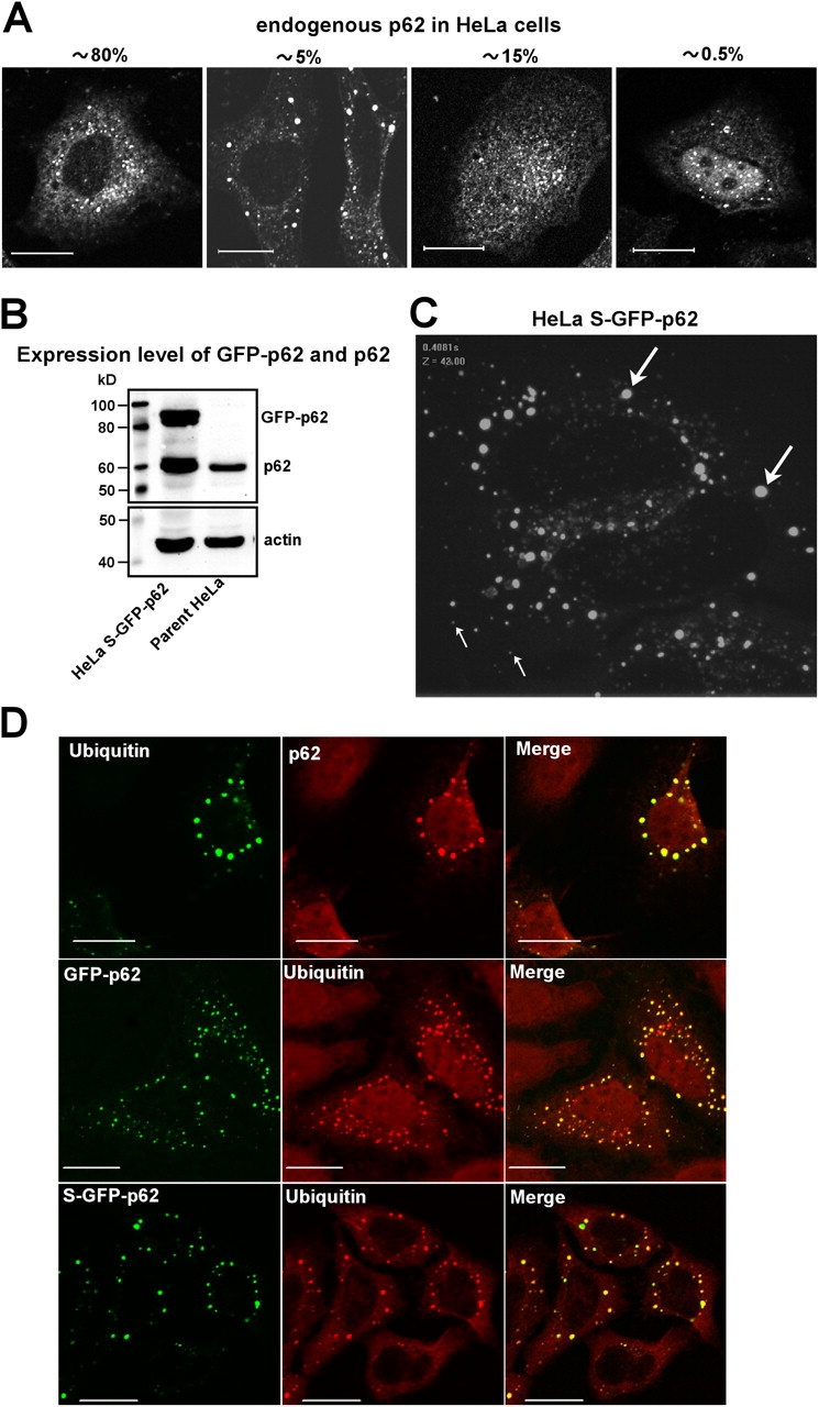
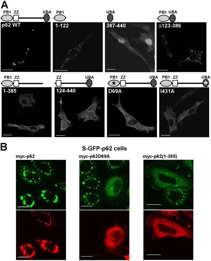
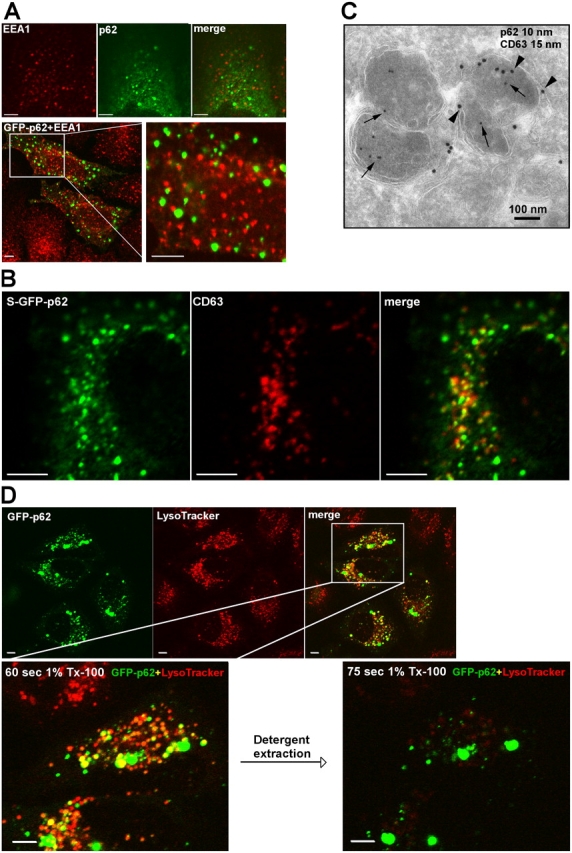
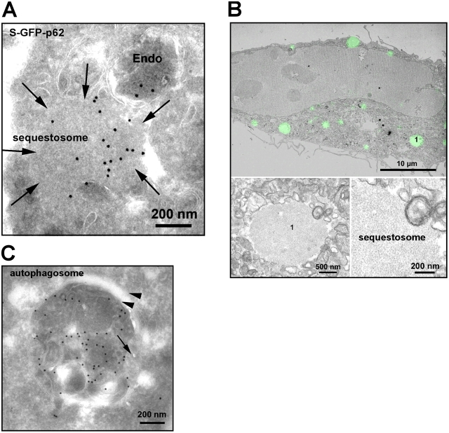
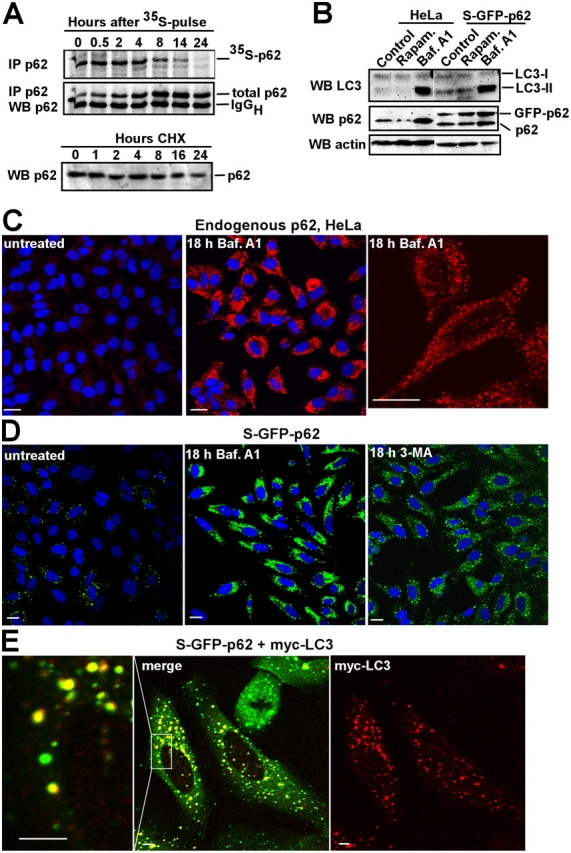
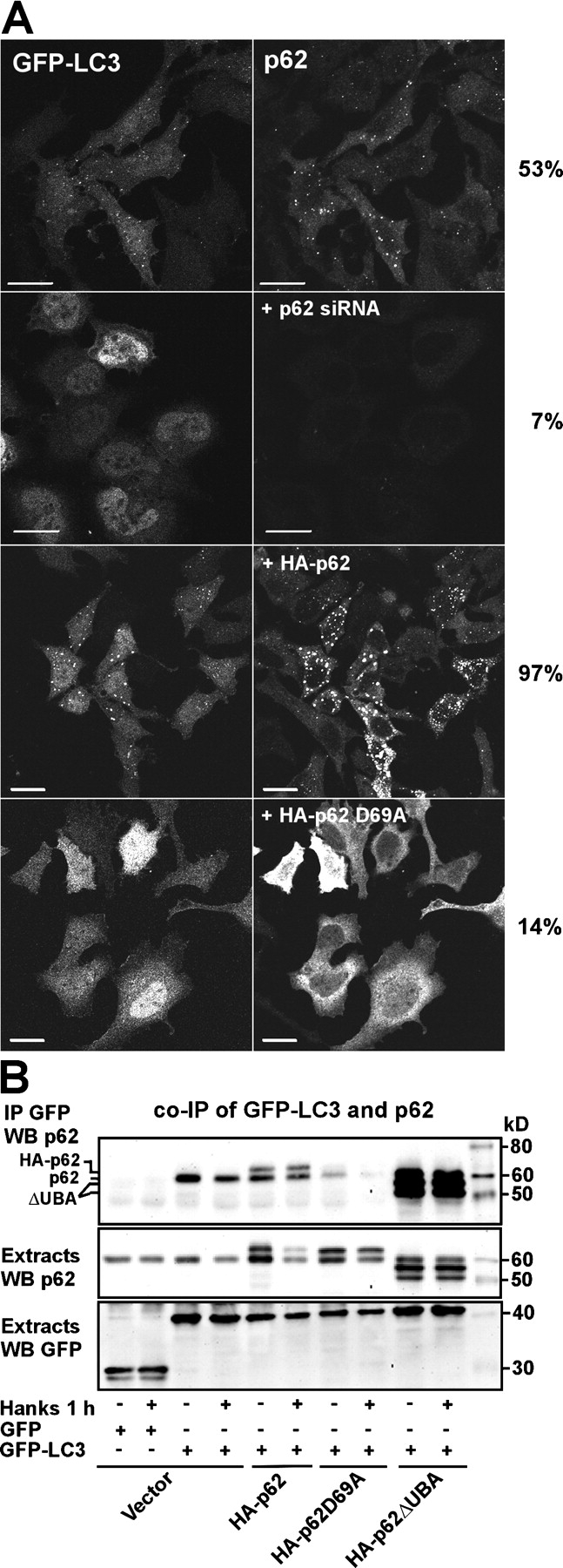
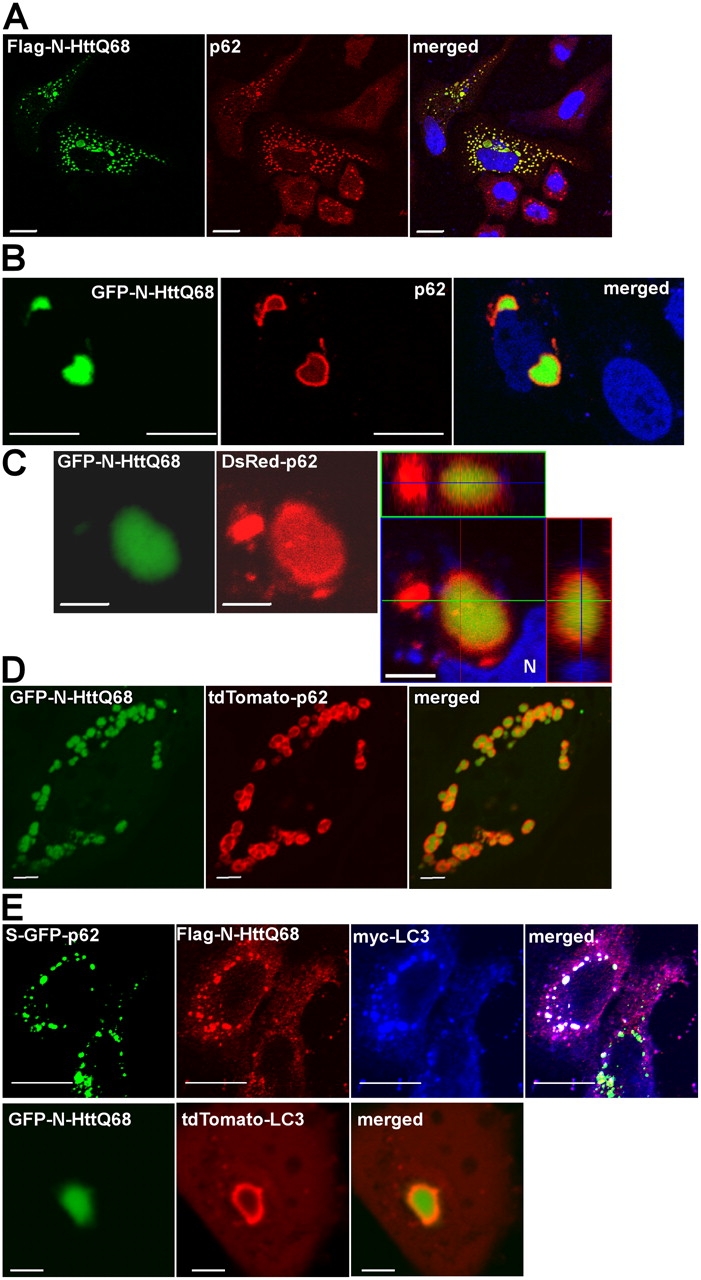
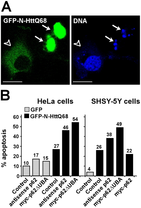
Autophagic degradation of ubiquitinated protein aggregates is important for cell survival, but it is not known how the autophagic machinery recognizes such aggregates. In this study, we report that polymerization of the polyubiquitin-binding protein p62/SQSTM1 yields protein bodies that either reside free in the cytosol and nucleus or occur within autophagosomes and lysosomal structures. Inhibition of autophagy led to an increase in the size and number of p62 bodies and p62 protein levels. The autophagic marker light chain 3 (LC3) colocalized with p62 bodies and co-immunoprecipitated with p62, suggesting that these two proteins participate in the same complexes. The depletion of p62 inhibited recruitment of LC3 to autophagosomes under starvation conditions. Strikingly, p62 and LC3 formed a shell surrounding aggregates of mutant huntingtin. Reduction of p62 protein levels or interference with p62 function significantly increased cell death that was induced by the expression of mutant huntingtin. We suggest that p62 may, via LC3, be involved in linking polyubiquitinated protein aggregates to the autophagy machinery.


Figures

Figure 1.
Figure 1.
The p62 bodies in the cytoplasm of HeLa cells are ubiquitin-containing protein aggregates. (A) HeLa cells were fixed and stained for p62. The different patterns of p62 distribution were scored in >200 cells as cytoplasmic with small bodies, cytoplasmic with large and intense bodies, equal nuclear and cytoplasmic staining, and nuclear enriched. The percentage of cells in the respective groups is indicated. (B) Expression level of GFP-p62 in HeLa cells stably expressing GFP-p62 (S–GFP-p62) compared with the level of endogenous p62 in the parent HeLa cells. (C) Cells stably expressing GFP-p62 display a cytoplasmic punctuate localization of GFP-tagged p62 similar to endogenous p62. Video microscopy of live cells demonstrated that the small, faint bodies (thin arrows) displayed directed migration, whereas the majority of the larger, intense bodies (thick arrows) were nonmigratory (Videos 1 and 2, available at http://www.jcb.org/cgi/content/full/jcb.200507002/DC1). A still image from Video 1 is shown here. (D) Bodies containing either endogenous p62 or transiently or stably transfected GFP-p62 all contain polyubiquitin. HeLa cells were fixed and stained with p62 and polyubiquitin (clone FK1) mAbs directly coupled with AlexaFluor555 (red) and AlexaFluor488 (green), respectively. Cells expressing GFP-p62 were only stained for polyubiquitin (red). Bars, 20 μm.
Figure 2.
Figure 2.
Both the PB1 and UBA domains are needed for p62 to form cytoplasmic bodies. (A) The indicated deletion constructs were fused COOH terminal to GFP and expressed in NIH3T3 fibroblasts. Asterisks indicate point mutations in the PB1 (D69A) and UBA (I431A) domains, respectively. (B) S–GFP-p62 cells were transiently transfected with the indicated myc-tagged p62 constructs, fixed, and stained for the myc tag. Bars, 20 μm.
Figure 3.
Figure 3.
p62 bodies are found both as membrane-free protein aggregates and as membrane-confined autophagosomal and lysosomal structures. (A) Localization of bodies containing endogenous p62 or transiently expressed GFP-p62 relative to EEA1-positive early endosomes. Endogenous p62 were stained green using p62 antibodies directly labeled with AlexaFluor488, and EEA1 was stained red with EEA1 mAbs directly labeled with AlexaFluor555. Alternatively, transiently expressed GFP-p62 was expressed in HeLa cells, and EEA1 was stained red with EEA1 mAb (bottom). The boxed area is shown to the right at a higher magnification. (B) Colocalization of GFP-p62 and CD63 (stained red using a mAb) in HeLa cells stably expressing GFP-p62. (C) Immunoelectron micrograph of S–GFP-p62 stained with a GFP pAb (10-nm gold particle, arrows) and monoclonal CD63 (15-nm gold particle, arrowheads). (D) Rapid detergent extraction of GFP-p62 from LysoTracker-positive acidic organelles. HeLa cells transiently expressing GFP-p62 were labeled with LysoTracker for 60 min. The detergent extractions were imaged in a time series with 15-s time intervals after adding 1% Triton X-100. The boxed area is shown in the bottom two images at a higher magnification before and after detergent extraction. Bars (A, B, and D), 20 μm.
Figure 4.
Figure 4.
p62 is found both in autophagosomes and in cytosolic aggregates/sequestosomes. (A) Immuno-EM of GFP-p62. S–GFP-p62 cells were labeled with rabbit anti-GFP (Abcam) followed by protein A–gold (15 nm). We observed labeling in membrane-free cytosolic structures and sequestosomes (arrows) as well as in endosomes (Endo). (B) Correlative immunofluorescence/EM of HeLa cells treated with 10 μM PSI for 5 h displaying typical sequestosomes. The insets show two magnifications of the sequestosome, which is labeled 1. (C) Representative image of a p62-containing autophagosome. HeLa cells transfected with GFP-p62 were immunogold labeled as in A. Note the cisternal-like membrane (arrowheads) surrounding the GFP-positive material. The arrow indicates a fused endosome.
Figure 5.
Figure 5.
p62 bodies are degraded by autophagy. (A) Comparison of p62 degradation using pulse-chase labeling with 35S-methionine and immunoblotting after cycloheximide (CHX) treatment. HeLa cells were pulsed with 35S-methionine and incubated for the indicated times in nonradioactive medium. After immunopurification, the amount of radioactive p62 was determined by autoradiography, and the total amount of p62 was determined by an immunoblot of the same membrane. The p62 level in total cellular lysates after different times of 10 μg/ml cycloheximide treatment determined by immunoblotting (bottom). (B) The levels of p62 and LC3-II change with autophagic activity. HeLa or S–GFP-p62 cells were either left untreated or rapamycin (10 μg/ml) or bafilomycin A1 (Baf. A1; 10 μg/ml) was added for 18 h. Immunoblots were sequentially probed using LC3, p62, and actin antibodies. (C and D) The amount of p62 located to cytoplasmic bodies increases upon inhibition of autophagy. HeLa cells or S–GFP-p62 HeLa cells were left untreated or bafilomycin A1 was added for 18 h, the cells were fixed, and p62 was either stained red using a p62 mAb (C) or imaged directly (D). Nuclei were visualized using the Draq5 DNA stain. The settings for imaging were identical for the treated cells and the untreated control. (E) The majority of S–GFP-p62 cytoplasmic bodies are stained with antibodies recognizing transiently expressed LC3. S–GFP-p62 cells were transiently transfected with myc-LC3. Myc-LC3 was stained red using an anti-myc tag mAb. The boxed area indicates the part of the cell that is shown to the left at a higher magnification. Bars, 5 μm.
Figure 6.
Figure 6.
p62 is required for the formation of GFP-LC3–positive punctuated structures in HeLa cells. (A) HeLa cells transiently transfected with GFP-LC3 alone or cotransfected with siRNA against p62, HA-p62, or HA-p62 D69A were either left in normal medium or starved for amino acids for 1 h. The cells were fixed, and p62 was stained using a p62 mAb. More than 200 randomly selected cells for each condition were scored for the cytoplasmic pattern of GFP-LC3 as either diffuse or punctuate. The frequency of GFP-LC3–positive cells with punctuate localization are indicated to the right. (B) Endogenous p62 as well as coexpressed myc-tagged wild-type p62 or a UBA deletion mutant of p62 coimmunoprecipitated with GFP-LC3 from HeLa cell extracts. GFP or GFP-LC3 was immunoprecipitated from total cellular extracts after cotransfecting the indicated constructs. The cell cultures were either left untreated or starved for amino acids for 1 h as indicated. Copurified endogenous or ectopically expressed myc-tagged p62 constructs were detected using the p62 mAb. Bars, 20 μm.
Figure 7.
Figure 7.
p62 forms a shell around huntingtin protein aggregates. (A) HeLa cells transiently expressing a Flag-tagged NH2-terminal fragment (amino acids 1–171) of huntingtin containing a 68-polyglutamine expansion, Flag–N-HttQ68. After 48 h, the cells were fixed, and Flag–N-HttQ68 was stained green using a Flag mAb. Endogenous p62 was stained red using the p62 pAb. (B) Endogenous p62 forms a shell around aggregated GFP–N-HttQ68. Transiently transfected HeLa cells were fixed 48 h after transfection, and endogenous p62 was detected using a p62 mAb (red). (C and D) The p62 shell surrounding GFP–N-HttQ68 was detected independently of the use of antibodies. (C) GFP–N-HttQ68 and DsRed-tagged p62 were cotransfected into HeLa cells, and images of live cells were obtained after 48 h of expression. The left panel shows one confocal plane of a representative GFP–N-HttQ68 and DsRed-p62 double-positive structure, and the right panel shows the Z-stack of planes with side views of the aggregate at the side and at the top. N, nucleus. (A–C) Nuclei were detected using Draq5. (D) GFP–N-HttQ68 and tdTomato-tagged p62 were cotransfected into HeLa cells, and images of live cells were obtained after 24 h of expression. (E) Transiently expressed Flag–N-HttQ68 and myc-LC3 colocalize in GFP-p62–positive structures. S–GFP-p62 cells were fixed 48 h after transfection, Flag–N-HttQ68 was stained red using a Flag tag mAb, and myc-LC3 was stained far red (blue) using a chicken myc tag pAb (top). The bottom panel shows a live cell image of a GFP–N-HttQ68 aggregate surrounded by a shell containing tdTomato-LC3. Bars, 5 μm.
Figure 8.
Figure 8.
Interfering with p62 protein level or function increases cell death induced by mutant huntingtin. (A) GFP-positive cells were scored as live or apoptotic by the appearance of nuclear Draq5 staining. After 48 h of expression, GFP-positive cells attached to the surface with a normal nuclear appearance (open arrowheads) and were scored as live, whereas rounded, partially detached cells with a condensed or fragmented nuclei (arrows) were scored as dead. More than 200 GFP-positive cells in randomly selected microscope views were scored. Bars, 20 μm. (B) The effect of coexpressing either a p62 antisense construct or a deletion construct of p62 lacking the UBA domain on N-HttQ68–induced cell death in HeLa and SHSY-5Y neuroblastoma cells.

Similar articles

Cited by

References

    1. Abeliovich, H., W.A. Dunn Jr., J. Kim, and D.J. Klionsky. 2000. Dissection of autophagosome biogenesis into distinct nucleation and expansion steps. J. Cell Biol. 151:1025–1034. - PMC - PubMed
    1. Arrasate, M., S. Mitra, E.S. Schweitzer, M.R. Segal, and S. Finkbeiner. 2004. Inclusion body formation reduces levels of mutant huntingtin and the risk of neuronal death. Nature. 431:805–810. - PubMed
    1. Avila, A., N. Silverman, M.T. Diaz-Meco, and J. Moscat. 2002. The Drosophila atypical protein kinase C-ref(2)p complex constitutes a conserved module for signaling in the toll pathway. Mol. Cell. Biol. 22:8787–8795. - PMC - PubMed
    1. Bence, N.F., R.M. Sampat, and R.R. Kopito. 2001. Impairment of the ubiquitin-proteasome system by protein aggregation. Science. 292:1552–1555. - PubMed
    1. Cariou, B., D. Perdereau, K. Cailliau, E. Browaeys-Poly, V. Bereziat, M. Vasseur-Cognet, J. Girard, and A.F. Burnol. 2002. The adapter protein ZIP binds Grb14 and regulates its inhibitory action on insulin signaling by recruiting protein kinase Czeta. Mol. Cell. Biol. 22:6959–6970. - PMC - PubMed

Publication types

MeSH terms