. 2005 Nov 21;171(4):729-38.
doi: 10.1083/jcb.200508010. Epub 2005 Nov 14.

Growth factor-induced shedding of syndecan-1 confers glypican-1 dependence on mitogenic responses of cancer cells

Affiliations

Growth factor-induced shedding of syndecan-1 confers glypican-1 dependence on mitogenic responses of cancer cells

Kan Ding et al. J Cell Biol. .

Abstract

The cell surface heparan sulfate proteoglycan (HSPG) glypican-1 is up-regulated by pancreatic and breast cancer cells, and its removal renders such cells insensitive to many growth factors. We sought to explain why the cell surface HSPG syndecan-1, which is also up-regulated by these cells and is a known growth factor coreceptor, does not compensate for glypican-1 loss. We show that the initial responses of these cells to the growth factor FGF2 are not glypican dependent, but they become so over time as FGF2 induces shedding of syndecan-1. Manipulations that retain syndecan-1 on the cell surface make long-term FGF2 responses glypican independent, whereas those that trigger syndecan-1 shedding make initial FGF2 responses glypican dependent. We further show that syndecan-1 shedding is mediated by matrix metalloproteinase-7 (MMP7), which, being anchored to cells by HSPGs, also causes its own release in a complex with syndecan-1 ectodomains. These results support a specific role for shed syndecan-1 or MMP7-syndecan-1 complexes in tumor progression and add to accumulating evidence that syndecans and glypicans have nonequivalent functions in vivo.


Figures

Figure 1.
Figure 1.
Glypican dependence of pancreatic carcinoma cell responses to FGF2. (A) PANC-1 cells maintained in serum-free medium were treated with or without 10 ng/ml FGF2 along with [3H]thymidine for 24 h. Cultures received 1 U/ml PIPLC (third bar) or 8 mU/ml heparinase III (fourth bar) 1 h before FGF addition and throughout the remainder of the assay. DNA was precipitated, and 3H incorporation was measured. Data are means ± SD of triplicates. (B) Glyp-TM+ samples were two independent clones of PANC-1 cells stably transfected with a transmembrane variant of glypican-1 (Kleeff et al., 1998). Sham-transfected samples were from two independent control PANC-1 clones transfected with vector only. Both types of clones were assayed as in A except that the concentration of FGF2 was 1 ng/ml. (A and B) Y axis is measured in counts per minute. (C) One of the Glyp-TM+ and one of the control clones from B were tested for MAPK activation after exposure to 2 ng/ml FGF2 for 1 h. For the right two lanes (PIPLC), 1 U/ml enzyme was added 1 h before FGF2 addition as well as during FGF2 incubation. Cells were lysed and subjected to Western blotting for p42/44ERK. (D) Mean values ± SD (error bars) of band intensities for each of the duplicate determinations shown in C; a similar picture is obtained if the data are expressed normalized to a loading control (not depicted). The reduction in MAPK activation by PIPLC in sham-transfected cells is statistically significant (P < 0.05; t test). Y axis is measured in arbitrary units. The results in A–C were also confirmed with FGF2 isolated from bovine brain (not depicted).
Figure 2.
Figure 2.
PIPLC releases only a fraction of cell surface heparan sulfate. PANC-1 cells metabolically labeled with [35S]sulfate were treated with or without PIPLC, and the supernatant was collected. Cells of each type were then incubated with or without TPCK-treated trypsin for 10 min on ice followed by the addition of trypsin inhibitor and collection of the supernatant. In this way, labeled fractions released by no enzyme, PIPLC, trypsin, and trypsin after PIPLC were obtained. To quantify protein-associated GAGs in these fractions, aliquots were digested with heparinase III, chondroitinase ABC, or no enzyme followed by TCA precipitation. Specific release of heparan sulfate (solid bars) by PIPLC was defined as the difference between chondroitinase-resistant radioactivity released from cells exposed and cells not exposed to PIPLC. The analogous calculations were performed to measure the specific release of heparan sulfate by trypsin or by trypsin after pretreatment with PIPLC. Similarly, the specific release of chondroitin sulfate (dotted bars) was calculated as heparinase-resistant radioactivity in the same fractions. Data are means ± SD (error bars) for duplicate experiments. Similar overall results were obtained when heparan sulfate was calculated by subtracting the amount of radioactivity precipitated by TCA after heparinase digestion from that precipitated from undigested supernatants. Likewise, similar results were obtained when chondroitin sulfate was calculated by subtracting the amount of radioactivity precipitated by TCA after chondroitinase digestion from that precipitated from undigested supernatants.
Figure 3.
Figure 3.
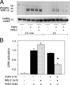
PIPLC blocks long-term but not short-term MAPK activation. (A) Where indicated, PANC-1 cells were incubated with 1 U/ml PIPLC for 1 h. Serum-free medium containing 1 ng/ml FGF2 (with 1 U/ml PIPLC where used) was added, and incubation continued for either 15 min or 1 h. Cells were lysed, and activated MAPK (p42/44ERK) was detected by immunoblotting. As a control for protein loading, immunoblotting was performed with an anti–β-tubulin mAb. Results from duplicate cultures are shown. (B) Cells were treated with FGF2 and analyzed as in A except that some samples were also exposed to FGF2 during the 15 min before the 1-h PIPLC incubation. When such cells were subsequently reexposed to FGF, short-term (15 min) MAPK activation was substantially PIPLC sensitive (asterisk; P < 0.02; t test). Data are duplicates ± SEM (error bars). Y axis is measured in arbitrary units.
Figure 4.
Figure 4.
FGF2 induces shedding of syndecan-1, and an “unsheddable” syndecan-1 makes FGF2 responses PIPLC resistant. (A) PANC-1 cells were treated with or without 2 ng/ml FGF2 for 30 min. After washing, trypsin was used to specifically release syndecan-1 ectodomains. Half of each sample was digested with 8 mU/ml heparinase III and 0.1 U/ml chondroitinase ABC. Syndecan-1 ectodomains were detected by Western blotting with mAb B-B4. Exposure times for the first and second lanes were longer than for the third and fourth lanes. (B) PANC-1 cells were stably transfected with expression constructs for wild-type mouse syndecan-1, an engineered variant of mouse syndecan-1 that replaces the cleavage sequence required for shedding with a heterologous one, or empty expression vector. Multiple clones of each type were expanded and examined by immunocytochemistry (not depicted) and Western blotting using mouse-specific syndecan-1 mAb 281.2. Western blot results from three representative clones are shown. Lanes 1 and 2, sham transfected; lanes 3 and 4, wild-type syndecan-1; lanes 5 and 6, cleavage mutant syndecan-1. (C) PANC-1 cells stably transfected with wild-type or cleavage mutant mouse syndecan-1 (from B) were treated with 2 ng/ml FGF2 for 30 min. After rinsing, cells were treated with trypsin as in A, and the released material was digested with 8 mU/ml heparinase III and 0.1 U/ml chondroitinase ABC. Syndecan-1 core protein was measured as in B. (A–C) Arrowheads show positions of molecular mass standards (in kD). (D) The three clones shown in B were tested for MAPK activation 1 h after the addition of 1 ng/ml FGF2. For both the sham-transfected and wild-type syndecan-1–transfected clones, pretreatment with 1 U/ml PIPLC (for 1 h) dramatically reduced FGF signaling (P < 0.02 in both cases; asterisks), whereas in the clone-expressing cleavage mutant syndecan-1, no significant reduction was seen. Data are from triplicate cultures for each condition and are normalized to loading controls. Error bars represent SEM. Y axis is measured in arbitrary units.
Figure 5.
Figure 5.
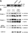
Inhibition of metalloproteinases protects cells from PIPLC inhibition of the FGF2 response. (A) Metalloproteinase inhibitors block FGF2-induced shedding of syndecan-1. PANC-1 cells were treated with or without 1μM GM6001 or 500 ng/ml TIMP-3 for 1 h and with 2 ng/ml FGF2 for 30 min. Syndecan-1 remaining on cell surfaces was released with trypsin, concentrated, digested with heparinase and chondroitinase, and quantified by Western blotting with mAb B-B4 as in Fig. 4. Arrows show molecular mass markers. (B) Metalloproteinase inhibition makes long-term FGF2 responses of tumor cells PIPLC insensitive, whereas the FGF2 responses of a nontumor cell line are already insensitive to PIPLC. PANC-1 cells, MDA-MB-468 breast carcinoma cells, and C2C12 mouse myoblasts were treated for 1 h with 1 μM GM6001, 500 ng/ml TIMP-3, or no protease inhibitor as indicated. Cells were then cultured for 1 h in the presence of 1 ng/ml FGF2 or no growth factor (control). FGF2 + PIPLC cells were also exposed to 1 U/ml PIPLC during both the first and second hours of incubation. Cell lysates were probed for p42/44ERK activation as in Figs. 1–4.
Figure 6.
Figure 6.
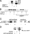
MMP7 is activated by FGF2, causes syndecan-1 shedding, and is sufficient to make FGF responses PIPLC sensitive. (A) Exogenous MMP7 causes shedding of syndecan-1. PANC-1 cells were treated with or without 1 μg/ml MMP7 for 30 min. The media were collected, concentrated, and digested with 8 mU/ml heparinase III and 0.1 U/ml chondroitinase ABC followed by SDS-PAGE and Western blotting with mAb B-B4. Syndecan-1 is seen as a high molecular mass smear that is converted by GAGases to a band of ∼80 kD apparent size. (B) When PANC-1 cells are pretreated with MMP7, even short-term responses to FGF2 become PIPLC sensitive. The responses of PANC-1 cells to a 15-min treatment with FGF2 were measured as in Fig. 3 A except that where indicated, cells were treated with 1 μg/ml MMP7 30 min before the addition of PIPLC (if used) and throughout the 15-min exposure to FGF2. In MMP7-treated cells, MAPK activation was strongly decreased by PIPLC (P < 0.01). (C) FGF2 induces MMP7 activation and release from the cell surface. PANC-1 cells were exposed to 2 ng/ml FGF2 for 30 min. The medium was removed, and cell surface MMP7 was released with 0.3 mg/ml heparin (in PBS). After concentrating as in A, samples were subjected to SDS-PAGE and Western blotting using a mixture of antibodies specific for human pro-MMP7 and activated MMP7. The asterisk shows the increase in released active MMP7 in response to FGF2 treatment. (A and C) Arrows on the right show positions of molecular mass standards (in kD). (D) FGF2 releases complexes of syndecan-1 and active MMP7. PANC-1 cells were treated with 5 ng/ml FGF2 for 30 min. Medium was collected and immunoprecipitated with antisyndecan-1 antibody, and precipitates were subjected to SDS-PAGE and Western blotting for activated MMP7. Controls consisted of immunoprecipitates from cells not treated with FGF2 or precipitation omitting antisyndecan antibody. The top two bands (asterisks) in the antibody-containing samples are IgG heavy and light chains. The lowest band is active MMP7 (arrow).
Figure 7.
Figure 7.
MMP7 is required for syndecan-1 shedding. (A) Syndecan-1 shedding activity can be removed from the cell surface by treatment with heparin. PANC-1 cells were treated with or without 0.3 mg/ml heparin for 30 min. After washing twice with PBS, cells were exposed to 5 ng/ml FGF2 for 30 min. Medium was collected and subjected to heparinase and chondroitinase digestion as in Fig. 6 A. Samples were concentrated and subjected to SDS-PAGE and Western blotting for syndecan-1, the core protein of which appears at ∼80 kD (arrows show positions of molecular mass markers in kD). Results from two independent tests are shown. (B) Pretreatment with heparin rescues the PIPLC dependence of long-term FGF signaling. PANC-1 cells were treated with heparin as in A and were tested for the PIPLC dependence of long-term (1 h) FGF2 signaling as in Fig. 3 A. Whereas PIPLC greatly diminished the MAPK response of control cells (P < 0.01; t test), the responses of heparin-treated cells were unchanged (P > 0.4). (C) Pretreatment with a neutralizing antibody to MMP7 renders long-term FGF signaling PIPLC insensitive. PANC-1 cells were tested for the PIPLC dependence of long-term (1 h) FGF2 signaling as in Fig. 3 A, with antiactivated MMP7 or nonimmune antibody (both at 4 μg/ml) added 1 h before FGF2. (D) Activation of MMP7 by FGF2 does not require the action of a GM6001-sensitive metalloproteinase. PANC-1 cells were exposed to 1 μM GM600l for 1 h. Then, either 5 ng/ml FGF2 or control medium was added along with 1 μM GM6001 for 30 min. Cell surface MMP7 was harvested by extraction with 0.3 mg/ml heparin (in PBS), and samples were concentrated as in A. The presence of activated MMP7 was detected by Western blotting as in Fig. 6 D. Band densities are averaged from triplicate determinations ± SD (error bars) from equal numbers of cells and reveal a greater than twofold increase in activated MMP7 (P < 0.05; t test). (C and D) Y axis is measured in arbitrary units.

Similar articles

Cited by

References

    1. Alexander, C.M., F. Reichsman, M.T. Hinkes, J. Lincecum, K.A. Becker, S. Cumberledge, and M. Bernfield. 2000. Syndecan-1 is required for Wnt-1-induced mammary tumorigenesis in mice. Nat. Genet. 25:329–332. - PubMed
    1. Asundi, V.K., R. Erdman, R.C. Stahl, and D.J. Carey. 2003. Matrix metalloproteinase-dependent shedding of syndecan-3, a transmembrane heparan sulfate proteoglycan, in Schwann cells. J. Neurosci. Res. 73:593–602. - PubMed
    1. Aviezer, D., and A. Yayon. 1994. Heparin-dependent binding and autophosphorylation of epidermal growth factor (EGF) receptor by heparin-binding EGF-like growth factor but not by EGF. Proc. Natl. Acad. Sci. USA. 91:12173–12177. - PMC - PubMed
    1. Barbareschi, M., P. Maisonneuve, D. Aldovini, M.G. Cangi, L. Pecciarini, F. Angelo Mauri, S. Veronese, O. Caffo, A. Lucenti, P.D. Palma, et al. 2003. High syndecan-1 expression in breast carcinoma is related to an aggressive phenotype and to poorer prognosis. Cancer. 98:474–483. - PubMed
    1. Bellaiche, Y., I. The, and N. Perrimon. 1998. Tout-velu is a Drosophila homologue of the putative tumour suppressor EXT-1 and is needed for Hh diffusion. Nature. 394:85–88. - PubMed

Publication types

MeSH terms