

A simplified severity scale for age-related macular degeneration: AREDS Report No. 18
- PMID: 16286620
- PMCID: PMC1473206
- DOI: 10.1001/archopht.123.11.1570
A simplified severity scale for age-related macular degeneration: AREDS Report No. 18
Abstract
Objective: To develop a simplified clinical scale defining risk categories for development of advanced age-related macular degeneration (AMD).
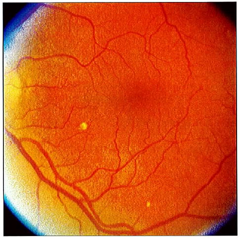
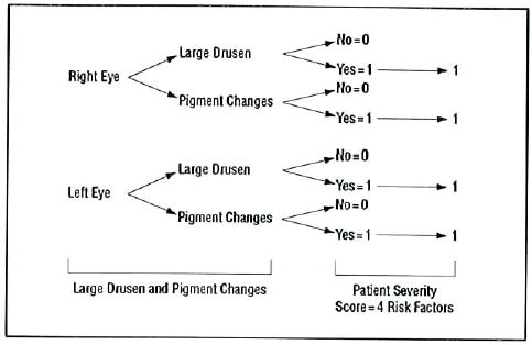
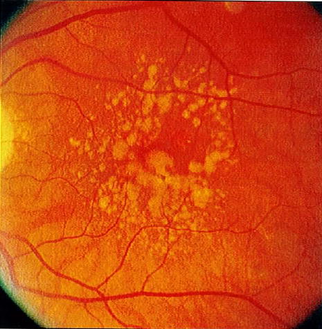
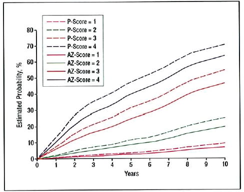
Methods: Following development of a detailed scale for individual eyes based on gradings of fundus photographs in the Age-Related Eye Disease Study, rates of progression to advanced AMD were assessed in cross-tabulations of presence or absence in each eye of 2 easily identified retinal abnormalities, drusen and pigment abnormalities. Large drusen and any pigment changes were particularly predictive of developing advanced AMD.
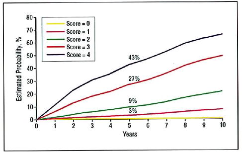
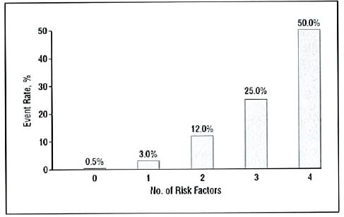
Results: The scoring system developed for patients assigns to each eye 1 risk factor for the presence of 1 or more large (> or = 125 microm, width of a large vein at disc margin) drusen and 1 risk factor for the presence of any pigment abnormality. Risk factors are summed across both eyes, yielding a 5-step scale (0-4) on which the approximate 5-year risk of developing advanced AMD in at least one eye increases in this easily remembered sequence: 0 factors, 0.5%; 1 factor, 3%; 2 factors, 12%; 3 factors, 25%; and 4 factors, 50%. For persons with no large drusen, presence of intermediate drusen in both eyes is counted as 1 risk factor.
Conclusion: This simplified scale provides convenient risk categories for development of advanced AMD that can be determined by clinical examination or by less demanding photographic procedures than used in the Age-Related Eye Disease Study.
Figures
Comment in
-
The Age-Related Eye Disease Study severity scale for age-related macular degeneration: AREDS Report No. 17.Arch Ophthalmol. 2005 Nov;123(11):1484-98. doi: 10.1001/archopht.123.11.1484. Arch Ophthalmol. 2005. PMID: 16286610 Free PMC article.
-
Age-Related Eye Disease Study severity scale and simplified severity scale for age-related macular degeneration.Arch Ophthalmol. 2005 Nov;123(11):1598-9. doi: 10.1001/archopht.123.11.1598. Arch Ophthalmol. 2005. PMID: 16286625 No abstract available.
References
-
- The Age-Related Eye Disease Study Research Group. The Age-Related Eye Disease Study system for classifying age-related macular degeneration from stereoscopic color fundus photographs: AREDS Report Number 6. Am J Ophthalmol. 2001;132:688–681. - PubMed
-
- Klein R, Davis MD, Magli YL, Segal P, Klein BE, Hubbard L. The Wisconsin Age-Related Maculopathy Grading System. Ophthalmology. 1991;98:1128–1134. - PubMed
-
- Bird AC, Bressler NM, Bressler SB, et al. An international classification and grading system for age-related maculopathy and age-related macular degeneration: the International ARM Epidemiological Study Group. Surv Ophthalmol. 1995;39:367–374. - PubMed
-
- Early Treatment Diabetic Retinopathy Study Research Group. Fundus photographic risk factors for progression of diabetic retinopathy: ETDRS Report Number 12. Ophthalmology. 1991;98:823–833. - PubMed
Publication types
MeSH terms
Grants and funding
LinkOut - more resources
Full Text Sources
Other Literature Sources
Medical
Molecular Biology Databases
