. 2005 Nov 22;102(47):17130-5.
doi: 10.1073/pnas.0508480102. Epub 2005 Nov 14.

Calcineurin regulates bone formation by the osteoblast

Affiliations

Calcineurin regulates bone formation by the osteoblast

Li Sun et al. Proc Natl Acad Sci U S A. .

Abstract

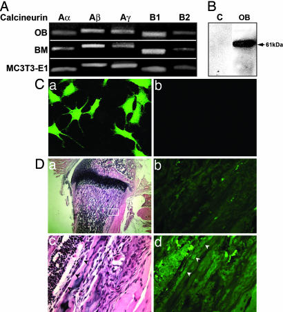
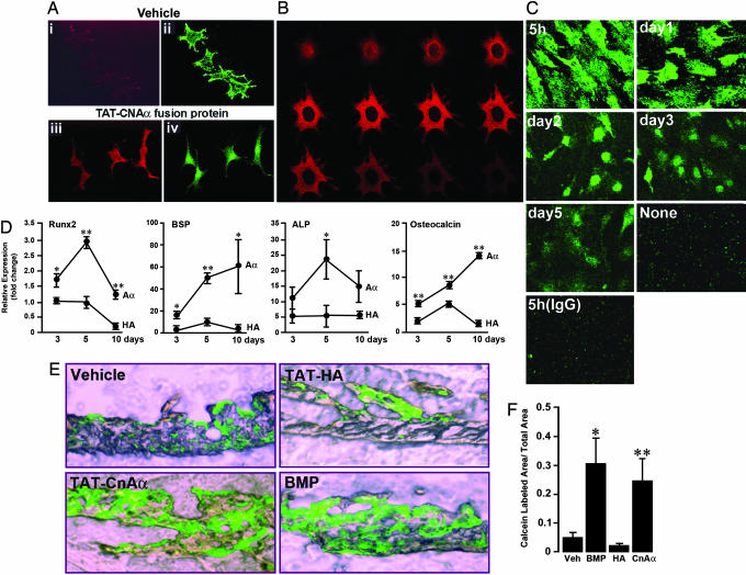
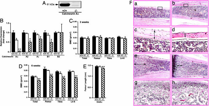
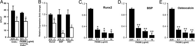
Two of the most commonly used immunosuppressants, cyclosporine A and tacrolimus (FK506), inhibit the activity of a ubiquitously expressed Ca(2+)/calmodulin-sensitive phosphatase, calcineurin. Because both drugs also cause profound bone loss in humans and in animal models, we explored whether calcineurin played a role in regulating skeletal remodeling. We found that osteoblasts contained mRNA and protein for all isoforms of calcineurin A and B. TAT-assisted transduction of fusion protein TAT-calcineurin Aalpha into osteoblasts resulted in the enhanced expression of the osteoblast differentiation markers Runx-2, alkaline phosphatase, bone sialoprotein, and osteocalcin. This expression was associated with a dramatic enhancement of bone formation in intact calvarial cultures. Calcineurin Aalpha(-/-) mice displayed severe osteoporosis, markedly reduced mineral apposition rates, and attenuated colony formation in 10-day ex vivo stromal cell cultures. The latter was associated with significant reductions in Runx2, bone sialoprotein, and osteocalcin expression, paralleled by similar decreases in response to FK506. Together, the gain- and loss-of-function experiments indicate that calcineurin regulates bone formation through an effect on osteoblast differentiation.


Figures

Fig. 1.
Fig. 1.
Expression of calcineurin A and B isoforms in osteoblasts. (A) RT-PCR bands showing the expression of calcineurin isoforms Aα, Aβ, Aγ, B1, and B2 in MC3T3.E1 cells, primary osteoblasts (OB), and freshly isolated bone marrow cells (BM). (B) Western blot showing a 61-kDa band in primary calvarial osteoblasts (30 μg of protein); C, control lane. (C) Immune localization of calcineurin Aα in MC3T3.E1 osteoblasts that were incubated with antibody PP2BAα (a) or nonimmune rabbit serum (b). (D) In situ labeling of osteoblasts for calcineurin Aα in the sections of wild-type mouse bones. (a)A6-μm section stained with hematoxylin and eosin at low power, with the region of interest (box) shown at high power in c, where osteoblasts are indicated by arrowheads. The same section is shown in epifluorescence after calcineurin Aα immune labeling, indicating that the osteoblasts are immunelabeled (arrowheads) (d). In an adjacent section, a comparable region in epifluorescence (b) is shown with the primary antibody omitted.
Fig. 2.
Fig. 2.
Effect of overexpressing calcineurin Aα as a TAT fusion protein on osteoblast differentiation and bone formation. (A) Immunohistochemical detection of calcineurin Aα (CNAα) and TAT, respectively, with anti-CNAα antiserum (PPB2Aα, green) and anti-TAT antibody (red) in MC3T3.E1 osteoblasts that were transduced with TAT-CNAα fusion protein (200 nM) (Aiii and Aiv) or vehicle (Ai and Aii) for 10 min at 37°C. Note the similar distribution of red and green staining after transduction. (B) Confocal micrographs (1-μm-thick coronal sections of the same cell) demonstrating the mainly cytosolic localization of TAT-CNAα fusion protein in intact cells by using an anti-TAT antibody (red). Note that the nucleus is spared, indicating that the TAT construct does not permeate into the nuclear compartment. (C) Immunolabeling for HA (green) in primary osteoblasts treated with TAT-HA (200 nM for 10 min), demonstrating the persistence of TAT-HA for at least 5 days. No green staining was seen without transduction or with the use of an irrelevant IgG. (D) Enhanced expression of osteoblast differentiation markers, Runx-2 (early), BSP, ALP, and osteocalcin in MC3T3.E1 cells at 3, 5, and 10 days after transduction with TAT-CNAα (Aα) or TAT-HA, (control) (200 nM each for 10 min). The real-time PCR results are expressed as fold-change normalized to GAPDH and cell control (+SEM). Statistics by Student's t test; **, P < 0.01; *, P < 0.05, comparisons with HA at every time point. (E and F) Sections of calvaria from newborn mice incubated in the presence of calcein with vehicle, TAT-HA (200 nM), TAT-CNAα (200 nM), or BMP-2, 200 ng/ml. Quantitative estimates of calcein-labeled surfaces expressed relative to total surface area (F), measured morphometrically by using fovea pro (Reindeer Graphics). Mean ± SEM from five fields from three or more sections were analyzed by Student's t test, comparing each treatment with vehicle/TAT-HA; *, P < 0.05; **, P < 0.01.
Fig. 3.
Fig. 3.
Phenotypic characterization of the calcineurin Aα-/- mice. (A) Western blot of a whole-bone extract showing the absence of the 61-kDa calcineurin Aα band in null mice. (B) Real-time PCR on RNA isolated from bone-marrow-derived osteoblasts to examine the expression of the calcineurin A and B isoforms (shown) in calcineurin Aα+/+,Aα+/-, and Aα-/- mice. (C and D) Severe osteoporosis in the calcineurin Aα-/- mouse at 6 weeks. Comparisons of BMD (g/cm2) at femur, tibia, lumbar spine (L4-L6), and total body in 3- (C) and 6-week-old (D) calcineurin Aα+/+,Aα+/-, and Aα-/- mice by using a Lunar PIXImus small animal densitometer (Lunar, precision <1.5%). Student's t test was used to compare the BMDs of homozygotes and heterozygotes with wild-type littermates at each site. **, P < 0.01; *, P < 0.05. (E) Estimates of femur length (cm) in calcineurin Aα+/+, Aα+/-, and Aα-/- mice. Statistics by Student's t test; **, P < 0.01. (F) Histological comparisons of cortical thickness (a and b at magnification ×1 and c and d at magnification ×2), overall trabecular volume (e and f), and growth plate (g and h) between 6-week-old calcineurin Aα-/- and Aα+/+ mice. There was ≈40% reduction in diaphyseal cortical thickness (Table 1) and a mild reduction in trabecular bone (not quantifiable precisely at this age) in calcineurin Aα-/- mice. The respective articular surfaces showed no apparent difference (data not shown).
Fig. 4.
Fig. 4.
Effect of either calcineurin Aα gene deletion or the inhibition of calcineurin enzyme activity by FK506 on the number of CFU-F (A) and the expression of osteoblast genes (B-E), namely Runx2, BSP, and osteocalcin in mouse primary osteoblasts after 10 days of culture in the presence of ascorbate-5-phosphate (1 mM). Student's t test was used to compare with wild-type littermates or zero-dose. *, P < 0.06; **, P < 0.01.

Similar articles

Cited by

References

    1. Klee, C. B., Crouch, T. H. & Krinks, M. H. (1980) Proc. Natl. Acad. Sci. USA 76, 6270-6273. - PMC - PubMed
    1. Klee, C. B., Draetta, G. F. & Hubbard, M. J. (1988) Adv. Enzymol. Relat. Areas Mol. Biol. 61, 149-200. - PubMed
    1. Klee, C. B., Ren, H. & Wang, X. (1998) J. Biol. Chem. 273, 13367-13370. - PubMed
    1. Buttini, M., Limonta, S., Luyten, M. & Boddeke, H. (1995) Histochem. J. 27, 291-299. - PubMed
    1. Guerini, D. (1997) Biochem. Biophys. Res. Commun. 235, 271-275. - PubMed

Publication types

MeSH terms