. 2005 Nov 22;102(47):17107-12.
doi: 10.1073/pnas.0500721102. Epub 2005 Nov 14.

The ER aminopeptidase, ERAP1, trims precursors to lengths of MHC class I peptides by a "molecular ruler" mechanism

Affiliations

The ER aminopeptidase, ERAP1, trims precursors to lengths of MHC class I peptides by a "molecular ruler" mechanism

Shih-Chung Chang et al. Proc Natl Acad Sci U S A. .

Abstract

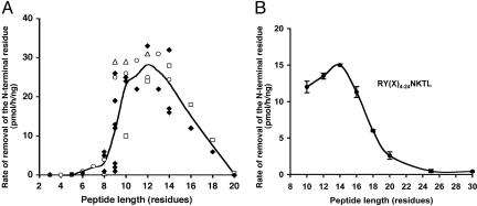
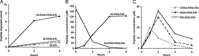
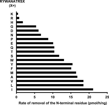
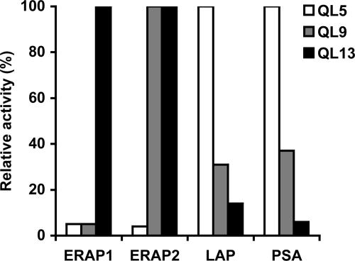
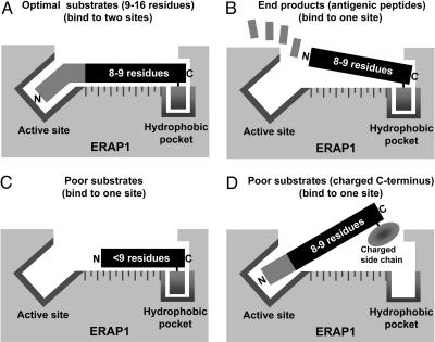
Endoplasmic reticulum aminopeptidase 1 (ERAP1) is an IFN-gamma-induced aminopeptidase in the endoplasmic reticulum that trims longer precursors to the antigenic peptides presented on MHC class I molecules. We recently reported that purified ERAP1 trimmed N-extended precursors but spared peptides of 8-9 residues, the length required for binding to MHC class I molecules. Here, we show another remarkable property of ERAP1: that it strongly prefers substrates 9-16 residues long, the lengths of peptides transported efficiently into the ER by the transporter associated with antigen processing (TAP) transporter. This aminopeptidase rapidly degraded a model 13-mer to a 9-mer and then stopped, even though the substrate and the product had identical N- and C-terminal sequences. No other aminopeptidase, including the closely related ER-aminopeptidase ERAP2, showed a similar length preference. Unlike other aminopeptidases, the activity of ERAP1 depended on the C-terminal residue of the substrate. ERAP1, like most MHC class I molecules, prefers peptides with hydrophobic C termini and shows low affinity for peptides with charged C termini. Thus, ERAP1 is specialized to process precursors transported by TAP to peptides that can serve as MHC class I epitopes. Its "molecular ruler" mechanism involves binding the hydrophobic C terminus of the substrate 9-16 residues away from the active site.


Figures

Fig. 1.
Fig. 1.
ERAP1 prefers substrates 9-16 residues long. (A) A variety of peptides were tested, including mature MHC class I-presented peptides (8-9 residues), 3- to 8-residue fragments of mature epitopes, and 9- to 20-residue N-extended precursors of MHC-binding peptides with the same epitope on their C termini and different N-terminal extensions (see Fig. 6 for identification of each peptide). Peptides (150 μM) were incubated with purified ERAP1 (3.5 μg/ml) at 37°C for 1 h. Each spot reflects the trimming of an individual peptide substrate. The line shows the mean of the degradation rates for different peptide substrates of the same length. The removal of their N-terminal residues was measured by RP-HPLC (see Materials and Methods). The trimming of the 14-mer, GLEQLESIINFEKL, the trimer, EKL, and all intervening peptides from 4-13 residues long, including SIINFEKL, the H2-Kb-binding antigenic peptide, are shown as open circle symbols. The degradation of N-extended FAPGNYPAL series is shown as open triangles, N-extended NANPDCKTIL series are shown as open squares, and other peptides are indicated as solid diamonds. (B) The maximum length of substrates was analyzed with a library of peptides of different lengths (10-30 residues) but identical N- and C-terminal sequences. The collection of peptides, RY(Xn)NKTL (100 μM), where X represents equimolar mixtures of A, D, E, F, G, H, I, K, L, M, P, Q, R, S, T or V, and n represents 4, 6, 8, 10, 12, 14, 19 or 24, was incubated individually with purified ERAP1 (3.5 μg/ml) at 37°C for 1 h and analyzed by the method of fluorescence detection (see Materials and Methods for details).
Fig. 2.
Fig. 2.
ERAP1 rapidly cleaves a 13-mer, but not a 9-mer or a 5-mer with identical N and C termini. (A) QLESQLESQLESL, QLESQLESL, and QLESL (150 μM) were incubated with purified ERAP1 (3.5 μg/ml). The peptide-containing supernatant was analyzed by RP-HPLC. (B) Quantitative conversion of the 13-mer to the 9-mer by ERAP1. QLESQLESQLESL was incubated with purified ERAP1 as described above, and an aliquot of the reaction was analyzed by HPLC at each time. (C) The 12-, 11- and 10-residue intermediates were generated from the 13-mer QLESQLESQLESL in a nonprocessive manner. LESQLESQLESL (12-mer), ESQLESQLESL (11-mer), and SQLESQLESL (10-mer) were clearly demonstrable before the product 9-mer was generated.
Fig. 3.
Fig. 3.
ERAP1 prefers peptides with a hydrophobic C terminus. The library of 19 peptides of RYWANATRSX (150 μM respectively), where X represents each of the naturally occurring 20 aa (except Cys), was incubated with purified ERAP1 (3.5 μg/ml) at 37°C for 1 h. Samples were analyzed by RP-HPLC with a linear gradient of 0.05% trifluoroacetic acid (TFA) to 50% acetonitrile in 0.03% TFA.
Fig. 4.
Fig. 4.
ERAP1 shows an opposite length preference from ERAP2 and cytosolic aminopeptidases, leucine aminopeptidase (LAP), and puromycin-sensitive aminopeptidase (PSA). QLESQLESQLESL (QL13), QLESQLESL (QL9), and QLESL (Q5) (100 μM, respectively) were incubated with purified ERAP1, ERAP2, LAP, or PSA (3.5 μg/ml, respectively) at 37°C for 1 h. The aminopeptidase activity against different peptide substrates was measured by the method of fluorescence detection.
Fig. 5.
Fig. 5.
Proposed model based on present observations to explain the substrate preference of ERAP1 (molecular ruler). (A) High-affinity binding of optimal substrates through the C-terminal hydrophobic residue and active site. (B) Release of antigenic products (eight to nine residues) after removal of additional N-terminal residues. Affinity is low because it is unable to reach to the active site. (C) Poor substrates (fewer than nine residues) have low affinity because they can interact only with the active site, or if they bind to the hydrophobic pocket, their N-terminal residues cannot reach the active site. (D) A poor substrate has low affinity because its C-terminal residue has a charged side chain.

References

    1. Rock, K. L. & Goldberg, A. L. (1999) Annu. Rev. Immunol. 17, 739-779. - PubMed
    1. Yewdell, J. W. & Bennink, J. R. (2001) Curr. Opin. Immunol. 13, 13-18. - PubMed
    1. Shastri, N., Schwab, S. & Serwold, T. (2002) Annu. Rev. Immunol. 20, 463-493. - PubMed
    1. Rock, K. L., York, I. A., Saric, T. & Goldberg, A. L. (2002) Adv. Immunol. 80, 1-70. - PubMed
    1. Madden, D. R. (1995) Annu. Rev. Immunol. 13, 587-622. - PubMed

Publication types

MeSH terms

Substances