. 2005 Dec;26(6):557-65.
doi: 10.1002/humu.20263.

Multiple epiphyseal dysplasia mutations in MATN3 cause misfolding of the A-domain and prevent secretion of mutant matrilin-3

Affiliations
Free PMC article

Multiple epiphyseal dysplasia mutations in MATN3 cause misfolding of the A-domain and prevent secretion of mutant matrilin-3

Sally L Cotterill et al. Hum Mutat. 2005 Dec.
Free PMC article

Abstract

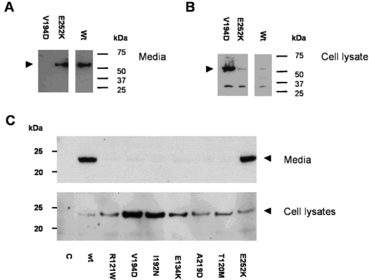
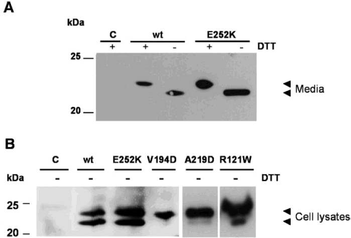
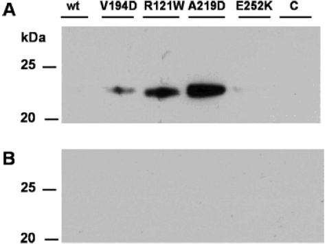
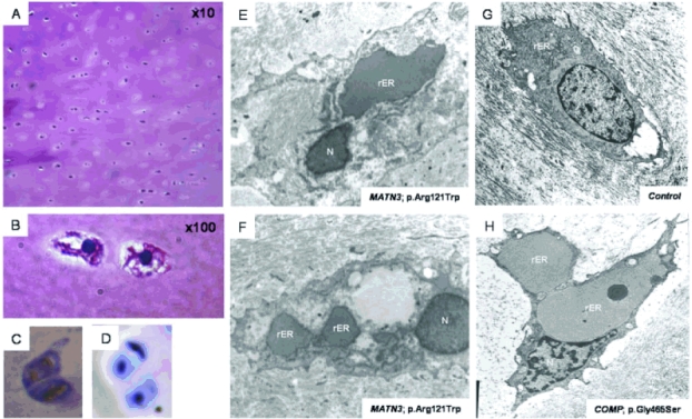
Multiple epiphyseal dysplasia (MED) is a relatively common skeletal dysplasia that can present in childhood with a variable phenotype of short stature and pain and stiffness in the large joints, and often progresses to early-onset osteoarthritis in adulthood. Mutations in the matrilin-3 gene (MATN3) have recently been shown to underlie some forms of autosomal dominant MED. To date all MED mutations in matrilin-3 cluster in the single A-domain, suggesting that they may disrupt the structure and/or function of this important domain. To determine the effects of MATN3 mutations on the structure and function of matrilin-3 we expressed both normal and mutant matrilin-3 in mammalian cells. Wild-type (wt) matrilin-3 was efficiently secreted into conditioned medium, whereas mutant matrilin-3 was retained and accumulated within the cell. Furthermore, when the mutant A-domains were examined individually, they existed primarily in an unfolded conformation. Co-immunoprecipitation experiments demonstrated that the mutant A-domains were specifically associated with ERp72, a chaperone protein known to be involved in mediating disulfide bond formation. Light microscopy of cartilage from an MED patient with a MATN3 mutation showed the presence of intracellular material within the chondrocytes, whilst the overall matrix appeared normal. On electron micrographs, the inclusions noted at the light microscopy level appeared to be dilated cisternae of rough endoplasmic reticulum and immunohistochemical analysis confirmed that the retained protein was matrilin-3. In summary, the data presented in this paper suggest that MED caused by MATN3 mutations is the result of an intracellular retention of the mutant protein.


Figures

Figure 1
Figure 1
SDS-PAGE and Western blot analysis of conditioned media and cell lysates from CHO cells stably transfected with full-length matrilin-3 (A and B) and single A-domain (C) constructs. Gels were run under reducing conditions, and molecular weights are shown in kDa. A and B: Media and cell lysate samples from cells expressing full-length wt, a MED mutation (pVal194Asp), and the polymorphism (pGlu252Lys) are shown. Proteins were detected with an anti-FLAG antibody, and the full-length protein is indicated with an arrowhead.C:Untransfected cells (C) and cells transfected with normal (wt) and polymorphic (pGlu252Lys) A-domain are shown.The A-domain missense mutations shown are pArg121Trp, p.Val194Asp, p.Ile192Asn, pGlu134Lys, p.Ala219Asp, and p.Thr120Met. Proteins were detected with an anti-c-myc antibody, and the A-domain is indicated with an arrowhead.
Figure 2
Figure 2
SDS-PAGE and Western blot analysis of conditionedmedia (A) and cell lysates (B) from CHO cells stably transfected with A-domain constructs under reducing (+) and non-reducing (−) conditions. Samples from cells expressing a normal A-domain construct (wt), a polymorphic construct (p.Glu252Lys), and constructs with the p.Val194Asp, p.Ala219Asp, and p.Arg121Trp mutations are shown. Samples from untransfected cells were used as a control (C).The recombinant protein was detected using an anti-c-myc antibody, andmolecular weights are shown in kDa.
Figure 3
Figure 3
A: ERp72 was immunoprecipitated from the cell lysates of recombinant cells, and the immunoprecipitates were analyzed byWestern blot.The c-myc antibody detected recombinant A-domain in the R121W and A219D immunoprecipitates and to a lesser extent in theV194D immunoprecipitate. No band was detectable in the normal (wt), polymorphic (E252K), or control (C) lanes. B: Immunoprecipitation in the absence of anti-ERp72 antibody was performed as a control for nonspecific binding to theProteinA-agarose beads.Untransfected CHO cells were used as the control (C).
Figure 4
Figure 4
Light and electron micrographs of cartilage from the iliac crest biopsy of a 14-year-old patient with the p.Arg121Trp mutation. A: H&E staining of hyaline cartilage showing a normal matrix at × 10 magnification. B: Magnification (× 100) of the same section of cartilage showing a chondrocyte with apparent staining of the cytoplasm suggesting the presence of protein material, which is distinct from the nucleus (dark blue). C: Immunohistochemistry of the cartilage showing that the protein retained in the patient's chondrocyte stains with an antibody to matrilin-3 (brown), whereas there appears to be no intracellular staining for matrilin- 3 in the chondrocyte of an aged matched control sample (D) (the cell nucleus is stained blue by DAPI). E and F: Electron micrographs of the patients chondrocytes showing that the rough endoplasmic reticulum cisternae are swollen with accumulated protein that has a granular appearance, when compared to a control chondrocyte (G). For comparison an electron micrograph of a chondrocyte from a pseudoachondroplasia patient with a COMP mutation (p.Gly465Ser) is also shown and in this case the enlarged rER completely occupies the cytoplasm (H). rER: rough endoplasmic reticulum; N: nucleus. [Color figure can be viewed in the online issue,which is available at http://www.interscience.wiley.com.]

References

    1. Ballhausen D, Bonafe L, Terhal P, Unger SL, Bellus G, Classen M, Hamel BC, Spranger J, Zabel B, Cohn DH, Cole WG, Hecht JT, Superti-Furga A. Recessive multiple epiphyseal dysplasia (rMED): phenotype delineation in eighteen homozygotes for DTDST mutation R279W. J Med Genet. 2003;40:65–71. - PMC - PubMed
    1. Belluoccio D, Schenker T, Baici A, Trueb B. Characterization of human matrilin-3 (MATN3) Genomics. 1998;53:391–394. - PubMed
    1. Borochowitz ZU, Scheffer D, Adir V, Dagoneau N, Munnich A, Cormier-Daire V. Spondylo-epi-metaphyseal dysplasia (SEMD) matrilin 3 type: homozygote matrilin 3 mutation in a novel form of SEMD. J Med Genet. 2004;41:366–372. - PMC - PubMed
    1. Briggs MD, Chapman KL. Pseudoachondroplasia and multiple epiphyseal dysplasia: mutation review, molecular interactions, and genotype to phenotype correlations. Hum Mutat. 2002;19:465–478. - PubMed
    1. Briggs MD, Hoffman SM, King LM, Olsen AS, Mohrenweiser H, Leroy JG, Mortier GR, Rimoin DL, Lachman RS, Gaines ES, Cekleniak JA, Knowlton RG, Cohn DH. Pseudoachondroplasia and multiple epiphyseal dysplasia due to mutations in the cartilage oligomeric matrix protein gene. Nat Genet. 1995;10:330–336. - PubMed

Publication types

MeSH terms

Substances