

B. subtilis ykuD protein at 2.0 A resolution: insights into the structure and function of a novel, ubiquitous family of bacterial enzymes
- PMID: 16287140
- PMCID: PMC2792008
- DOI: 10.1002/prot.20702
B. subtilis ykuD protein at 2.0 A resolution: insights into the structure and function of a novel, ubiquitous family of bacterial enzymes
Abstract
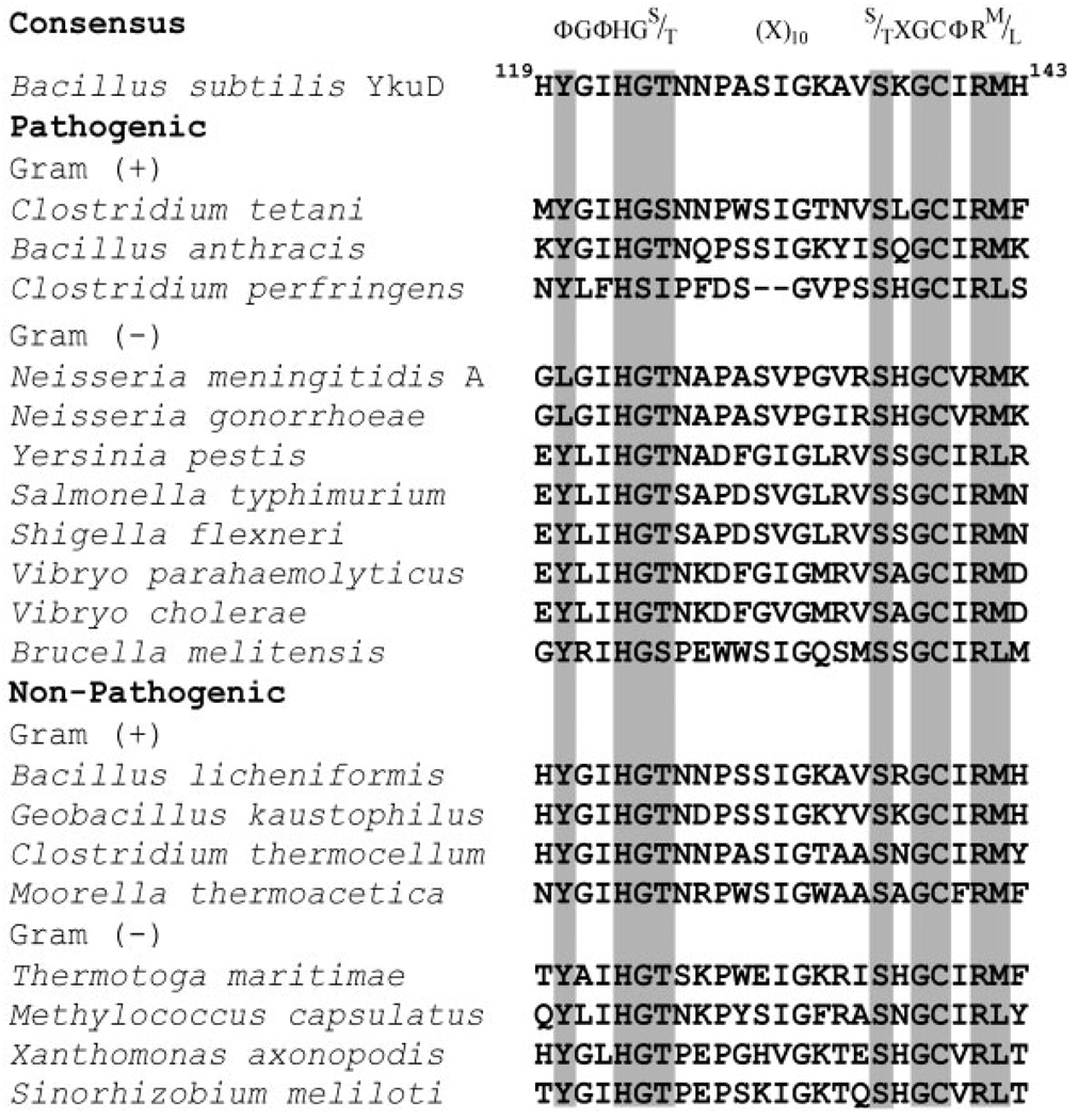
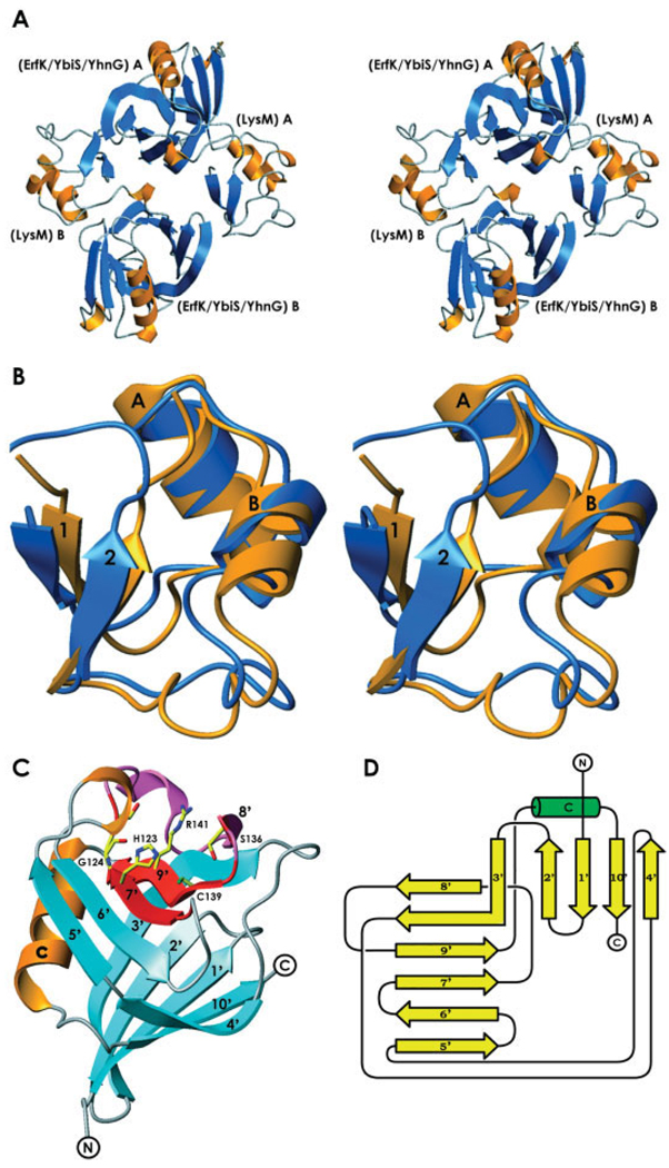
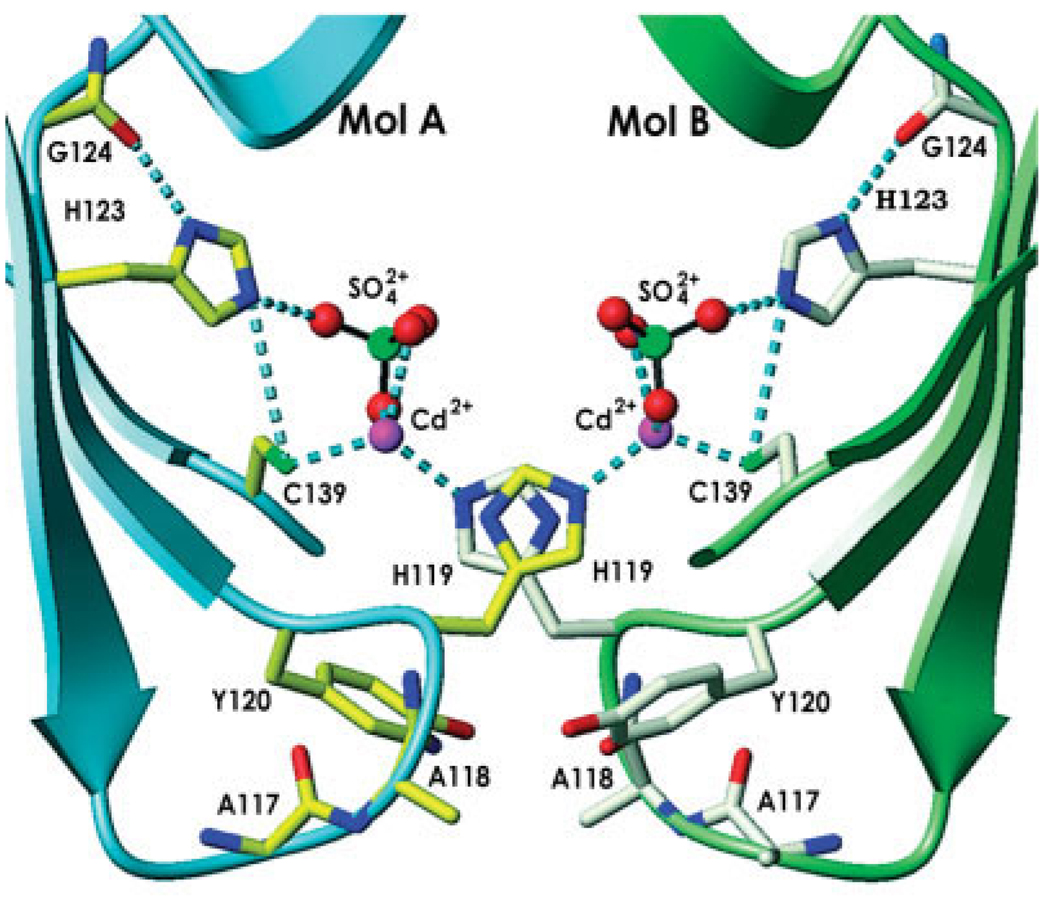
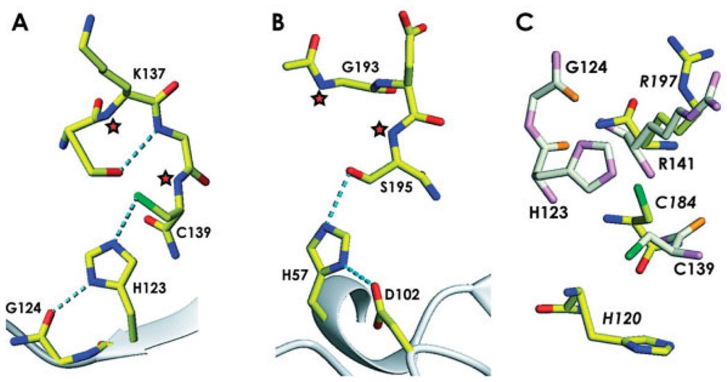
The crystal structure of the product of the Bacillus subtilis ykuD gene was solved by the multiwavelength anomalous dispersion (MAD) method and refined using data to 2.0 A resolution. The ykuD protein is a representative of a distinctly prokaryotic and ubiquitous family found among both pathogenic and nonpathogenic Gram-positive and Gram-negative bacteria. The deduced amino acid sequence reveals the presence of an N-terminal LysM domain, which occurs among enzymes involved in cell wall metabolism, and a novel, putative catalytic domain with a highly conserved His/Cys-containing motif of hitherto unknown structure. As the wild-type protein did not crystallize, a double mutant was designed (Lys117Ala/Gln118Ala) to reduce excess surface conformational entropy. As expected, the structure of the LysM domain is similar to the NMR structure reported for an analogous domain from Escherichia coli murein transglycosylase MltD. The molecular model also shows that the 112-residue-long C-terminal domain has a novel tertiary fold consisting of a beta-sandwich with two mixed sheets, one containing five strands and the other, six strands. The two beta-sheets form a cradle capped by an alpha-helix. This domain contains a putative catalytic site with a tetrad of invariant His123, Gly124, Cys139, and Arg141. The stereochemistry of this active site shows similarities to peptidotransferases and sortases, and suggests that the enzymes of the ykuD family may play an important role in cell wall biology.
2005 Wiley-Liss, Inc.
Figures
References
-
- Bateman A, Bycroft M. The structure of a LysM domain from E. coli membrane-bound lytic murein transglycosylase D (MltD) J Mol Biol. 2000;299:1113–1119. - PubMed
-
- Spaink HP. Specific recognition of bacteria by plant LysM domain receptor kinases. Trends Microbiol. 2004;12:201–204. - PubMed
-
- Derewenda ZS. Rational protein crystallization by mutational surface engineering. Structure. 2004;12:529–535. - PubMed
-
- Stols L, Gu M, Dieckman L, Raffen R, Collart FR, Donnelly MI. A new vector for high-throughput, ligation-independent cloning encoding a tobacco etch virus protease cleavage site. Protein Expr Purif. 2002;25:8–15. - PubMed
Publication types
MeSH terms
Substances
Grants and funding
LinkOut - more resources
Full Text Sources
Other Literature Sources
Molecular Biology Databases
