. 2005 Nov-Dec;16(6):1433-41.
doi: 10.1021/bc0501698.

Near-infrared fluorescent RGD peptides for optical imaging of integrin alphavbeta3 expression in living mice

Affiliations

Near-infrared fluorescent RGD peptides for optical imaging of integrin alphavbeta3 expression in living mice

Zhen Cheng et al. Bioconjug Chem. 2005 Nov-Dec.

Abstract

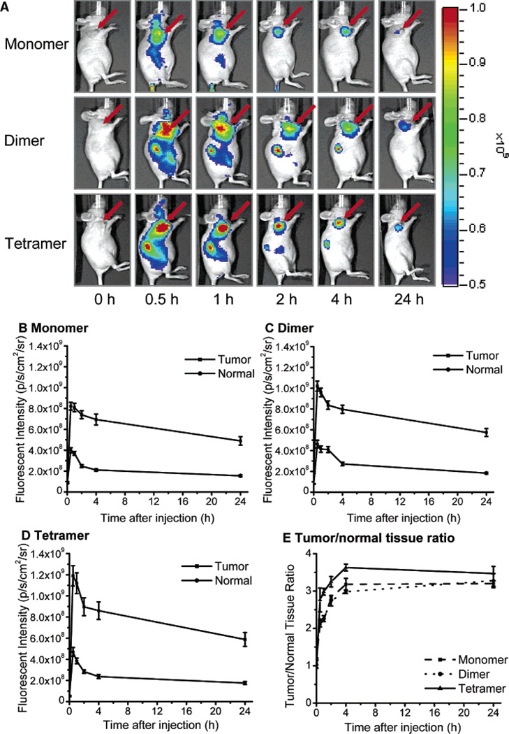
Near-infrared fluorescence optical imaging is a powerful technique for studying diseases at the molecular level in preclinical models. We recently reported that monomeric RGD peptide c(RGDyK) conjugated to the NIR fluorescent dye specifically targets integrin receptor both in cell culture and in living subjects. In this report, Cy5.5-conjugated mono-, di-, and tetrameric RGD peptides were evaluated in a subcutaneous U87MG glioblastoma xenograft model in order to investigate the effect of multimerization of RGD peptide on integrin avidity and tumor targeting efficacy. The binding affinities of Cy5.5-conjugated RGD monomer, dimer, and tetramer for alpha(v)beta(3) integrin expressed on U87MG cell surface were determined to be 42.9 +/- 1.2, 27.5 +/- 1.2, and 12.1 +/- 1.3 nmol/L, respectively. All three peptide-dye conjugates had integrin specific uptake both in vitro and in vivo. The subcutaneous U87MG tumor can be clearly visualized with each of these three fluorescent probes. Among them, tetramer displayed highest tumor uptake and tumor-to-normal tissue ratio from 0.5 to 4 h postinjection. Tumor-to-normal tissue ratio for Cy5.5-conjugated RGD monomer, dimer, and tetramer were found to be 3.18 +/- 0.16, 2.98 +/- 0.05, and 3.63 +/- 0.09, respectively, at 4 h postinjection. These results suggest that Cy5.5-conjugated monomeric, dimeric, and tetrameric RGD peptides are all suitable for integrin expression imaging. The multmerization of RGD peptide results in moderate improvement of imaging characteristics of the tetramer, compared to that of the monomer and dimeric counterparts.


Figures

Figure 1
Figure 1
Schematic structures of Cy5.5-conjugated RGD monomer Cy5.5-c(RGDyK), dimer Cy5.5-E[c(RGDyK)]2, and tetramer Cy5.5-E{E[c(RGDyK)]2}2.
Figure 2
Figure 2
Competitive integrin receptor assay of unlabeled echistatin (■), monomer Cy5.5-c(RGDyK) (▲), dimer Cy5.5-E[c(RGDyK)]2 (●), and tetramer Cy5.5-E{E[c(RGDyK)]2}2 (◆) to U87MG human glioma cells. All experiments were done in triplicate. The IC50 values for echistatin, Cy5.5-monomer, dimer, and tetramer were determined to be 1.2 ± 0.1, 12.1 ± 1.3, 27.5 ± 1.2, and 42.9 ± 1.2 nmol/L, respectively.
Figure 3
Figure 3
Specific binding and endocytosis of the Cy5.5-labeled cyclic RGD peptides. NIRF images of U87MG were obtained after the cells were incubated for 30 min at 37 °C in the presence of 10 nmol/L monomer Cy5.5-c(RGDyK), dimer Cy5.5-E[c(RGDyK)]2, tetramer Cy5.5-E{E[c(RGDyK)]2}2 with (D, E, F) or without (A, B, C) blocking dose of nonfluorescent RGD peptide c(RGDyK) (10 µmol/L). Complete blocking of NIR fluorescence of U87MG cells demonstrating high αvβ3 integrin specificity of these NIR dye-labeled RGD peptides.
Figure 4
Figure 4
(A) In vivo fluorescence imaging of athymic nude mice bearing subcutaneous U87MG glioblastoma xenografts after intravenous injection of 500 pmol Cy5.5-RGD monomer, dimer, or tetramer. The location of the tumor was indicated by an arrow. Fluorescence signal from Cy5.5 was pseudo-colored red. The tetramer and dimer displayer higher tumor uptake and tumor/normal contrast than that of monomer from 0.5 to 4 h pi. (B–D) In vivo targeting characters of Cy5.5-RGD monomer, dimer, and tetramer, respectively. Both tetramer and dimer had significantly higher accumulation in tumor than monomer (P < 0.05) at early time points (from 0.5 to 4 h) pi. But the differences in tumor uptakes among the three probes were minimal at 24 h pi. (E) Tumor contrast (tumor-to-normal tissue ratio) as a function of time post-administration of Cy5.5-RGD monomer (■), dimer (●), and tetramer (▲). The fluorescence intensity for the region of interest was recorded as photons per second per centimeter squared per steradian (p/ s/cm2/sr). The tumor contrast of Cy5.5-RGD-tetramer from 0.5 to 4 h postinjection is significantly higher than that of Cy5.5-RGD-dimer and monomer (P < 0.05).
Figure 5
Figure 5
Representative NIRF images of mice bearing subcutaneous U87MG tumor on the right foreleg demonstrating blocking of Cy5.5-RGD conjugates (500 pmol) uptake in the tumors by coinjection with 10 mg/kg of c(RGDyK). Pseudo-color fluorescence images of tumor-bearing mice were acquired 1 h after intravenous injection of Cy5.5-RGD conjugates (experiment) or Cy5.5-RGD conjugates + c(RGDyK) (block). (B) Tumor contrast (tumor-to-normal tissue ratio) calculated from region of interest measurement at 1 h postadministration of Cy5.5-RGD conjugates.
Figure 6
Figure 6
Representative images of dissected organs of mice bearing U87MG tumor sacrificed 24 h after intravenous injection of Cy5.5-RGD monomer (A), dimer (B), and tetramer (C) at a dose of 500 pmol per mouse. (D) Biodistribution of the probes at 24 h postinjection. (E) Tumor-to-normal organ ratios.

Similar articles

Cited by

References

    1. Massoud TF, Gambhir SS. Molecular imaging in living subjects: seeing fundamental biological processes in a new light. Genes Dev. 2003;17:545–580. - PubMed
    1. Sevick-Muraca EM, Houston JP, Gurfinkel M. Fluorescence-enhanced, near infrared diagnostic imaging with contrast agents. Curr. Opin. Chem. Biol. 2002;6:642–650. - PubMed
    1. Ntziachristos V, Bremer C, Weissleder R. Fluorescence imaging with near- infrared light: new technological advances that enable in vivo molecular imaging. Eur. Radiol. 2003;13:195–208. - PubMed
    1. Tung CH. Fluorescent peptide probes for in vivo diagnostic imaging. Biopolymers. 2004;76:391–403. - PubMed
    1. Weissleder R, Mahmood U. Molecular imaging. Radiology. 2001;219:316–333. - PubMed

Publication types