. 2005 Nov 14:3:73.
doi: 10.1186/1477-7819-3-73.

Aberrant E-cadherin staining patterns in invasive mammary carcinoma

Affiliations

Aberrant E-cadherin staining patterns in invasive mammary carcinoma

Malini Harigopal et al. World J Surg Oncol. .

Abstract

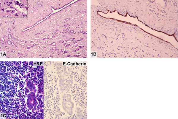
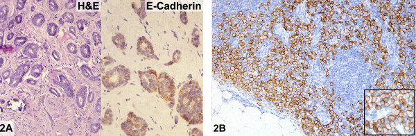
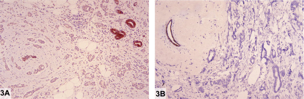
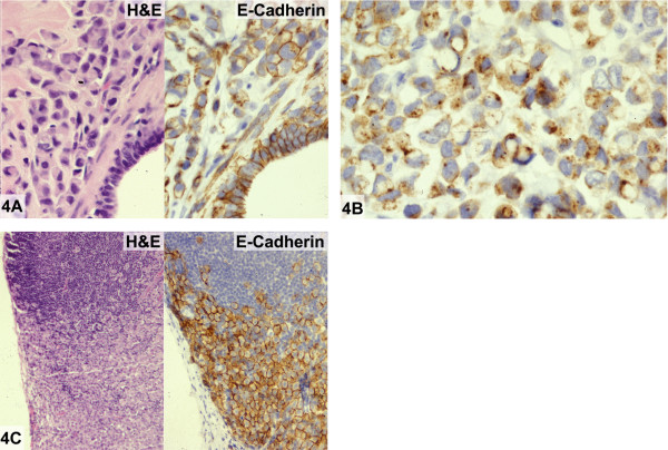
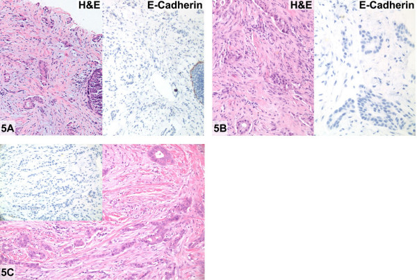
Background: E-cadherin, a cell surface protein involved in cell adhesion, is present in normal breast epithelium, benign breast lesions, and in breast carcinoma. Alterations in the gene CDH1 on chromosome 16q22 are associated with changes in E-cadherin protein expression and function. Inactivation of E-cadherin in lobular carcinomas and certain diffuse gastric carcinomas may play a role in the dispersed, discohesive "single cell" growth patterns seen in these tumors. The molecular "signature" of mammary lobular carcinomas is the loss of E-cadherin protein expression as evidenced by immunohistochemistry, whereas ductal carcinomas are typically E-cadherin positive.

Patients and methods: We report on E-cadherin immunostaining patterns in five cases of invasive mammary carcinoma.

Results: These were five exceptional instances in which the E-cadherin immunophenotype did not correspond to the apparent histologic classification of the lesion. These cases which are exceedingly rare in our experience are the subject of this report.

Conclusion: Findings such as those illustrated in this study occur in virtually all biologic phenomena and they do not invalidate the very high degree of correlation between the expression of E-cadherin and the classification of breast carcinomas as ductal or lobular type on the basis of conventional histologic criteria.


Figures

Figure 1
Figure 1
Absence of E-cadherin in invasive "ductal" carcinoma (case 1). A: Moderately differentiated invasive ductal carcinoma. Focally, tumor cells are seen growing in a linear fashion and contain intracytoplasmic mucin vacuoles (inset) (Hematoxylin and eosin). B: Invasive ductal carcinoma exhibiting complete absence of reactivity for E-cadherin. Normal duct serves as an internal positive control (Immunoperoxidase stain for E-cadherin). C: Metastatic carcinoma with glandular differentiation (Hematoxylin and eosin, left) showing complete absence of E-cadherin immunoreactivity (Immunoperoxidase stain for E-cadherin, right).
Figure 2
Figure 2
E-cadherin reactivity in metastatic "lobular" carcinoma (case 2). A: Well-differentiated invasive ductal carcinoma (Hematoxylin and eosin, left) exhibiting weak cell membrane positivity for E-cadherin (Immunoperoxidase stain for E-cadherin, right). B: Nodal metastasis demonstrating strong cell membrane immunoreactivity for E-cadherin; higher magnification of tumor cells (inset) (Immunoperoxidase stain for E-cadherin).
Figure 3
Figure 3
Weak and absent E-cadherin reactivity in two concurrent invasive "ductal" carcinomas (case 3). A: Larger invasive ductal carcinoma exhibiting weak staining for E-cadherin. Normal duct serves as an internal positive control (Immunoperoxidase stain for E-cadherin). B: Smaller invasive ductal carcinoma showing complete absence of staining for E-cadherin. Normal duct serves as an internal positive control (Immunoperoxidase stain for E-cadherin).
Figure 4
Figure 4
E-cadherin reactivity in invasive "lobular" carcinoma (case 4). A: Invasive carcinoma with pleomorphic lobular features showing predominantly alveolar and solid growth patterns and intermediate-high nuclear grade (Hematoxylin and eosin, left). Strong cell membrane E-cadherin reactivity in tumor cells and in normal adjacent ductal epithelium (Immunoperoxidase for E-cadherin, right). B: Some tumor cells demonstrate a punctate staining pattern within the cell membrane or in the cytoplasm (Immunoperoxidase for E-cadherin). C: Metastatic carcinoma with lobular characteristics involving an ipsilateral axillary lymph node (Hematoxylin and eosin, left). Tumor cells showing strong cell membrane reactivity for E-cadherin (Immunoperoxidase for E-cadherin, right).
Figure 5
Figure 5
Invasive lobular carcinoma with glandular "ductal" differentiation (case 5). A: Moderately differentiated invasive "ductal" carcinoma and carcinoma in situ on core biopsy (Hematoxylin and eosin, right) showing complete absence of E-cadherin immunoreactivity. (Immunoperoxidase stain for E-cadherin, left). B: No immunoreactivity for E-cadherin (Immunoperoxidase stain for E-cadherin, left) confirms invasive lobular carcinoma with glandular differentiation in the excision biopsy (Hematoxylin and eosin, right). C: Invasive lobular carcinoma with gland formation (Hematoxylin and eosin, right and immunoperoxidase stain for E-cadherin, left).

Similar articles

Cited by

References

    1. Acs G, Lawton TJ, Rebbeck TR, LiVolsi VA, Zhang PJ. Differential expression of E-cadherin in lobular and ductal neoplasms of the breast and its biologic and diagnostic implications. Am J Clin Pathol. 2001;115:85–98. doi: 10.1309/FDHX-L92R-BATQ-2GE0. - DOI - PubMed
    1. Bratthauer GL, Moinfar F, Stamatakos MD, Mezzetti TP, Shekitka KM, Man YG, Tavassoli FA. Combined E-cadherin and high molecular weight cytokeratin immunoprofile differentiates lobular, ductal, and hybrid mammary intraepithelial neoplasia. Hum Pathol. 2002;33:620–627. doi: 10.1053/hupa.2002.124789. - DOI - PubMed
    1. Gamallo C, Palacios J, Suarez A, Pizarro A, Navarro P, Quintanilla M, Cano A. Correlation of E-cadherin expression with differentiation with differentiation grade and histologic type in breast carcinoma. Am J Pathol. 1993;142:987–993. - PMC - PubMed
    1. Goldstein NS, Bassi D, Watts JC, Layfield LJ, Yaziji H, Gown AM. E-cadherin reactivity of 95 noninvasive ductal and lobular lesions of the breast. Implications for the interpretation of problematic lesions. Am J Clin Pathol. 2001;115:534–42. doi: 10.1309/B0DD-4M7H-GJG1-7KCW. - DOI - PubMed
    1. Jacobs TW, Pliss N, Kouria G, Schnitt SJ. Carcinomas in situ of the breast with indeterminate features: role of E-cadherin staining in categorization. Am J Surg Pathol. 2001;25:229–36. - PubMed

LinkOut - more resources