. 2005 Dec;25(23):10205-19.
doi: 10.1128/MCB.25.23.10205-10219.2005.

The hematopoietic transcription factor AML1 (RUNX1) is negatively regulated by the cell cycle protein cyclin D3

Affiliations

The hematopoietic transcription factor AML1 (RUNX1) is negatively regulated by the cell cycle protein cyclin D3

Luke F Peterson et al. Mol Cell Biol. 2005 Dec.

Abstract

The family of cyclin D proteins plays a crucial role in the early events of the mammalian cell cycle. Recent studies have revealed the involvement of AML1 transactivation activity in promoting cell cycle progression through the induction of cyclin D proteins. This information in combination with our previous observation that a region in AML1 between amino acids 213 and 289 is important for its function led us to investigate prospective proteins associating with this region. We identified cyclin D3 by a yeast two-hybrid screen and detected AML1 interaction with the cyclin D family by both in vitro pull-down and in vivo coimmunoprecipitation assays. Furthermore, we demonstrate that cyclin D3 negatively regulates the transactivation activity of AML1 in a dose-dependent manner by competing with CBFbeta for AML1 association, leading to a decreased binding affinity of AML1 for its target DNA sequence. AML1 and its fusion protein AML1-ETO have been shown to shorten and prolong the mammalian cell cycle, respectively. In addition, AML1 promotes myeloid cell differentiation. Thus, our observations suggest that the direct association of cyclin D3 with AML1 functions as a putative feedback mechanism to regulate cell cycle progression and differentiation.


Figures

FIG. 1.
FIG. 1.
In vitro association of AML1 with cyclin D proteins. A. Cyclin D3 protein was produced by an in vitro transcription/translation system and used for in vitro pull-down assays with bacterially produced GST, GST-Rb, and GST-AML1(88-381) immobilized on glutathione-agarose beads. B. Two hundred micrograms of protein from 293T cells transfected with pRc-CMV-cyclin D3-HA was incubated with 1 or 5 μg of bacterially produced GST, GST-Rb, and GST-AML1(88-381) immobilized on glutathione-agarose beads and subjected to Western blot analysis with anti-HA antibody.
FIG. 2.
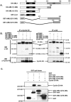
FIG. 2.
Cyclin D interacts with AML1 in vivo. A. Schematic depiction of HA-tagged AML1 proteins and GST-AML1 fusion proteins used in the experiments. runt, Runt homology domain; AD, activation domain. B. HA-tagged AML1 proteins were expressed in the presence of cyclin D3 by cotransfection in 293T cells. One hundred micrograms of protein lysate was subjected to immunoprecipitation (IP) and Western blotting (WB) with the indicated antibodies. IgH, immunoglobulin heavy chain. C. Analysis of domains of AML1 associating with cyclin D3. GST alone and GST-AML1 fusion proteins, GST-Runt, GST-AML1(213-395), and GST-AML1(315-395), were coexpressed with cyclin D3-HA in 293T cells and subjected to glutathione-agarose bead pull-down assays. The blot was sequentially probed with antibodies against the HA tag to demonstrate the association of AML1 with cyclin D3 and probed with antibodies against GST to demonstrate the expression of the fusion proteins.
FIG. 3.
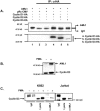
FIG. 3.
Cyclin D proteins are associated with AML1 in vivo. A. 293T cells were transfected with AML1 and cyclin D expression constructs as indicated in the figure. Cell lysates were immunoprecipitated (IP) with anti-HA antibodies (α-HA). The immunoblot was sequentially probed with the anti-AML1 antibodies (top panel) and anti-HA antibodies (bottom panel). Lane 1, AML1-positive control (10 μg of protein lysate from 293T cells expressing AML1). B. K562 cells were treated with or without 100 nM PMA for 3 days. Western blot analysis for endogenous AML1 and cyclin D3 expression. C. Western blot analysis of immunoprecipitates of AML1 from K562 and Jurkat cells for endogenous cyclin D3.
FIG. 4.
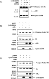
FIG. 4.
Effect of AML1 on cyclin D3 kinase activity. A. 293T cells were transfected with 1 μg of pRc-CMV-cyclin D3-HA and 0, 1, or 3 μg of pCMV5-AML1. Two-hundred-microgram samples of protein extracts were subjected to anti-HA immunoprecipitation. The immunoprecipitates were used in a kinase assay with 1 μg GST-Rb and [γ-32P]ATP. Twenty-microgram samples of transfected cell protein extracts were analyzed by Western blotting to examine the expression of AML1 and cyclin D3. B. HCT116 cells were transfected with 1 μg of each indicated expression construct. Western blot analysis for phosphor-Rb Ser780, total Rb, AML1, and cyclin D3 expression following 24 h of culture and serum starvation. C. As described for panel B, but cells were cultured for 48 h with 10% serum.
FIG. 5.
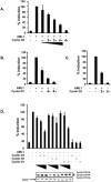
FIG. 5.
Inhibitory effect of cyclin D3 on AML1 transactivation. A. CV-1 cells were transfected with 6 μg of the luciferase reporter p(Mono)4TK81-luc, 600 ng of pCMV5-CBFβ, and 30 ng of pRL-CMV in every transfection. Six hundred ng of pCMV5-AML1 was used with an increasing amount of pRcCMV-cyclin D3-HA from 0.6 to 2.4 μg (depicted by + to 4+). The total DNA content was adjusted with the pRc-CMV empty vector to 10.5 μg. The promoter activity is presented as a percentage of induction relative to the transfection containing p(Mono)4TK81-luc, pCMV5-AML1, and pCMV5-CBFβ. Transfection efficiency was normalized according to cotransfected pRL-CMV Renilla luciferase activity. B. K562 cells were transfected with 10 μg of p(Mono)4TK81-luc, 2 μg of pCMV5-CBFβ, and 0.5 μg of pNull-Renilla in every transfection. Two μg of pCMV5-AML1 was added in the absence or presence of 2 or 8 μg of pRcCMV-cyclin D3-HA. The total DNA content was adjusted with herring sperm DNA to 25 μg. All the other parameters were the same as described for panel A. The averages and standard deviations were generated from results of duplicates of two independent experiments (four sets of data). C. Cyclin D3 inhibits AML1 activation of the B-cell-specific promoter BLK. K562 cells were transfected with 10 μg of pBLK-luc and 2 μg of pCMV5-CBFβ. Two micrograms of AML1 with or without 4 μg of cyclin D3 expression constructs was added. Values were calculated as described above. The results represent the averages with standard deviations from two independent duplicate experiments and were normalized to Renilla luciferase expression. D. Three members of cyclin D proteins inhibit AML1 activity. CV-1 cells were transfected as described for panel A with the variation of the addition of pRcCMV-cyclin D1-HA and pRcCMV-cyclin D2-HA. The transfection results depicted are normalized to the amount of Renilla luciferase expressed from pRL-CMV. The 100% induction is set for AML1 transactivation in the absence of cyclin D. The results shown are average results of duplicates of two independent experiments (four sets of results) with standard deviations. Western blot analysis for the expression of the cyclin D family members in one of the experiments is shown with EGFP as the loading control.
FIG. 6.
FIG. 6.
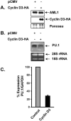
Effect of exogenous cyclin D3 on endogenous PU.1 gene expression. 416B cells were transfected by electroporation with 40 μg of pRc-CMV or pRc-CMV-cyclin D3-HA and harvested for RNA and protein 16 h posttransfection. A. Western blot analysis of the exogenously expressed cyclin D3-HA. B. Northern blot analysis of PU.1 expression in 10 μg of total RNA, and ethidium bromide-stained gel showing 28S and 18S rRNAs as loading controls. C. Real-time PCR depicting the level of PU.1 expressed as corrected for GAPDH expression in each sample, with a standard deviation from results of four separate reactions from two sets of experiments.
FIG. 7.
FIG. 7.
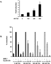
HDAC activity is not required for cyclin D3 inhibition of AML1. K562 cells were transfected with 10 μg of p(Mono)4TK81-luc, 2 μg of pCMV5-CBFβ with or without 2 μg of pCMV5-AML1, and/or 4 μg of pRcCMV-cyclin D3-HA. The cells were cultured for 6 h, and then TSA was added at the indicated concentrations and cultured for an additional 16 h. A. Stimulatory effect of TSA on basal reporter activity. Depiction of the control vector p(Mono)4TK81-luc with pCMV5-CBFβ after treatment of cells for 16 h with the indicated concentrations of TSA. The averages and standard deviations were generated from the results of duplicates of two independent experiments. B. Each set of bars represents a concentration of TSA where AML1 only was set at 100% and the level of induction in that set was calculated according to the luciferase activity of AML1 transfection alone. The average and standard deviation were generated from results of duplicates of two independent experiments.
FIG. 8.
FIG. 8.
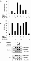
The presence of C/EBPα partially relieves cyclin D3 inhibition on AML1. A. K562 cells were transfected with the indicated expression constructs as described for Fig. 5 with or without 2 μg of pCMV5-C/EBPα in the presence or absence of 4 μg pRcCMV-cyclin D3-HA. The luciferase results were normalized to the expression of Renilla luciferase. The averages and standard deviations were generated from results from duplicates of two independent experiments. B. One hundred percent induction was set for AML1 or for AML1 and C/EBPα in the absence of cyclin D3. The averages and standard deviations were generated from results of duplicates of two independent experiments. C. Cyclin D3 and C/EBPα do not compete with each other in their interaction with AML1. 293T cells were transfected with 5 μg of pRSV-cyclin D3 and 5 μg of pcDNA6-HA-AML1 with the addition of 5 or 15 μg of pMSV-C/EBPα and equilibrated to 25 μg with the empty vector. HA-tagged AML1 protein was immunoprecipitated (IP) with anti-HA antibody (αHA), and Western blotting (WB) was performed with the indicated antibodies (top panels). Straight Western blotting of 10 μg of each sample is shown (bottom panels).
FIG. 9.
FIG. 9.
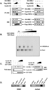
Cyclin D3 competes with CBFβ for AML1 association and disrupts AML1 DNA binding. A. Competitive association between cyclin D3 and CBFβ with AML1. 293T cells were cotransfected with 5 μg of pRSV-cyclin D3 and pcDNA6-HA-AML1, with the addition of 0.5 μg or 2.5 μg of p3XFlag-CBFβ (left panels) or cotransfected with 1 μg of p3XFlag-CBFβ and 5 μg pcDNA6-HA-AML1 with the addition of 5 μg or 10 μg of pRSV-cyclin D3 (right panels) and equilibrated to 25 μg with the empty vector. HA-tagged AML1 protein was immunoprecipitated (IP) with anti-HA (αHA) antibody, and Western blotting (WB) was performed with the indicated antibodies (top panels). A straight Western blot of 10 μg of each sample is shown (bottom panels). B. Cyclin D3 disrupts Runt and Runt-CBFβ DNA binding. GST-Runt and GST-CBFβ proteins were isolated from E. coli, and cyclin D3 was in vitro translated. Sixty ng of GST-Runt was mixed with 40 ng of GST-CBFβ. Reticulocyte lysates of cyclin D3-HA and/or control pRc-CMV was used to a maximum of 5 μl in the titrations, and the binding was performed on ice in the presence of the c-fms promoter AML1 binding site probe. The protein-DNA complexes were resolved on a 5% (29:1) 0.5× Tris-borate-EDTA gel for 1 h at 15 mA at 4°C and dried for autoradiography. Arrows indicate the shifted complex of GST-Runt and the GST-CBFβ/GST-Runt heterodimer. C. Dose-dependent competition between cyclin D3 and CBFβ for association with AML1. 293T cells were transfected with 10 μg of pcDNA6-HA-AML1 or 20 μg of pRSV-cyclin D3. A pull-down assay with GST-CBFβ was performed with HA-AML1 and increasing concentrations of cyclin D3 lysate. Western blot analysis was performed with the indicated antibodies (left panels). Straight Western blot with 15 μg of each sample before the addition of GST-CBFβ (right panels). D. Chromosome immunoprecipitation of AML1 and cyclin D3 on the MIP1α promoter. K562 and Jurkat cells were subjected to CHIP assays with α-AML1 or α-cyclin D3 or rabbit IgG and beads alone as controls. CHIP was done on 2 × 10⁶ cells with each antibody. PCR was performed with the primers described in Materials and Methods for the MIP1α promoter region containing an AML1 binding site. The input represents DNA isolated from 2 × 10⁵ cells.
FIG. 10.
FIG. 10.
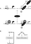
Models of the effect of cyclin D on the regulation of AML1 function. A. Cyclin D is able to compete with CBFβ for AML1 binding. The loss of association with CBFβ reduces AML1 transactivation activity through the lowering of its ability to bind DNA that could be due to conformational changes in the Runt domain. Alternatively, conformational changes in AML1 could also lead to variations in cofactors associated with carboxy-terminal domains of AML1. C/EBPα can partially rescue the negative effect of cyclin D. B. AML1 is known to promote cell cycle progression and differentiation in various hematopoietic cells. The positive regulation of cyclin D proteins could thereby regulate its expression directly by its association with AML1 during proliferative signals to prevent overproduction of cyclin D proteins. In addition, the proposed differentiation activity of AML1 could be counteracted by the displacement of CBFβ or, alternatively, the coactivator p300 by cyclin D proteins, known to cooperate with AML1 during differentiation.

Similar articles

Cited by

References

    1. Bates, S., and G. Peters. 1995. Cyclin D1 as a cellular proto-oncogene. Semin. Cancer Biol. 6:73-82. - PubMed
    1. Behre, G., P. Zhang, D. E. Zhang, and D. G. Tenen. 1999. Analysis of the modulation of transcriptional activity in myelopoiesis and leukemogenesis. Methods 17:231-237. - PubMed
    1. Bernardin, F., and A. D. Friedman. 2002. AML1 stimulates G1 to S progression via its transactivation domain. Oncogene 21:3247-3252. - PubMed
    1. Bernardin-Fried, F., T. Kummalue, S. Leijen, M. I. Collector, K. Ravid, and A. D. Friedman. 2004. AML1/RUNX1 increases during G1 to S cell cycle progression independent of cytokine-dependent phosphorylation and induces cyclin D3 gene expression. J. Biol. Chem. 279:15678-15687. - PubMed
    1. Bodrug, S. E., B. J. Warner, M. L. Bath, G. J. Lindeman, A. W. Harris, and J. M. Adams. 1994. Cyclin D1 transgene impedes lymphocyte maturation and collaborates in lymphomagenesis with the myc gene. EMBO J. 13:2124-2130. - PMC - PubMed

Publication types

LinkOut - more resources