. 2005 Dec;25(23):10261-72.
doi: 10.1128/MCB.25.23.10261-10272.2005.

Muscle-specific loss of apoptosis-inducing factor leads to mitochondrial dysfunction, skeletal muscle atrophy, and dilated cardiomyopathy

Affiliations

Muscle-specific loss of apoptosis-inducing factor leads to mitochondrial dysfunction, skeletal muscle atrophy, and dilated cardiomyopathy

Nicholas Joza et al. Mol Cell Biol. 2005 Dec.

Abstract

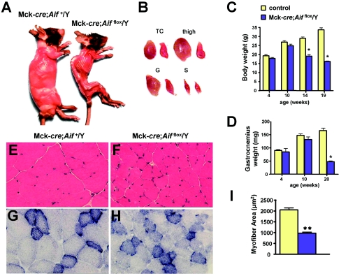
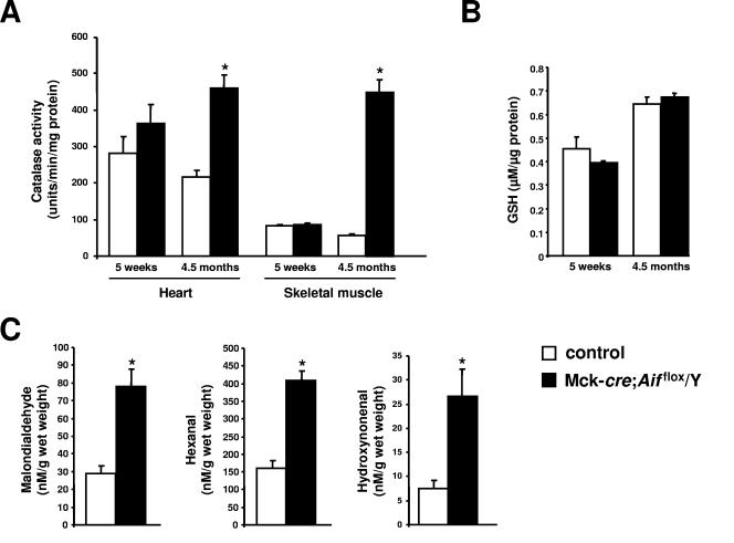
Cardiac and skeletal muscle critically depend on mitochondrial energy metabolism for their normal function. Recently, we showed that apoptosis-inducing factor (AIF), a mitochondrial protein implicated in programmed cell death, plays a role in mitochondrial respiration. However, the in vivo consequences of AIF-regulated mitochondrial respiration resulting from a loss-of-function mutation in Aif are not known. Here, we report tissue-specific deletion of Aif in the mouse. Mice in which Aif has been inactivated specifically in cardiac and skeletal muscle exhibit impaired activity and protein expression of respiratory chain complex I. Mutant animals develop severe dilated cardiomyopathy, heart failure, and skeletal muscle atrophy accompanied by lactic acidemia consistent with defects in the mitochondrial respiratory chain. Isolated hearts from mutant animals exhibit poor contractile performance in response to a respiratory chain-dependent energy substrate, but not in response to glucose, supporting the notion that impaired heart function in mutant animals results from defective mitochondrial energy metabolism. These data provide genetic proof that the previously defined cell death promoter AIF has a second essential function in mitochondrial respiration and aerobic energy metabolism required for normal heart function and skeletal muscle homeostasis.


Figures

FIG. 1.
FIG. 1.
Conditional inactivation of the mouse Aif gene. (A) Partial restriction map of the X-linked mouse Aif locus and schematic illustrations of the targeting vector, the modified Aif locus after homologous recombination (neo), the Aif gene after excision of the neo cassette after transient expression of Flp recombinase (flox), and the expected configuration of the mutant Aif gene after Cre-mediated excision of exon 7 (Δex7). Exon 7 is flanked by loxP sequences (red triangles); the neo cassette is flanked by frt sequences (blue circles). The 5′ flanking probe used for Southern blots is indicated by a horizontal black bar. The neo cassette and diphtheria toxin A (DTA) cassettes are not drawn to scale. The open triangles above the diagram of the wild-type allele indicate the positions of the primers (P1, P2, and P3) used for PCR genotyping. The dotted lines indicate the regions in which homologous recombination occurred. E, EcoRI. (B) By ∼E11.0, β-actin-cre; Aifflox/Y embryos are markedly reduced in size. A wild-type (Aif+/Y) embryo is shown as an age-matched control. Bar, 1 mm. (C) PCR analysis of tissue-specific recombination at the Aif locus in a 1-week-old Mck-cre; Aifflox/Y mouse. The Δex7 (null) allele is only present in heart and skeletal muscle (Skm). The presence of a PCR product representing the flox allele in these tissues likely reflects the presence of nonmyocyte cells. (D) Western blot analysis of AIF protein expression in heart, skeletal muscle (skm), and liver from control (lanes 1, 2, 4, and 6) and mutant Mck-cre;  Aifflox/Y (lanes 3, 5, and 7) mice. Lysates (30 μg) from 13-week-old mice were immunoblotted with anti-AIF and control antiactin antibodies.
FIG. 2.
FIG. 2.
Progressive skeletal muscle atrophy in Mck-cre; Aifflox/Y mutant mice. (A) Typical gross morphology of control Mck-cre; Aif+/Y and mutant Mck-cre; Aifflox/Y mice at 4.5 months of age. (B) Representative pictures of gross morphology of triceps (TC), thigh, gastrocnemius (G), and soleus (S) muscles from (left) control and (right) mutant mice. (C) Body weight measurements for 4-, 10-, 14-, and 19-week-old male Mck-cre; Aif+/Y and mutant Mck-cre; Aifflox/Y mice. (D) Weight measurements of one gastrocnemius muscle for 4-, 10-, and 20-week-old male Mck-cre; Aif+/Y and mutant Mck-cre; Aifflox/Y mice. It should be noted that female mutant mice (Mck-cre; Aifflox/flox) have a similar skeletal muscle phenotype. (E and F) Sections of triceps from (E) control and (F) mutant mice stained with hematoxylin and eosin. Magnification, ×300. (G and H) Representative sections of triceps from (G) control and (H) mutant mice stained for SDH activity. Magnification, ×300. (I) Quantitation of myofiber area from triceps of control and mutant mice. Approximately 50 fibers from 2 mice of each genotype were measured by using Oracle software. Mean values ± the standard error of the mean (SEM). *, P < 0.05; **, P < 0.01 (comparisons between genetic groups [Student’s t test]).
FIG. 3.
FIG. 3.
Dilated cardiomyopathy in Mck-cre; Aifflox/Y mutant mice. (A and B) Representative heart sections of 9-week-old Mck-cre; Aif+/Y (A) control and Mck-cre; Aifflox/Y (B) mutant littermates. LV, left ventricle; RV, right ventricle. Masson's trichrome staining was used. (C) Quantitation of heart weight/tibial-length ratios from 9-week-old male control (n = 7) and mutant (n = 7) littermates. (D and E) Representative heart sections from 9-week-old control (D) and mutant (E) littermates stained with Masson's trichrome. Note the increased cardiomyocyte cell size. Magnification, ×300. (F and G) Electron micrographs of left ventricular heart sections from 13-week-old control (F) and Mck-cre; Aifflox/Y mutant (G) littermates. Note the abnormal ultrastructure of mitochondria in mutant hearts. Magnification, ×10,000. (H) Increased cell size of cardiomyocytes from 9-week-old control and mutant littermates. A total of 100 individual myocytes from three different mice of each genotype were analyzed per group. (I) Increased mRNA expression (mean ± the SEM) of the cardiac hypertrophy markers ANF and BNP. 18S rRNA was used as a control. The results are from heart tissue isolated from seven control and six mutant littermates at 8 weeks of age. *, P < 0.01; **, P < 0.002 (for comparisons between genotypes [Student’s t test]).
FIG. 4.
FIG. 4.
Loss of Aif results in heart failure. (A) M-mode echocardiographic images of contracting hearts in 9-week-old Mck-cre; Aif+/Y control and Mck-cre; Aifflox/Y mutant littermates. Representative images are shown. (B) Representative Doppler images of heart contractility in 9-week-old control and mutant littermates. (C) Heart function of 4- and 9-week-old control and mutant littermates. Values were determined by M-mode and Doppler echocardiography. HR, heart rate (beats per minute [bpm]); AW, anterior wall thickness; PW, posterior wall thickness; LVEDD, left ventricle end diastolic dimension; LVESD, left ventricle end systolic dimension; %FS, percent fractional shortening; PAVc, peak aortic outflow velocity; VCFC, velocity of circumferential fiber shortening. Boldface values at 4 weeks indicate P < 0.01; boldface values at 9 weeks indicate P < 0.001 (Student’s t test). (D) Percent fractional shortening (%FS) in 9-week-old control and mutant littermates. Values (mean ± the SEM) were determined by echocardiography. (E) +dP/dT-max values in 9-week-old control and mutant littermates. Values (mean ± the SEM) were determined by invasive hemodynamics. In panels D and E, “**” indicates a P value of <0.01 between groups (Student’s t test).
FIG. 5.
FIG. 5.
AIF is required for mitochondrial respiration. (A) Respiratory chain complex activities of mitochondria isolated from heart, skeletal muscle, and liver tissue from 18-week-old control and Mck-cre; Aifflox/Y mutant littermates. Shown are relative values of activity (± the SEM) for each complex (I, II, III, IV, and V) of mutant tissues compared to control tissues (100%). *, P < 0.05; **, P < 0.0001 (B) Respiratory chain complex activities in heart and skeletal muscle from 5-week-old control and Mck-cre; Aifflox/Y mutant mice. Relative values of activity (± the SEM) for complexes I, III, and IV normalized to complex V activity and expressed relative to control tissues (100%) are shown. *, P < 0.05; **, P <0.001. (C) Diminished expression of respiratory chain complex I in Aif mutant tissues from 5-week-old mutant mice. Heart (H) and gastrocnemius (G) muscle mitochondria from wild-type (WT) and Mck-cre; Aifflox/Y (KO) mice were subjected to BN-PAGE to resolve respiratory chain complexes. Arrows indicate complexes I, III, IV, and V. (D) Abrogated expression of respiratory chain complex I (CI) subunits in the heart, gastrocnemius, and soleus muscles from Mck-cre; Aifflox/Y mutant mice compared to Mck-cre; Aif+/Y control mice. Tissue lysates from 18-week-old mice were subjected to Western blotting. Liver tissue was used as a control. Cytochrome c (Cyt c) expression is shown as a loading control.
FIG. 6.
FIG. 6.
Increased markers of oxidative stress in mutant tissues. (A) Levels of catalase activity in hearts and thigh muscle (skeletal muscle) from 5-week-old and 4.5-month-old mutant Mck-cre; Aifflox/Y and control Mck-cre; Aif+/Y mice. Three to five mice were assayed per group. Mean values ± the SEM are shown. *, P < 0.005. (B) Total glutathione levels in hearts from 5-week-old and 4.5-month-old mutant Mck-cre; Aifflox/Y and control Mck-cre; Aif+/Y mice. Mean values ± the SEM are shown. Values between mutant and control samples are not significant. (C) Quantification of lipid peroxidation products malondialdehyde, hexanal, and 4-hydroxynonenal in the hearts of 8-week-old mutant Mck-cre; Aifflox/Y and control Mck-cre; Aif+/Y mice. Mean values ± the SEM are shown. *, P < 0.01. n = six mice assayed per group.
FIG. 7.
FIG. 7.
AIF mutant hearts undergo a metabolic switch toward glycolysis. (A and B) Ex vivo heart function of control and mutant (KO) littermates perfused with pyruvate or glucose. HR, heart rate (in beats per minute [bpm]); LVEDP, left ventricular end diastolic pressure; LVSP, left ventricular systolic pressure; LVDP, left ventricular developed pressure; LVDP, LVSP-LVEDP; +dP/dtmax, maximum first derivative of the change in left ventricular pressure; −dP/dtmin, minimum first derivative of the change in left ventricular pressure. Values are means ± the SEM are shown. Male Aif knockouts and littermate controls at 9 weeks of age were analyzed. n = five per group. *, P < 0.01 compared to all other groups; #, P < 0.01 compared to mutant hearts perfused with pyruvate.

References

    1. Benit, P., A. Slama, F. Cartault, I. Giurgea, D. Chretien, S. Lebon, C. Marsac, A. Munnich, A. Rotig, and P. Rustin. 2004. Mutant NDUFS3 subunit of mitochondrial complex I causes Leigh syndrome. J. Med. Genet. 41:14-17. - PMC - PubMed
    1. Bruning, J. C., M. D. Michael, J. N. Winnay, T. Hayashi, D. Horsch, D. Accili, L. J. Goodyear, and C. R. Kahn. 1998. A muscle-specific insulin receptor knockout exhibits features of the metabolic syndrome of NIDDM without altering glucose tolerance. Mol. Cell 2:559-569. - PubMed
    1. Buchholz, F., P. O. Angrand, and A. F. Stewart. 1998. Improved properties of FLP recombinase evolved by cycling mutagenesis. Nat. Biotechnol. 16:657-662. - PubMed
    1. Budihardjo, I., H. Oliver, M. Lutter, X. Luo, and X. Wang. 1999. Biochemical pathways of caspase activation during apoptosis. Annu. Rev. Cell Dev. Biol. 15:269-290. - PubMed
    1. Chomyn, A., and G. Attardi. 2003. MtDNA mutations in aging and apoptosis. Biochem. Biophys. Res. Commun. 304:519-529. - PubMed

Publication types

MeSH terms

LinkOut - more resources