. 2005 Dec;25(23):10273-85.
doi: 10.1128/MCB.25.23.10273-10285.2005.

Loss of SOD1 and LYS7 sensitizes Saccharomyces cerevisiae to hydroxyurea and DNA damage agents and downregulates MEC1 pathway effectors

Affiliations

Loss of SOD1 and LYS7 sensitizes Saccharomyces cerevisiae to hydroxyurea and DNA damage agents and downregulates MEC1 pathway effectors

Carole D Carter et al. Mol Cell Biol. 2005 Dec.

Abstract

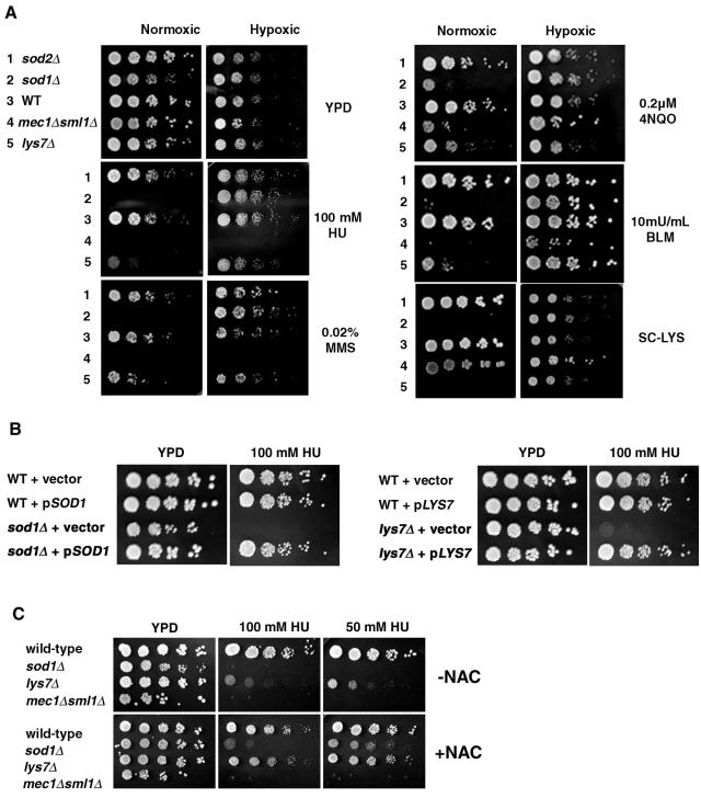
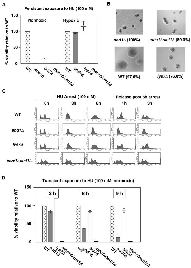
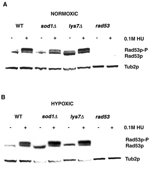
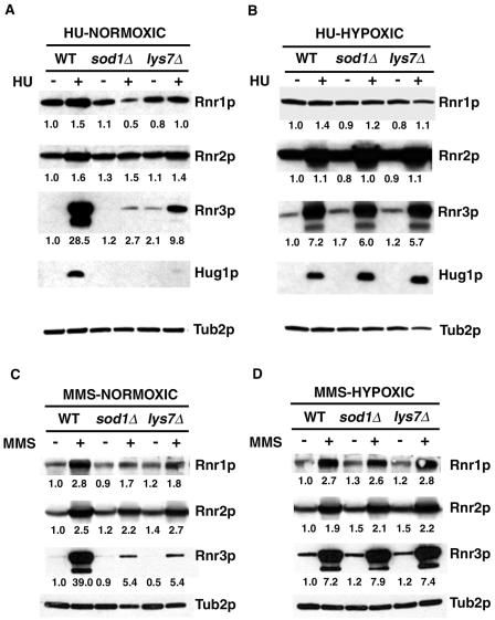
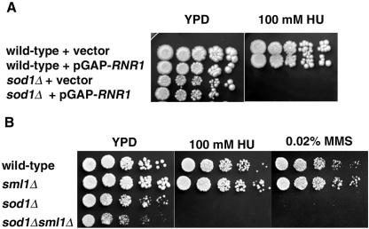
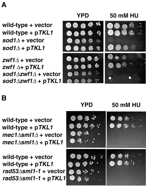
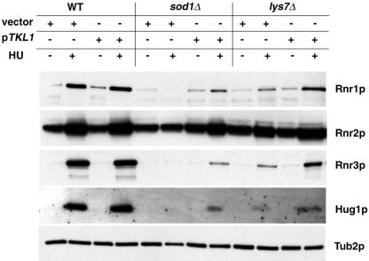
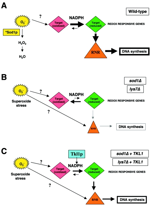
Aerobic metabolism produces reactive oxygen species, including superoxide anions, which cause DNA damage unless removed by scavengers such as superoxide dismutases. We show that loss of the Cu,Zn-dependent superoxide dismutase, SOD1, or its copper chaperone, LYS7, confers oxygen-dependent sensitivity to replication arrest and DNA damage in Saccharomyces cerevisiae. We also find that sod1Delta strains, and to a lesser extent lys7Delta strains, when arrested with hydroxyurea (HU) show reduced induction of the MEC1 pathway effector Rnr3p and of Hug1p. The HU sensitivity of sod1Delta and lys7Delta strains is suppressed by overexpression of TKL1, a transketolase that generates NADPH, which balances redox in the cell and is required for ribonucleotide reductase activity. Our results suggest that the MEC1 pathway in sod1Delta mutant strains is sensitive to the altered cellular redox state due to increased superoxide anions and establish a new relationship between SOD1, LYS7, and the MEC1-mediated checkpoint response to replication arrest and DNA damage in S. cerevisiae.


Figures

FIG. 1.
FIG. 1.
Sensitivity to replication arrest and DNA damage is oxygen dependent in the absence of SOD1 or LYS7. (A) Three microliters of fivefold serial dilutions of sod2Δ, sod1Δ, wild-type (BY4741), mec1Δ sml1Δ (U953-61D), or lys7Δ strains was spotted onto YPD or YPD containing 100 mM HU, 0.02% MMS, 0.2 μM 4NQO, or 10 mU/ml BLM or minimal medium lacking lysine (SC-LYS) and incubated under normoxic or hypoxic conditions at 30°C for 3 to 5 days. (B) Wild-type (BY4741), sod1Δ, and lys7Δ strains were transformed with vector alone, pLJ175 (pSOD1/CEN/URA3), or pLS113 (pLYS7/CEN/URA3). Three microliters of fivefold serial dilutions of each transformant was spotted onto YPD with or without 100 mM HU and incubated under normoxic conditions at 30°C for 2 to 3 days. (C) Wild-type (BY4741), sod1Δ, lys7Δ, and mec1Δ sml1Δ (U953-61D) strains were spotted onto YPD or YPD containing 100 mM or 50 mM HU with or without 100 mM NAC and incubated under normoxic conditions at 30°C for 3 to 5 days. WT indicates wild-type strains.
FIG. 2.
FIG. 2.
Strains lacking SOD1 and LYS7 arrest, but sod1Δ strains fail to produce viable colonies on HU medium. (A) Wild-type (WT; BY4741), sod1Δ, lys7Δ, and mec1Δ sml1Δ (U953-61D) strains were plated on YPD containing 100 mM HU, and plates were incubated under hypoxic or normoxic conditions for 3 to 6 days. lys7Δ strains showed small colonies only upon prolonged incubation for 8 days at 30°C. (B) Terminal phenotypes of strains from panel A grown on HU-containing medium under normoxic conditions were recorded using a light microscope. The percentage of cells with the depicted morphology is indicated in parentheses for each strain. (C) DNA content analysis by flow cytometry. Wild-type (BY4741), sod1Δ, lys7Δ, and mec1Δ sml1Δ (U953-61D) strains were grown under normoxic conditions to logarithmic phase (column 1) and treated with 100 mM HU for 3 hours (column 2) or 6 hours (column 3). Strains arrested with 100 mM HU for 6 hours were released into drug-free medium, and an aliquot of cells was removed every 30 min for up to 4 hours. The DNA contents of cells from 1 (column 4) and 3 hours (column 5) post-HU release are shown. Cells from these samples were analyzed by flow cytometry, and DNA fluorescence units per cell were measured. (D) Wild-type (BY4741), sod1Δ, lys7Δ, and mec1Δ sml1Δ (U953-61D) strains were grown under normoxic conditions to logarithmic phase and treated with 100 mM HU for 6 hours. Cells were washed, and appropriate dilutions were plated on YPD, allowed to recover under normoxic conditions, and counted after 2 to 3 days of incubation at 30°C. For panels A and D, at least 2,500 cells per strain were plated per experiment. For each experiment, the percent viability of wild-type strains was considered to be 100%. Plating efficiency for wild-type cells was 36% on YPD containing HU versus YPD alone. Numbers are averages from three experiments, and error bars represent standard deviation.
FIG. 3.
FIG. 3.
Rad53p is expressed and phosphorylated following replication arrest and DNA damage. Wild-type (WT; BY4741), sod1Δ, lys7Δ, and rad53Δ sml1-1 (U960-5C) strains were grown in YPD in the presence or absence of HU (100 mM, 3 hours) under normoxic (A) or hypoxic (B) conditions as indicated. WCE were subjected to Western blotting using a Rad53p C-terminal polyclonal antibody, and a Tub2p polyclonal antibody was used as a loading control.
FIG. 4.
FIG. 4.
sod1Δ and lys7Δ strains show altered levels of MEC1 checkpoint pathway effector protein Rnr3p and of Hug1p after replication arrest or DNA damage. Wild-type (BY4741), sod1Δ, or lys7Δ strains were grown for 3 hours in YPD with or without 100 mM HU (A and B) or 0.02% MMS (C and D) under normoxic (A and C) or hypoxic (B and D) conditions. WCE were subjected to Western blotting using Rnr1p, Rnr2p, Rnr3p, Hug1p, or Tub2p polyclonal antibodies. The value below each band indicates the ratio of the signal intensity of each protein band to the signal intensity of the Tub2p band compared to the ratio of these bands in the control lane (untreated WT) for each blot. Signal intensities were compared using ImageQuant TL software (Amersham Biosciences, Piscataway, NJ). Quantitation of proteins from a second set of blots gave similar normalized values. No values are given for Hug1p, since the HUG1 transcript is not present in the absence of replication arrest or DNA damage (6).
FIG. 5.
FIG. 5.
Overexpression of RNR1 and deletion of SML1 do not suppress sod1Δ phenotypes. (A) Three microliters of fivefold serial dilutions of wild-type (EG103) or sod1Δ (KS101) strains transformed with either vector alone (pBAD54) or RNR1 expressed from a constitutive promoter (pBAD790) was spotted on YPD and YPD containing 100 mM HU. At least two independent transformants were tested for each strain, and expression of RNR1 was verified by Western blot analysis (data not shown). (B) Three microliters of fivefold serial dilutions of wild-type (YMB3233), sml1Δ (YMB3234), sod1Δ (YMB3235), and sod1Δ sml1Δ strains (YMB3236) was spotted onto YPD, YPD containing 100 mM HU, or 0.02% MMS and then incubated under normoxic conditions at 30°C for 2 to 3 days. At least two independent strains were tested for each genotype.
FIG. 6.
FIG. 6.
Overexpression of TKL1 in sod1Δ strains, but not in mec1 or rad53 strains, restores HU resistance. (A) Three microliters of fivefold serial dilutions of wild-type (1783), sod1Δ (KS105), zwf1Δ (KS113), or sod1Δ zwf1Δ (KS117) strains transformed with vector alone or pKS10 (pTKL1/2μm/LEU2) was spotted onto YPD with or without 50 mM HU and incubated under normoxic conditions at 30°C for 2 days. At least two independent transformants were tested for each strain. Expression of pKS10 (pTKL1/2μm/LEU2) in lys7Δ strains also complemented the HU sensitivity of these strains (data not shown). (B) Three microliters of fivefold serial dilutions of wild-type (W1588-4A), mec1Δ sml1Δ (U953-61D), or rad53Δ sml1-1 (U960-5C) strains transformed with vector alone or pKS10 (pTKL1/2μm/LEU2) was spotted onto YPD with or without 50 mM HU and incubated under normoxic conditions at 30°C for 2 days.
FIG. 7.
FIG. 7.
Overexpression of TKL1 in sod1Δ and lys7Δ strains partially restores expression of the MEC1 pathway effector protein Rnr3p and of Hug1p. Wild-type (WT; BY4741), sod1Δ, or lys7Δ strains transformed with vector alone or with pTKL1 (pKS10) were grown under hypoxic conditions overnight in minimal medium lacking leucine. Cultures were diluted, grown to logarithmic phase, and then grown for 3 hours in YPD with or without 100 mM HU under normoxic conditions. WCE were subjected to Western blotting. Blots were probed with polyclonal antibodies to Rnr1p, Rnr2p, Rnr3p, Hug1p, and Tub2p.
FIG. 8.
FIG. 8.
Model for relationship between superoxide anions, NADPH levels, and RNR activity. (A) In wild-type strains, superoxide anions are scavenged by active Sod1p (*Sod1p), and sufficient levels of NADPH are provided through the pentose phosphate shunt to maintain redox-sensitive proteins in a reduced state. Redox-sensitive cellular targets, such as thioredoxins and glutaredoxins, transfer reductive capacity to RNR, and sufficient levels of active RNR are therefore available for DNA synthesis and repair. (B) In sod1Δ and lys7Δ strains, increases in superoxide anions and lowered NADPH levels shift the equilibrium of redox-sensitive cellular targets to more oxidized forms, resulting in lower RNR activity, decreased DNA synthesis and repair, and sensitivity to replication arrest and DNA damaging agents. Activity of RNR and induction of the MEC1 pathway effector Rnr3p and of Hug1p may also be decreased by the redox sensitivity of MEC1 pathway components. (C) Overexpression of TKL1 in sod1Δ or lys7Δ strains increases the available NADPH in a ZWF1-dependent manner and shifts the equilibrium of redox-sensitive cellular targets towards their reduced forms, thereby increasing RNR activity and suppressing the replication arrest and DNA damage phenotypes of sod1Δ and lys7Δ strains. In addition, induction of Rnr3p and Hug1p is partially restored in the sod1Δ and lys7Δ strains.

Similar articles

Cited by

References

    1. Adams, A., D. E. Gottschling, C. A. Kaiser, and T. Stearns. 1997. Methods in yeast genetics. Cold Spring Harbor Laboratory, Cold Spring Harbor, N.Y.
    1. Aruoma, O. I., B. Halliwell, B. M. Hoey, and J. Butler. 1989. The antioxidant action of N-acetylcysteine: its reaction with hydrogen peroxide, hydroxyl radical, superoxide, and hypochlorous acid. Free Radic. Biol. Med. 6:593-597. - PubMed
    1. Assfalg, M., L. Banci, I. Bertini, P. Turano, and P. R. Vasos. 2003. Superoxide dismutase folding/unfolding pathway: role of the metal ions in modulating structural and dynamical features. J. Mol. Biol. 330:145-158. - PubMed
    1. Barzilai, A., G. Rotman, and Y. Shiloh. 2002. ATM deficiency and oxidative stress: a new dimension of defective response to DNA damage. DNA Repair 1:3-25. - PubMed
    1. Basrai, M. A., J. Kingsbury, D. Koshland, F. Spencer, and P. Hieter. 1996. Faithful chromosome transmission requires Spt4p, a putative regulator of chromatin structure in Saccharomyces cerevisiae. Mol. Cell. Biol. 16:2838-2847. - PMC - PubMed

Publication types

MeSH terms

Substances