. 2005 Dec;25(23):10286-300.
doi: 10.1128/MCB.25.23.10286-10300.2005.

The neuroplasticity-associated arc gene is a direct transcriptional target of early growth response (Egr) transcription factors

Affiliations

The neuroplasticity-associated arc gene is a direct transcriptional target of early growth response (Egr) transcription factors

Lin Li et al. Mol Cell Biol. 2005 Dec.

Abstract

Early growth response (Egr) transcription factors (Egr1 to Egr4) are synaptic activity-inducible immediate early genes (IEGs) that regulate some aspects of synaptic plasticity-related to learning and memory, yet the target genes regulated by them are unknown. In particular, Egr1 is essential for persistence of late-phase long-term potentiation (L-LTP), for hippocampus-dependent long-term memory formation, and for reconsolidation of previously established memories. Here, we show that Egr1 and Egr3 directly regulate the plasticity-associated activity-regulated cytoskeletal-related (Arc) gene, a synaptic activity-induced effector molecule which is also required for L-LTP and hippocampus-dependent learning and memory processing. Moreover, Egr1-deficient and Egr3-deficient mice lack Arc protein in a subpopulation of neurons, while mice lacking both Egr1 and Egr3 lack Arc in all neurons. Thus, Egr1 and Egr3 can indirectly modulate synaptic plasticity by directly regulating Arc and the plasticity mechanisms it mediates in recently activated synapses.


Figures

FIG. 1.
FIG. 1.
Arc expression is induced by Egr1 and Egr3 in vitro and is coexpressed with Egr1 and Egr3 in the hippocampal neurons (n) after seizure in vivo. (A) qRT-PCR analysis of Arc expression in primary (1°) myotubes differentiated 10 DIV and infected for 24 h with adenoviruses expressing EGFP and transcriptionally active Egr3 (Egr3) and adenoviruses expressing EGFP and Egr3Tr. Arc was induced 11.4 ± 1.4-fold in myotubes expressing Egr3 relative to myotubes expressing Egr3Tr. (B) In primary hippocampal neurons (n.) differentiated 3 DIV and infected for 24 h with EGFP and Egr1 (Egr1) or Egr3 and EGFP (Egr3), Arc expression was induced 250.1 ± 76.1-fold and 220.0 ± 23.7-fold, respectively, relative to neurons infected with a virus expressing only EGFP. (C) In primary cortical neurons differentiated 3 DIV and infected for 24 h with Egr1- or Egr3-expressing adenovirus, Arc expression was induced 8.0 ± 2.0-fold and 15.4 ± 2.6-fold, respectively, relative to neurons infected with a virus expressing only EGFP. (D) After KA-induced seizure, Egr1, Egr3, and Arc are coinduced, each with a stereotypic time course. The transient Arc protein levels are most closely correlated with the transient levels of Egr3 protein. By contrast, Egr1 protein has a shorter transient time course after seizure. Two hours after seizure, Egr1 (E) is robustly induced and coexpressed with Arc (E′), similar to Egr3 (F), which is also coexpressed with Arc (F′) in hippocampal dentate gyrus granule neurons. Scale = 50 μm. *, P < 0.01, Student's t test relative to conditions without asterisks. Values represent means ± standard deviations of three or four experimental replicates for each condition.
FIG. 2.
FIG. 2.
Egr1 and Egr3 bind the Arc promoter in vivo after seizure and transactivate the Arc promoter through a response element in the proximal promoter. (A) Egr1 and Egr3 bind to a region of the Arc promoter that is proximal to the transcription start site. The position of the distal (Pr1 and Pr2) and proximal (Pr3 and Pr4) primers used for PCR and qPCR are shown relative to the TATA box and transcription start site (+1) of the mouse Arc gene (numbering according to reference 36). For each primer pair, endpoint PCR and qPCR are shown with ChIP libraries generated from unseized and seized mouse hippocampus by using Egr1 (α1)- and Egr3 (α3)-specific antibodies or nonimmune IgG. Neither Egr1 nor Egr3 were bound to a distal region of the Arc promoter (Pr1-Pr2) before or after seizure. However, they were bound to a proximal region of the Arc promoter (Pr3-Pr4) in vivo after seizure and enriched 21.9- ± 5.5- and 41.7- ± 22.9-fold in ChIP libraries generated with Egr1- and Egr3-specific antibodies, respectively, relative to nonimmune IgG. (B) Luciferase reporter plasmids containing portions of the proximal Arc promoter −775 to −87 nucleotides upstream of the transcription start site were significantly activated by Egr1 and Egr3 relative to the CMV expression vector alone. All of the promoter fragments down to −87 nucleotides upstream of the transcription start site were activated by Egr1 and Egr3, suggesting that the ERE was located within this region of the proximal promoter. *, P < 0.01, Student's t test relative to conditions without asterisks; values represent means ± standard deviations of three or four experimental replicates.
FIG. 3.
FIG. 3.
Egr1 and Egr3 bind to a high-affinity response element that is necessary and sufficient to transactivate Arc gene expression. (A) The proximal Arc promoters from mice, rats, and humans were aligned to the TATA box to identify conserved regions that might represent Egr binding sites. Two GC rich domains were identified (E1 and E2). The E2 domain appeared to contain a highly conserved response element (ERE; gray). (B) Nuclear proteins from unseized and seized mouse hippocampi were able to bind to probes representing the E2 domain but not the E1 domain by EMSA. The nuclear proteins were induced by seizure and the protein DNA complexes specifically contained Egr1 and Egr3 but not other Egr proteins as demonstrated by supershifting of the Egr-containing complexes by Egr-specific antibodies (asterisk). Moreover, no additional binding proteins were observed, as only Egr1- and Egr3-containing complexes were bound to the E2 domain. (C) To determine whether Egr proteins were bound to the putative ERE in the E2 domain, EMSA was used with a probe that contained a mutation expected to disrupt Egr protein binding within the core binding domain (E2m1) and a probe that contained a mutation outside of the core binding domain not expected to disrupt Egr protein binding. (D) The mutation within E2m1 completely abrogated Egr protein binding with nuclear proteins obtained from seized mouse hippocampus. Egr1 and Egr3 proteins were capable of binding to the probe containing mutations outside of the core binding domain (E2m2) similar to the wild-type sequence. (E, left panel) When the E2m1 mutation was introduced into the −775Arc luciferase reporter construct, activation by either Egr1 or Egr3 was completely abrogated. (E, center panel) Egr3 appeared to consistently activate the Arc promoter better than Egr1, but this was also observed on the minimal 4× ERE reporter, which has been previously shown to result from increased levels of Egr3 expression/stability relative to Egr1 from the CMV expression plasmids. (E, right panel) Egr1- and Egr3-dependent activation of −775Arc promoter was subject to repression by the Egr corepressor Nab2 which completely abrogated Arc transactivation. *, P < 0.01, Student's t test relative to conditions without asterisks, values represent means ± standard deviations of three experimental replicates.
FIG. 4.
FIG. 4.
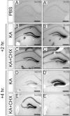
Arc is transiently expressed as a protein synthesis-independent immediate early gene and its expression is modulated by a protein synthesis-dependent mechanism. In caged wild-type adult mice treated with PBS, Egr3 (A) and Arc (A′) expression were very low in the hippocampus. Two hours after KA-induced seizure, Egr3 (B) and Arc (B′) were markedly upregulated. Unlike Egr3 mRNA (B), which was localized within the neuron somata and nuclei, Arc mRNA (B′, arrow) was also characteristically transported into the dendrites of hippocampal dentate gyrus neurons. When mice were treated with CHX 15 min prior to seizure to inhibit protein synthesis, there was no detectable difference in Egr3 (C) or Arc (C′) expression compared to mice not receiving CHX. By 4 h after KA-induced seizure in the absence of CHX treatment, Egr3 (D) and Arc (D′) expression were still elevated above basal (PBS treatment) levels. In CHX-pretreated mice 4 h after KA-induced seizure, Egr3 (E) expression was superinduced, consistent with its regulation as an immediate early gene; however Arc (E′) expression was completely abrogated. Thus, Arc expression is independent of protein synthesis within at least 2 h after KA-induced seizure but by 4 h after seizure, Arc expression is completely regulated by a protein synthesis-dependent mechanism. Scale = 0.5 μm.
FIG. 5.
FIG. 5.
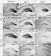
The protein synthesis-dependent phase of Arc gene expression requires Egr3 after seizure-induced synaptic activation. Similar to Arc (A), Egr3 (A′), and Egr1 (A′, inset), which were markedly upregulated in wild-type mice independent of new protein synthesis 2 h after seizure, Arc (B) and Egr3 (B′) were appropriately induced in Egr1-deficient mice and Arc (C) and Egr1 (C′) were appropriately induced in Egr3-deficient mice, indicating that similar seizure induced activity was generated the Egr gene-deficient mice to induce qualitatively equivalent levels of protein synthesis-independent Arc expression. However 4 h after seizure, when Arc expression was dependent upon new protein synthesis, Arc (D) and Egr3 (D′) were still upregulated and (D′, inset) Egr1 had returned to basal levels. In Egr1-deficient mice, Arc (E) and Egr3 (E′) were also elevated similar to wild-type mice. However, in Egr3-deficient mice, Arc (F) expression was markedly diminished and Egr1 (F′) was slightly elevated above basal levels. Thus, protein synthesis-dependent Arc expression requires Egr3 after seizures in the hippocampus. Scale = 0.5 μm.
FIG. 6.
FIG. 6.
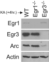
Prolonged elevation of Egr3 protein in the hippocampus after seizure explains why Egr3, but not Egr1, regulates the protein synthesis-dependent phase of Arc expression. Hippocampal lysates obtained four hours after seizure, when Arc expression depends upon protein synthesis in wild-type hippocampus, contain very low levels of Egr1 and high levels of Egr3 protein because of the difference in kinetics between the two proteins after seizure induction. Similarly, Egr3 protein levels remain high in the hippocampus of Egr1-deficient mice, and consequently, Arc protein levels are comparable to wild-type levels. By contrast, in the hippocampus of Egr3-deficient mice, Egr1 levels are very low, as they are in wild-type mice 4 h after seizure, and without Egr1 or Egr3, Arc protein levels are very low compared to those in wild-type or Egr1-deficient mice. These results indicate that Egr1 is dispensable for regulating the protein synthesis-dependent phase of Arc expression in this seizure paradigm. In Egr1-deficient mice, there are high levels of Egr3 which are sufficient to regulate Arc expression whereas in Egr3-deficient mice there is an insufficient amount of Egr1 protein to compensate for Egr3 loss leading to correspondingly very low levels of Arc protein.
FIG. 7.
FIG. 7.
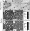
Arc, Egr1, and Egr3 are coexpressed in hippocampal dentate gyrus granule neurons and cortical neurons during physiologic synaptic activity. Four hours after mice were subjected to a novel environment, Egr1 (A), Arc (B), and Egr3 (C) were coexpressed in a subpopulation of hippocampal dentate gyrus granule neurons. Some Arc-expressing neurons coexpressed Egr1 (white-filled arrowheads), some coexpressed Egr3 (open arrowheads), and some coexpressed both Egr1 and Egr3 (black-filled arrowheads). In the somatosensory cortex, in Arc protein (D)-expressing neurons, Egr1 protein (E) was present in 94.6 ± 4.1% of the neurons (F). Arc protein (G)-expressing neurons also expressed Egr3 (H) in 96.6 ± 2.4% of the neurons (I). (A through C and D through H) Scale = 50 μm. (F and I) Values represent means ± standard deviations of neuron counts from six 0.1-mm2 high power fields from two wild-type 21-day-old mice exposed to a novel environment.
FIG. 8.
FIG. 8.
Arc expression requires Egr1 and Egr3 in the context of physiologic synaptic activity. (A) In 21-day-old mice, four hours after exposure to a novel environment, Arc was expressed at high levels by many cortical and hippocampal neurons. In the hippocampus, a subpopulation of neurons (arrow) expressed high levels of Arc (E), and in the frontal cortex (somatosensory cortex shown), pyramidal neurons in superficial and deep layers of cortex preferentially expressed Arc (I). In Egr1-deficient mice (B) and Egr3-deficient mice (C), Arc expression was decreased compared to that in wild-type mice, which was reflected by a decreased number of Arc-expressing hippocampal dentate gyrus granule neurons (F and G, arrows) and a decrease in the number and intensity of Arc-expressing neurons in cortex (J and K). (D) In Egr1/3 dKO mice, Arc expression was very low compared to that in wild-type mice. In the hippocampal dentate gyrus, no Arc-expressing neurons were identified (H) and similarly in cortex, Arc expression was barely detectable (L). Boxes in panels A through D orient the magnified regions shown in panels E through L. (A through D) Scale = 1 mm. (E through H) Scale = 100 μm. (I through L) Scale = 50 μm.
FIG. 9.
FIG. 9.
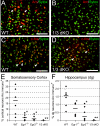
Arc protein is decreased in Egr-deficient cortical and hippocampal neurons. Many cortical neurons (somatosensory cortical neurons shown here) contain high levels of Arc protein in wild-type (A) but not Egr1/3 dKO (B) mice after identical treatment to novel environmental stimuli. The cortical neurons were apparently activated similarly because the level of c-fos, an activity regulated immediate early protein, was similar in wild-type (C) relative to Egr1/3 dKO (D) cortical neurons (red labeling corresponds to Arc or c-fos protein and green corresponds to SYTOX green staining for DNA [scale bar = 100 μm]). (E) In wild-type somatosensory cortex, 17.5% of neurons were immunoreactive for Arc, whereas 2.2%, 1.3%, and 0% of neurons were immunopositive for Arc in Egr1-deficient, Egr3-deficient, and Egr1/3 dKO mice, respectively. (F) In wild-type mice, 1.6% of hippocampal dentate gyrus neurons were immunoreactive for Arc protein. By contrast, in Egr1-deficient and Egr3-deficient mice, 0.8% and 0.2% of hippocampal dentate gyrus neurons, respectively, were immunoreactive for Arc protein. In hippocampal dentate gyrus neurons from Egr1/3 dKO mice, no immunoreactive neurons (0%) were identified. (E and F) Values cited are shown as horizontal bars which represent the medians of six measurements from three animals and two similar tissue sections analyzed for each genotype. *, P < 0.05; **, P < 0.005, Student's t test relative to wild type.

References

    1. Abraham, W. C., M. Dragunow, and W. P. Tate. 1991. The role of immediate early genes in the stabilization of long-term potentiation. Mol. Neurobiol. 5:297-314. - PubMed
    1. Albert, Y., J. Whitehead, L. Eldredge, J. Carter, X. Gao, and W. G. Tourtellotte. 2005. Transcriptional regulation of myotube fate specification and intrafusal muscle fiber morphogenesis. J. Cell Biol. 169:257-268. - PMC - PubMed
    1. Andrews, N. C., and D. V. Faller. 1991. A rapid micropreparation technique for extraction of DNA-binding proteins from limiting numbers of mammalian cells. Nucleic Acids Res. 19:2499. - PMC - PubMed
    1. Banker, G., and K. Goslin. 1998. Culturing nerve cells, 2nd ed. MIT Press, Cambridge, Mass.
    1. Bozon, B., S. Davis, and S. Laroche. 2003. A requirement for the immediate early gene zif268 in reconsolidation of recognition memory after retrieval. Neuron 40:695-701. - PubMed

Publication types

MeSH terms

Substances