. 2005 Dec;25(23):10329-37.
doi: 10.1128/MCB.25.23.10329-10337.2005.

The reaper-binding protein scythe modulates apoptosis and proliferation during mammalian development

Affiliations

The reaper-binding protein scythe modulates apoptosis and proliferation during mammalian development

Fabienne Desmots et al. Mol Cell Biol. 2005 Dec.

Abstract

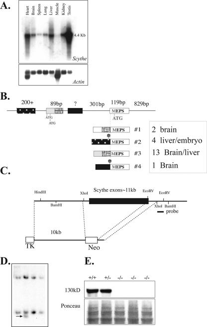
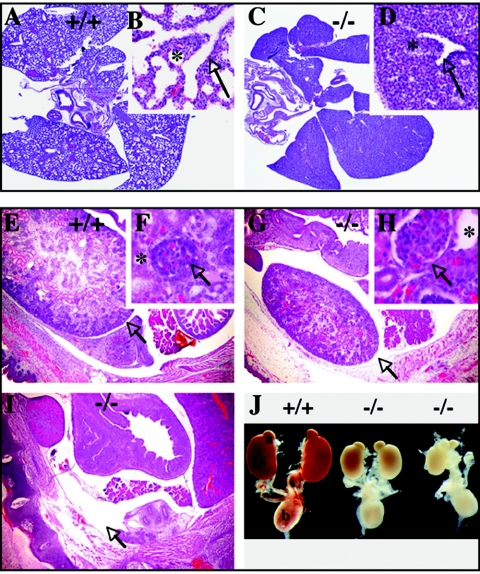
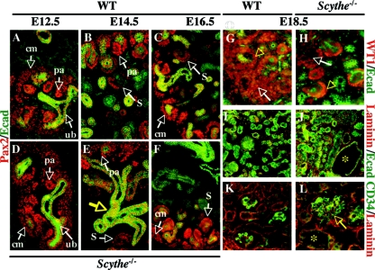
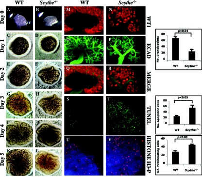
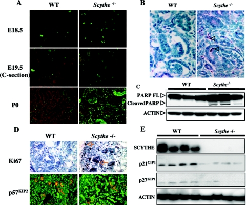
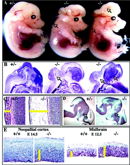
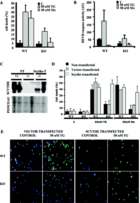
Scythe (BAT3 [HLA-B-associated transcript 3]) is a nuclear protein that has been implicated in apoptosis, as it can modulate Reaper, a central apoptotic regulator in Drosophila melanogaster. While Scythe can markedly affect Reaper-dependent apoptosis in Xenopus laevis cell extracts, the function of Scythe in mammals is unknown. Here, we report that inactivation of Scythe in the mouse results in lethality associated with pronounced developmental defects in the lung, kidney, and brain. In all cases, these developmental defects were associated with dysregulation of apoptosis and cellular proliferation. Scythe-/- cells were also more resistant to apoptosis induced by menadione and thapsigargin. These data show that Scythe is critical for viability and normal development, probably via regulation of programmed cell death and cellular proliferation.


Figures

FIG. 1.
FIG. 1.
Inactivation of mouse Scythe. (A) Northern blot analysis of Scythe mRNA expression in various adult tissues (top). Actin reprobing of the blot confirmed equal RNA loading (bottom). (B) Schematic representation of the Scythe 5′ UTR sequence and the alternative splices found by reverse transcriptase PCR for several tissues, including liver and brain. The sizes of some 5′ UTR and coding exons are shown schematically positioned on genomic DNA, and the spliced transcripts are indicated below the genomic DNA structure. The relative abundance of a particular transcript is indicated by the number of times it was isolated by PCR from a particular tissue, i.e., transcript no. 3 was the most common 5′ version of the Scythe. (C) Structure of the Scythe targeting vector that replaced almost the entire Scythe ORF. The probe used for the Southern blot analysis is indicated. TK, thymidine kinase. (D) Southern blot analysis after BamHI digestion of ES cells. (E) Scythe (130 kDa) is present in WT (+/+) and heterozygous (+/−) cells but is absent in Scythe-null (−/−) mouse embryo fibroblasts.
FIG. 2.
FIG. 2.
Lung and kidney abnormalities occur in E18.5 Scythe/ embryos. (A to D) Compared to the WT (+/+) (A and B), abnormal lung development was present in Scythe mutants (−/−) (C and D). Lungs were analyzed at the saccular stage of development. In the WT, bronchial tree formation in the lungs was complete (arrow) and normal alveolization was observed (*) (B). Lungs were smaller and markedly less developed in Scythe/ embryos (C and D), with decreased branching of terminal bronchioles (arrow) and nearly complete lack of alveolization (*). (Inset panels B and D show higher magnifications of panels A and C, respectively). (E to J) Abnormalities observed in E18.5 Scythe/ kidneys. Scythe mutants showed smaller kidneys (G) or no kidney (I), with enlarged glomeruli (H, arrow) and dilated tubules (H, asterisk) compared to those of the WT (E and F, same symbols). Sections were stained with hematoxylin and eosin. Aside from kidney defects in Scythe/ animals, the remainder of the urogenital system was normal (J). a, adrenal gland; k, kidney; b, bladder.
FIG. 3.
FIG. 3.
Analysis of Scythe/ kidneys. (A to F) Immunostaining of Pax2 (red) and E-cadherin (Ecad) (green) with control (A, B, and C) and mutant (D, E, and F) embryos was done at E12.5, E14.5, and E16.5. At E12.5, the ureteric bud (ub) was surrounded by condensed mesenchyme (A and D), and ureteric bud branching and formation of pretubular aggregates (pa) could be seen for both WT and Scythe/ embryos. Pax2 was expressed in condensed mesenchymal cells (cm), pretubular aggregates, and comma- and S-shaped bodies (S) in both WT and Scythe/ embryos at E14.5 and E16.5, but nephrogenic differentiation was delayed in Scythe/ embryos (B, C, E, and F). The structure of the epithelium (Ecad staining) showed branching abnormalities (E, yellow arrow) in Scythe/ kidneys. (G to L) At E18.5, WT1 (red) and E-cadherin (green) expression was detected in the condensing blastema (arrows), the proximal part of S-shaped bodies, and the podocytes of functional glomeruli (arrowheads) for both WT and Scythe/ kidney (G and H) but to a lesser extent in Scythe/ kidney. Laminin (red) distribution in Scythe/ kidneys at E18.5 showed a pattern similar to that of the WT, while E-cadherin staining (green) showed enlarged tubules (asterisk) in Scythe/ kidneys (I and J). The endothelial marker CD34 (green) is expressed similarly in the WT and in Scythe/, although enlarged glomeruli (arrow) and tubules (asterisk) are seen with Scythe/ kidney (K and L).
FIG. 4.
FIG. 4.
Branching, apoptosis, and proliferation defects in Scythe/ kidney explants. (A to L) Embryonic kidney rudiments were dissected from E12.5 WT and Scythe/ embryos and grown in culture for 5 days (A to L). The growth and branching morphogenesis of Scythe/ explants was impaired compared to that of the WT. (M to R) After 4 days in culture, explants were immunostained with E-cadherin (ECAD) (green) and WT1 (red) antibodies. E-cadherin stained ureteric bud branches, and WT1 is expressed in the condensed mesenchyme. (S and T) TUNEL analysis revealed a substantial increase in apoptosis for Scythe mutants (n = 3; t test, P of <0.05) compared to the WT. (U and V) Anti-phosphorylated histone H3 (H3-P) was used to identify mitotic cells and showed a 1.5-fold increase in Scythe/ kidney explants (n = 3; t test, P of <0.01) compared to WT controls. Histograms correspond to the quantitative analysis of branch points, apoptotic cells, and proliferating cells for three different experiments.
FIG. 5.
FIG. 5.
Increased apoptosis and cellular proliferation occurs in Scythe/ kidneys. (A) TUNEL assays of kidney sections at E18.5, E19.5, and P0 showed increased apoptosis in Scythe/ kidneys. C-section, cesarean section. (B) Staining with anti-cleaved caspase 3 (arrowheads) showed increased apoptosis in cortical mesenchyme from E18.5 Scythe/ kidneys. (C) Proteins were isolated from three WT and three Scythe/ E18.5 kidneys, and PARP cleavage was analyzed by Western blot. Actin protein levels were used as a loading control. FL, full length. (D) Ki67 and p57KIP2 immunohistochemistry showed increased proliferation in Scythe/ kidneys at E18.5 compared to the WT control. (E) Western blot analysis of cyclin-dependent kinase inhibitors p21CIP1 and p27KIP1 from multiple individual WT and Scythe/ E18.5 kidneys showed expression patterns consistent with increased proliferation. Scythe and actin protein levels were used as controls.
FIG. 6.
FIG. 6.
Scythe loss affects neural development. (A) Excencephaly (arrows) was observed for Scythe/ embryos at E14.5. (B) Improper expansion of the midbrain and the hindbrain (arrows) was observed for these E14.5 embryos, as indicated by Nissl staining. (C) Edema was also present. (D and E) Tuj1 and Ki67 staining showed an expanded ventricular zone at E12.5 in Scythe/ embryos (D) and an increase in proliferating cells in the Scythe/ neopallial cortex at E14.5 and midbrain at E12.5 (E).
FIG. 7.
FIG. 7.
Scythe/ cortical neurons are resistant to certain apoptotic stimuli. Scythe/ neurons are resistant to apoptosis after menadione or thapsigargin treatment, as determined using TUNEL assays (A) and caspase activity towards a DEVD substrate (B). Reintroduction of Scythe protein to Scythe/ cells using pHA-Scythe or vector alone was confirmed by Western blot analysis (C) and restored susceptibility towards menadione and thapsigargin (D). TUNEL analysis of WT and Scythe/ cortical neurons after treatment with thapsigargin and menadione in nontransfected, pHA-Scythe-transfected, or vector-alone-transfected cells (D and E). TUNEL-positive apoptotic cells are green, and DAPI-counterstained cells are blue. Me, menadione; TG, thapsigargin; KO, knockout; C, control; AU, arbitrary units; NT, nontransfected; Scythe-T, transfected.

Similar articles

Cited by

References

    1. Ball, E. M., and G. P. Risbridger. 2001. Activins as regulators of branching morphogenesis. Dev. Biol. 238:1-12. - PubMed
    1. Banerji, J., J. Sands, J. L. Strominger, and T. Spies. 1990. A gene pair from the human major histocompatibility complex encodes large proline-rich proteins with multiple repeated motifs and a single ubiquitin-like domain. Proc. Natl. Acad. Sci. USA 87:2374-2378. - PMC - PubMed
    1. Bard, J. B., A. Gordon, L. Sharp, and W. I. Sellers. 2001. Early nephron formation in the developing mouse kidney. J. Anat. 199:385-392. - PMC - PubMed
    1. Bouchard, M. 2004. Transcriptional control of kidney development. Differentiation 72:295-306. - PubMed
    1. Brodbeck, S., and C. Englert. 2004. Genetic determination of nephrogenesis: the Pax/Eya/Six gene network. Pediatr. Nephrol. 19:249-255. - PubMed

Publication types

MeSH terms