. 2005 Dec;25(23):10365-78.
doi: 10.1128/MCB.25.23.10365-10378.2005.

Sumoylation of p45/NF-E2: nuclear positioning and transcriptional activation of the mammalian beta-like globin gene locus

Affiliations

Sumoylation of p45/NF-E2: nuclear positioning and transcriptional activation of the mammalian beta-like globin gene locus

Yu-Chiau Shyu et al. Mol Cell Biol. 2005 Dec.

Abstract

NF-E2 is a transcription activator for the regulation of a number of erythroid- and megakaryocytic lineage-specific genes. Here we present evidence that the large subunit of mammalian NF-E2, p45, is sumoylated in vivo in human erythroid K562 cells and in mouse fetal liver. By in vitro sumoylation reaction and DNA transfection experiments, we show that the sumoylation occurs at lysine 368 (K368) of human p45/NF-E2. Furthermore, p45 sumoylation enhances the transactivation capability of NF-E2, and this is accompanied by an increase of the NF-E2 DNA binding affinity. More interestingly, we have found that in K562 cells, the beta-globin gene loci in the euchromatin regions are predominantly colocalized with the nuclear bodies promyelocytic leukemia protein (PML) oncogenic domains that are enriched with the PML, SUMO-1, RNA polymerase II, and sumoylatable p45/NF-E2. Chromatin immunoprecipitation assays further showed that the intact sumoylation site of p45/NF-E2 is required for its binding to the DNase I-hypersensitive sites of the beta-globin locus control region. Finally, we demonstrated by stable transfection assay that only the wild-type p45, but not its mutant form p45 (K368R), could efficiently rescue beta-globin gene expression in the p45-null, erythroid cell line CB3. These data together point to a model of mammalian beta-like globin gene activation by sumoylated p45/NF-E2 in erythroid cells.


Figures

FIG. 1.
FIG. 1.
Sumoylation of p45/NF-E2 in vitro. (A) Schematic representation of the domain organization of p45. The region (positions 1 to 114) interacting with CBP, the PY motif interacting with the WW domain, and the bZIP domain are indicated. Also shown are the phosphorylation site at S169 and the sumoylation site at K368; the sequence around K368 is compared with the consensus motif of sumoylation (ψKXE). (B) Sumoylation reaction in vitro. The reaction was carried out as described in Materials and Methods. The GST-p45 substrate and the SUMO-1-GST-p45 generated in the reaction were analyzed by immunoblotting (IB) with anti-p45 (upper panel) or anti-SUMO-1 (lower panel). (C) Requirement of K368 for in vitro sumoylation. The products of sumoylation and/or subsequent desumoylation reactions using GST-p45 and GST-p45 (K368R) as the substrates were analyzed by immunoblotting with anti-p45. SUMO-1-GST-p45 could only be generated from the reaction in lane 5.
FIG. 2.
FIG. 2.
Sumoylation of p45/NF-E2 in vivo. (A) Sumoylation of p45 in transfected K562 cells. The transfections of K562 cells were carried out using pEF-p45 and pCMV-His-SUMO-1 as described in Materials and Methods. The whole-cell extracts were then prepared and incubated with Ni-NTA agarose to isolate His-tagged protein(s). The retained fractions were analyzed by immunoblotting (IB) with anti-p45. (B) Requirement of K368 for sumoylation of human p45/NF-E2 in vivo. 293 cells were transfected with different combinations of the expression plasmids. The whole-cell extracts (WCE) were then prepared, IP with anti-Flag, and immunoblotted (IB) with anti-p45. Note that sumoylated p45 could only be detected in the 293 extract containing myc-p45 and FlagDsRed-SUMO-1 GG (lane 1, upper panel). The WCE prior to IP were also analyzed by Western blotting with anti-p45 (middle panel) and anti-SUMO-1 (lower panel), respectively. The arrowheads point to the positions of the sumoylated p45 on the gel. (C) Sumoylation of endogenous p45 in K562 cells. The nuclear extract was prepared from K562 cells, fractionated by precipitation with 20% to 50% ammonium sulfate, and analyzed by immunoblotting with anti-p45 (left panel) or with anti-SUMO-1 (middle panel). The 20% ammonium sulfate fraction was also immunoprecipitated with rabbit preimmune serum (left lane, right panel) or rabbit anti-p45 (right lane, right panel) and then hybridized with mouse anti-SUMO-1. (D) Sumoylation of endogenous p45 in mouse E14.5 fetal liver. The whole-cell extract was prepared from mouse E14.5 fetal livers as described in Materials and Methods and then analyzed by Western blotting using anti-p45 (lane 1) or anti-SUMO-1 (lane 2) as the probe. The extract was also immunoprecipitated with rabbit preimmune serum (lane 3) or rabbit anti-p45 (lane 4). The immunoprecipitates were then analyzed by Western blotting using mouse anti-SUMO-1 antibody as the probe.
FIG. 3.
FIG. 3.
Enhancement of transactivation capability of p45/NF-E2 by sumoylation. (A) Reporter transactivation by myc-p45 (black bars) and myc-p45 (K368R) (white bars), respectively. 293 cells were transfected with 2 μg of pRBGP2-Luc (map shown on top) and different amounts of the expression plasmid pEF-myc-p45 or pEF-myc-p45 (K368R). The luciferase activities were measured and calculated. Their relative values are shown in the bar histogram. Data shown are expressed as means ± standard errors of the means from at least three independent assays, each carried out in duplicate. The amounts of myc-p45 and myc-p45 (K368R) in different transfectants were compared by immunoblotting. (B) Enhancement of p45-mediated transactivation by coexpressed UBC9. 293 cells were transfected with 2 μg of pRBGP2-Luc, 2 μg of pEF-myc-p45, or 2 μg of pEF-myc-p45 (K368R) and different amounts of the expression plasmid pcDNA-UBC9. Note that the transactivation mediated by myc-p45 (black bars), but not myc-p45 (K368R) (white bars), was enhanced with coexpression of UBC9. The amounts of myc-p45 and myc-p45 (K368R) in different transfectants were compared by immunoblotting. (C) Enhancement of p45-mediated transactivation by coexpressed SUMO-1. 293 cells were transfected with 2 μg of pRBGP2-Luc, 2 μg of pEF-myc-p45, and different amounts of the expression plasmid pCMV-SUMO-1 GG or pCMV-SUMO-1 AA. Note that the transactivation mediated by myc-p45 was enhanced by coexpression of SUMO-1 GG (black bars) but not by that of SUMO-1-AA (white bars). (D) Requirement of K368 for enhancement of p45-mediated transactivation by SUMO. Parallel transfections were done as described above for panel C except that pEF-myc-p45 was replaced with pEF-myc-p45 (K368R). The amounts of myc-p45 (K368R) in different transfectants were estimated by immunoblotting. We have also analyzed the transactivation capability of p45/NF-E2 in K562 cells as affected by coexpression of UBC9 or SUMO-1. Results similar to those seen with 293 cells were obtained, but the effects were smaller due to the low activity of the cytomegalovirus promoter in K562 cells (data not shown).
FIG. 4.
FIG. 4.
Effects of p45 sumoylation on NF-E2/DNA binding. EMSA was carried out using extracts prepared from 293 cells cotransfected with pEF-p45 plus pEF-His-Maf G (lanes 4, 5, 11, 13, and 15) or with pEF-p45 plus pEF-His-Maf G plus pCMV-SUMO-1 (lanes 6, 7, 8 to 10, 12, 14, and 16). Nuclear extracts were prepared from 293 or transfected 293 cells and used for EMSA with different oligonucleotides (oligo) as the probes. Lane 1, probe only; lane 2, probe plus anti-p45; lane 3, probe plus 293 extract. The samples in lanes 5, 7, and 9 were the same as those in lanes 4, 6, and 8 except that anti-p45 was included in the binding reactions. The sample in lane 10 was the same as that in lane 8 except that anti-SUMO-1 was included in the binding reaction. The amounts of p45 protein in the different extracts used for EMSA were estimated by Western blotting (IB-p45). The filled and blank arrowheads point to the positions of the NF-E2/DNA complex and AP1/DNA complex, respectively, on the gel. Note the slightly slower migration of a portion of the NF-E2/DNA complex(es) in the SUMO (+) sample lanes (for example, compare lane 15 to lane 16). This portion is likely the complex formed between the DNA oligonucleotide and NF-E2 containing sumoylated p45.
FIG. 5.
FIG. 5.
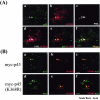
Immunofluorescence staining of K562 cells. (A) K562 cells were immunostained for the nuclear distributions of p45 (panel a), SUMO-1 (panel b), and PML (panel c). The merged patterns of panels b and c, a and b, and a, b, and c are shown in panels d, e, and f, respectively. The scale bars are 2 μm. (B) K562 pools overexpressing myc-p45 or myc-p45 (K368R) were immunostained with anti-myc (panels a, c, d, and f) or anti-SUMO-1 (panels b, c, e, and f), respectively. Note the colocalization of p45 and SUMO-1 in K562-myc-p45 but not in K562-myc-p45 (K368R).
FIG. 6.
FIG. 6.
Immuno-DNA-FISH. Immuno-DNA-FISH experiments using HeLa cells (A) and K562 cells (B, C, and D) were carried out as described in Materials and Methods. The merged patterns of DAPI staining and DNA-FISH of the β-globin locus are shown in panels a. Note the colocalization patterns of β-globin locus-p45 body-SUMO-1 body, β-globin locus-PML body-SUMO-1 body, and β-globin locus-PML body-RNAP II body for the K562 cells shown in panels B, C, and D, respectively.
FIG. 6.
FIG. 6.
Immuno-DNA-FISH. Immuno-DNA-FISH experiments using HeLa cells (A) and K562 cells (B, C, and D) were carried out as described in Materials and Methods. The merged patterns of DAPI staining and DNA-FISH of the β-globin locus are shown in panels a. Note the colocalization patterns of β-globin locus-p45 body-SUMO-1 body, β-globin locus-PML body-SUMO-1 body, and β-globin locus-PML body-RNAP II body for the K562 cells shown in panels B, C, and D, respectively.
FIG. 7.
FIG. 7.
ChIP assay. ChIP was used to assay the association of myc-p45 and myc-p45 (K368) with the β-like globin locus in transfected K562 cells. (A) Map of the globin locus and the regions analyzed for p45/NF-E2 binding. (B, C, D, E, F, and G) DNAs precipitated from K562 cell transfected with pEF-myc (gray-shaded bars), pEF-myc-p45 (black bars), and pEF-myc-p45 (K368R) (white bars), respectively, were analyzed by PCR using primers specific for HS1 (B), HS2 (C), HS3 (D), and HS4 (E) of β-LCR, two intergenic regions (region a [panel F] and region b [panel G]), and the β-actin gene. The samples of the input (In) and those precipitated with the use of anti-p45 (p45), anti-myc (myc), and preimmune serum (pre), respectively, are individually indicated underneath the gel panels. The DNA amounts used for PCR were determined by the intensities of the β-actin signals. The PCR signals were first normalized to those from the β-actin region. The different target/β-actin ratios were then further normalized again the target/β-actin ratios of the input samples and used to plot the histographs. The relative intensities of the positive signals were given as the increases (fold) over those from the preimmune samples. Each histogram consists of averages of data derived from two to three sets of PCR analysis conducted with the use of chromatin DNAs precipitated from at least two different K562 pools. The differences (fold) are given as means ± standard errors of the means.
FIG. 8.
FIG. 8.
Rescue of adult globin gene expression in CB3 cells. RNAs and protein extracts were isolated from MEL (lanes 1 and 2), CB3 (lanes 7 and 8), and CB3 pools stably integrated with pEF-p45 (lanes 3 to 6) or pEF-p45 (K368R) (lanes 9 to 12), with or without prior induction by DMSO for 72 h. The RNAs were subjected to analysis by reverse transcription-PCR for estimation of the gene expression levels of adult βmajor-globin and α-globin and embryonic ε-globin. Glyceraldehyde-3-phosphate dehydrogenase (G3PDH) was used as the control. Western blotting (IB) was used to estimate the levels of p45 and tubulin proteins. Note that, as already been observed previously by others (7, 34), the expression level of the βmajor-globin gene is often higher in CB3(p45) than in MEL without DMSO induction.
FIG. 9.
FIG. 9.
Model of transcriptional activation of human β-like globin genes by p45/NF-E2 sumoylation. It is proposed that sumoylation of p45 plays an important role in the activation of the human-like β-globin locus by regulation of the NF-E2/DNA binding affinity and its function to anchor the gene locus within the POD.

Similar articles

Cited by

References

    1. Amrolia, P. J., L. Ramamurthy, D. Saluja, N. Tanese, S. M. Jane, and J. M. Cunningham. 1997. The activation domain of the enhancer binding protein p45NF-E2 interacts with TAFII130 and mediates long-range activation of the alpha- and beta-globin gene loci in an erythroid cell line. Proc. Natl. Acad. Sci. USA 94:10051-10056. - PMC - PubMed
    1. Andrews, N. C. 1998. The NF-E2 transcription factor. Int. J. Biochem. Cell Biol. 30:429-432. - PubMed
    1. Andrews, N. C., H. Erdjument-Bromage, M. B. Davidson, P. Tempst, and S. H. Orkin. 1993. Erythroid transcription factor NF-E2 is a haematopoietic-specific basic-leucine zipper protein. Nature 362:722-728. - PubMed
    1. Andrews, N. C., K. J. Kotkow, P. A. Ney, H. Erdjument-Bromage, P. Tempst, and S. H. Orkin. 1993. The ubiquitous subunit of erythroid transcription factor NF-E2 is a small basic-leucine zipper protein related to the v-maf oncogene. Proc. Natl. Acad. Sci. USA 90:11488-11492. - PMC - PubMed
    1. Armstrong, J. A., and B. M. Emerson. 1996. NF-E2 disrupts chromatin structure at human beta-globin locus control region hypersensitive site 2 in vitro. Mol. Cell. Biol. 16:5634-5644. - PMC - PubMed

Publication types

MeSH terms