. 2005 Dec;25(23):10454-64.
doi: 10.1128/MCB.25.23.10454-10464.2005.

Generation and characterization of p38beta (MAPK11) gene-targeted mice

Affiliations

Generation and characterization of p38beta (MAPK11) gene-targeted mice

Victoria A Beardmore et al. Mol Cell Biol. 2005 Dec.

Abstract

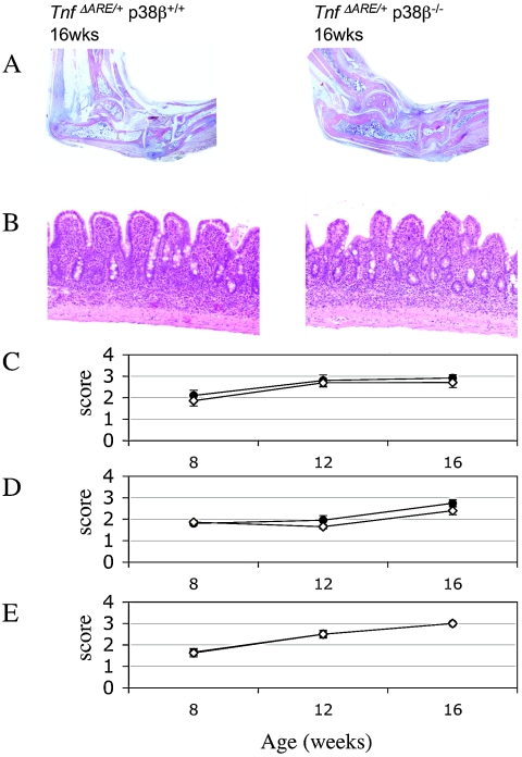
p38 mitogen-activated protein kinases (MAPKs) are activated primarily in response to inflammatory cytokines and cellular stress, and inhibitors which target the p38alpha and p38beta MAPKs have shown potential for the treatment of inflammatory disease. Here we report the generation and initial characterization of a knockout of the p38beta (MAPK11) gene. p38beta-/- mice were viable and exhibited no apparent health problems. The expression and activation of p38alpha, ERK1/2, and JNK in response to cellular stress was normal in embryonic fibroblasts from p38beta-/- mice, as was the activation of p38-activated kinases MAPKAP-K2 and MSK1. The transcription of p38-dependent immediate-early genes was also not affected by the knockout of p38beta, suggesting that p38alpha is the predominant isoform involved in these processes. The p38beta-/- mice also showed normal T-cell development. Lipopolysaccharide-induced cytokine production was also normal in the p38beta-/- mice. As p38 is activated by tumor necrosis factor, the p38beta-/- mice were crossed onto a TNFDeltaARE mouse line. These mice overexpress tumor necrosis factor, which results in development symptoms similar to rheumatoid arthritis and inflammatory bowel disease. The progression of these diseases was not however moderated by knockout of p38beta. Together these results suggest that p38alpha, and not p38beta, is the major p38 isoform involved in the immune response and that it would not be necessary to retain activity against p38beta during the development of p38 inhibitors.


Figures

FIG. 1.
FIG. 1.
Generation of p38β kinase-dead knockin mice. To generate p38β knockin mice a targeting vector was made to introduce an Asp168Ala point mutation in exon 7 of the p38β/MAPK11 gene (A). This vector was used to target ES cells which were screened for homologous recombination using a probe 3′ to the targeting vector. The neomycin resistance cassette was removed from correctly targeted cells by transient expression of CRE recombinase. Removal of the neomycin resistance cassette was confirmed by Southern blotting after EcoRI/SspI digestion of genomic DNA with a probe 5′ to the targeting vector. Southern blotting with this probe resulted in a 10.5-kb band for the wild-type locus, and a 3.5-kb band for a correctly targeted p38b locus (B). Heterozygous ES cells were used to generate mice which were genotypes using a PCR-based strategy, described in the Materials and Methods, that generated a 236-bp fragment for the targeted genes and 100-bp fragment for the wild-type gene (C). To analyze expression of the Asp168Ala protein, primary MEF cells were cultured from p38β+/+, p38β−/− and p38βki/ki mice. MEF protein lysates were immunoblotted for expression of p38β (D). To analyze expression of p38β mRNA in the same cells, total RNA was extracted and p38β mRNA levels determined by quantitative reverse transcription-PCR using 18s rRNA levels as a loading control. Two primer sets were used, designated p38β1 (black bars) and p38β2 (white bars), for which sequences are given in Table 1. Error bars represent the standard error of duplicate assays on three separate plates of cells per genotype (E). To analyze the stability of D168A, C211R, and D168A/C211R mutations of p38β compared to the wild-type sequence, HA-tagged p38β was transfected into HeLa cells. Lysates were immunoblotted using an HA tag antibody to detect p38β, and a ribosomal protein S6 kinase (RSK) antibody as a loading control (F).
FIG. 2.
FIG. 2.
Generation of p38β knockout mice. To generate p38β knockout mice a targeting vector was constructed to delete exons 2 to 7 in the p38β/MAPK11 gene (A). This vector was used to target ES cells which were screened for homologous recombination using a probe 3′ to the targeting vector (A). Southern blotting after EcoR1/SspI digestion of genomic DNA with this probe resulted in a 10.5kb band for the wild-type locus, and a 7kb band for a correctly targeted p38β locus (B). Heterozygous ES cells were used to generate mice which were genotyped using a PCR based strategy, described in the Materials and Methods, that generated a 900-bp fragment for the targeted genes and 500-bp fragment for the wild-type gene (C).
FIG. 3.
FIG. 3.
Expression of p38 isoforms in p38β+/+ and p38β−/− tissues. Lysates were prepared from the total brain, cerebellum, cortex, adrenals, thymus, spleen, liver, pancreas, kidney, lung, heart, and skeletal muscle of p38β+/+ and p38β−/− mice; 30 μg of soluble protein was run on 4 to 12% polyacrylamide gels and immunoblotted for total p38β (A), p38α (B), p38γ (C), and p38δ (D).
FIG. 4.
FIG. 4.
Characterization of p38 dependent signaling in p38β−/− MEF cells. Primary MEF cells were isolated from p38β+/+ and p38β−/− mice. Cells were serum starved for 16 h and then left unstimulated (control), incubated with 5 μM SB 203580 for 1 h, and then stimulated with 10 μg/ml anisomycin (aniso + SB) for a further hour or stimulated with 10 μg/ml anisomycin for 1 h (A and B) or for the times indicated (C) without any addition of SB203580. (A) Cells were then lysed and protein extracts were immunoblotted for p38α, p38β, phosphorylated p38α, ERK1/2, phosphorylated ERK1/2, JNK, or phosphorylated JNK. (B) Cells were lysed and MAPKAP-K2 or MSK1 was immunoprecipitated and assayed for activity as described in the Materials and Methods. One unit was defined as the incorporation of 1 nmol of phosphate into the substrate peptide in 1 min. Black bars represent results from p38β+/+ cells, while white bars represent results from p38β−/− cells. Error bars represent the standard errors of duplicate assays on three independent stimulations per condition. (C) Total RNA was isolated from cells and quantitative reverse transcription-PCR carried out as described in the Materials and Methods. Levels of c-jun, c-fos, junB, and nur77 were determined relative to 18s rRNA and fold stimulation calculated relative to the levels of the mRNA in unstimulated p38β+/+ cells. Black bars represent results from p38β+/+ cells, while white bars represent results from p38β−/− cells. Error bars represent the standard errors of duplicate assays on three independent stimulations per condition.
FIG. 5.
FIG. 5.
p38β is not required for T-cell development. Thymi and spleens were isolated from 6-week-old p38β+/+ and p38β−/− mice and analyzed as described in the Materials and Methods. (A) Total cell numbers, determined by counting isolated cell suspensions, were determined for both the thymus and spleen of p38β+/+ and p38β−/− mice. (B) Fluorescence-activated cell sorting plot of CD4 and CD8 stained Thy1-positive cells in the thymus. Results are representative plots from the analysis of three p38β+/+ and p38β−/− mice. (C) Quantification of the total numbers of double-negative cells from p38β+/+ and p38β−/− thymi. (D) p38β+/+ and p38β−/− mice were injected with either PBS or anti-CD3 antibody (50 ng). Mice were culled 48 h after injection and numbers of double-positive and CD4 and CD8 single-positive thymocytes were determined by counting and fluorescence-activated cell sorting analysis.
FIG. 6.
FIG. 6.
p38β is not required for cytokine production. A) Macrophages were derived from the bone marrow of p38β+/+ and p38β−/− mice. Cells were stimulated with 10 ng/ml of LPS for the times indicated and the induction of interleukin-6 (Il-6) and TNF-α mRNAs was measured by real-time PCR as described in the Materials and Methods. B) Mice were given an intraperitoneal injection of 37.5 μg of LPS in a sterile saline solution, or an injection equivalent volume of saline. Mice were sacrificed after 1 or 4 h and plasma levels of interleukins 1a, 1b, 6, 12p40, and 13, TNF-α, Mip1a, Mip1b, and RANTES were measured by a Luminex-based multiplex assay as described in the Materials and Methods. All mice were 10 to 12 weeks of age and error bars represent the standard deviation of measurement on five individual mice.
FIG. 7.
FIG. 7.
Inflammatory arthritis and inflammatory bowel disease in TnfΔARE/+/p38β+/+ and TnfΔARE/+/p38β−/− mice. Histologic examination (A) of ankle joints showed formation of the inflammatory pannus, and areas of cartilage and bone erosion are evident in both genotypes (B) of portions of ileum. Villous blunting and inflammatory infiltration are evident. Inflammatory bowel disease was evaluated histologically (C), while levels of arthritis were evaluated histologically (D) and clinically (E) as described in the Materials and Methods.

Similar articles

Cited by

References

    1. Adams, R. H., A. Porras, G. Alonso, M. Jones, K. Vintersten, S. Panelli, A. Valladares, L. Perez, R. Klein, and A. R. Nebreda. 2000. Essential role of p38alpha MAP kinase in placental but not embryonic cardiovascular development. Mol. Cell 6:109-116. - PubMed
    1. Allen, M., L. Svensson, M. Roach, J. Hambor, J. McNeish, and C. A. Gabel. 2000. Deficiency of the stress kinase p38alpha results in embryonic lethality: characterization of the kinase dependence of stress responses of enzyme-deficient embryonic stem cells. J. Exp. Med. 191:859-870. - PMC - PubMed
    1. Brancho, D., N. Tanaka, A. Jaeschke, J. J. Ventura, N. Kelkar, Y. Tanaka, M. Kyuuma, T. Takeshita, R. A. Flavell, and R. J. Davis. 2003. Mechanism of p38 MAP kinase activation in vivo. Genes Dev. 17:1969-1978. - PMC - PubMed
    1. Cheung, P. C., D. G. Campbell, A. R. Nebreda, and P. Cohen. 2003. Feedback control of the protein kinase TAK1 by SAPK2a/p38alpha. EMBO J. 22:5793-5805. - PMC - PubMed
    1. Darragh, J., A. Soloaga, V. A. Beardmore, A. Wingate, G. R. Wiggin, M. Peggie, and J. S. Arthur. 2005. MSKs are required for the transcription of the nuclear orphan receptors Nur77, nurr1 and nor1 downstream of MAP kinase signalling. Biochem. J. 390:749-759. - PMC - PubMed

Publication types

MeSH terms