. 2005 Dec;25(23):10479-91.
doi: 10.1128/MCB.25.23.10479-10491.2005.

Hypoxia-inducible factors 1alpha and 2alpha regulate trophoblast differentiation

Affiliations

Hypoxia-inducible factors 1alpha and 2alpha regulate trophoblast differentiation

Karen D Cowden Dahl et al. Mol Cell Biol. 2005 Dec.

Abstract

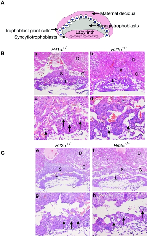
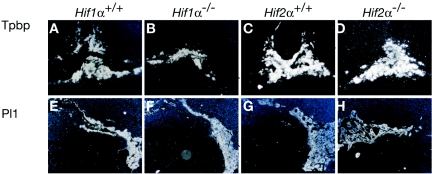
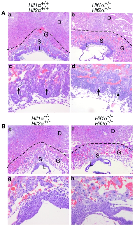
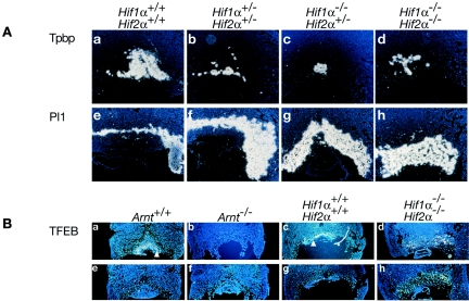
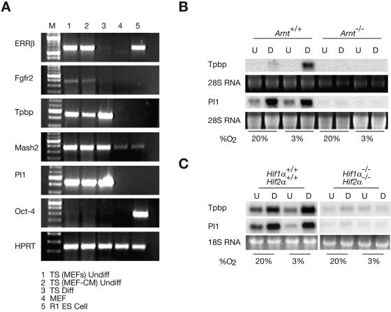
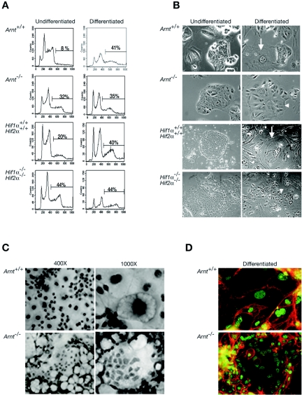
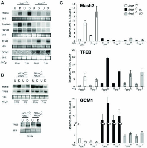
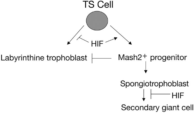
Placental development initially occurs in a low-oxygen (O2) or hypoxic environment. In this report we show that two hypoxia-inducible factors (HIFs), HIF1alpha and HIF2alpha, are essential for determining murine placental cell fates. HIF is a heterodimer composed of HIFalpha and HIFbeta (ARNT) subunits. Placentas from Arnt-/- and Hif1alpha-/- Hif2alpha-/- embryos exhibit defective placental vascularization and aberrant cell fate adoption. HIF regulation of Mash2 promotes spongiotrophoblast differentiation, a prerequisite for trophoblast giant cell differentiation. In the absence of Arnt or Hifalpha, trophoblast stem cells fail to generate these cell types and become labyrinthine trophoblasts instead. Therefore, HIF mediates placental morphogenesis, angiogenesis, and cell fate decisions, demonstrating that O2 tension is a critical regulator of trophoblast lineage determination. This novel genetic approach provides new insights into the role of O2 tension in the development of life-threatening pregnancy-related diseases such as preeclampsia.


Figures

FIG. 1.
FIG. 1.
Vascular defects in Hif1α/ placentas. (A) Diagram of placental architecture. Trophoblast giant cells (TGCs) are placental lactogen 1+ (Pl 1+), spongiotrophoblasts are Tpbp+, and the syncytiotrophoblasts of the labyrinthine are TFEB+. (B) E9.5 placental sections stained for H&E: Hif1α+/+ (a and c), Hif1α/ (b and d). (C) E9.5 placental sections stained for H&E: Hif2α+/+ (e and g) and Hif2α/ (f and h). Hif1α/ placentas show a decrease in the number of fetal vessels invading from the allantois (arrows in panels e, f, g, and h) into the labyrinthine layer. Original magnification, ×50 (a, b, e, and f) and ×200 (c, d, g, and h). D, maternal deciduum; L, labyrinthine trophoblast; S, spongiotrophoblast; G, TGCs.
FIG. 2.
FIG. 2.
Expression of cell type-specific markers in trophoblast tissue. E9.5 placentas analyzed by 35S in situ hybridization for Tpbp and Pl 1 in Hif1a+/+ (A and E), Hif1α/ (B and F), Hif2α+/+ (C and G), and Hif2α/ (D and H) embryos. Original magnification, ×50.
FIG. 3.
FIG. 3.
Vascular and architectural defects in Hif1α+/− Hif2α+/, Hif1α−/− Hif2α+/, and Hif1α/ Hif2α−/− placentas. The dotted lines indicate the extent of desidual invasion for each placental section. (A) E9.5 placental sections stained for H&E: Hif1α+/+ Hif2α+/+ (a and c) Hif1α+/ Hif2α+/− and (b and d). (B) E9.5 placental sections stained for H&E: Hif1α−/− Hif2α+/ (e and g) and Hif1α/ Hif2α/ (f and h). Arrows denote fetal blood vessels. No fetal vessels (g and h) were detected in the labyrinthine layer of Hif1α−/− Hif2α+/ and Hif1α−/− Hif2α/ placentas. Original magnification, ×50 (a, b, e, and f) and ×200 (c, d, g, and h). D, maternal deciduum; L, labyrinthine trophoblast; S, spongiotrophoblast; G, TGCs.
FIG. 4.
FIG. 4.
Expression of cell type-specific markers in trophoblast tissue. (A) E9.5 placentas analyzed by 35S in situ hybridization for Tpbp and Pl 1 in Hif1α+/+ Hif2α+/+ (a and e), Hif1α+/− Hif2α+/ (b and f), Hif1α−/− Hif2α+/ (c and g), and Hif1α/ Hif2α−/− (d and h) embryos. Original magnification, ×50. (B) E9.5 placentas analyzed by 35S in situ hybridization for expression of TFEB in Arnt+/+ (a and e), Arnt/ (b and f) Hif1α+/+ Hif2α+/+ (c and g), and Hif1α/ Hif2α/ (d and h) embryos. Antisense probes are shown in a, b, c, and d, and sense probes are shown in e, f, g, and h. Original magnification, ×50. Arrowheads indicate cells expressing TFEB.
FIG. 5.
FIG. 5.
Generation and differentiation of trophoblast stem (TS) cells. (A) Wild-type undifferentiated TS cells on MEFs or in MEF-conditioned media (lanes 1 and 2), differentiated TS cells (lane 3), MEFs alone, and R1 ES cells were analyzed by RT-PCR for ERRβ, FGFR2, Tpbp, Mash2, Pl 1, Oct-4, and HPRT. (B) Arnt+/+ and Arnt/ TS cells were differentiated and examined by Northern blot assay for expression of Tpbp and Pl 1 under 20% O2 and 3% O2. U, undifferentiated (undiff) cells; D, differentiated (diff) cells. (C) Hif1α+/+ Hif2α+/+ and Hif1α−/− Hif2α/ TS cells were differentiated and examined by Northern blot for Tpbp and Pl 1 expression.
FIG. 6.
FIG. 6.
HIF-deficient TS cells differentiate into polyploid cells. (A) DNA content of undifferentiated and differentiated TS cells. Arnt+/+, Arnt/, Hif1α+/+ Hif2α+/+, and Hif1α−/− Hif2α/ TS cells were analyzed for DNA content by PI incorporation and flow cytometry. Percentage of cells with >4 N DNA content is indicated. (B) Morphology of undifferentiated and differentiated TS cells. TS cells were photographed using a phase-contrast microscope (×100 magnification). Undifferentiated Arnt+/+, Arnt/, Hif1α+/+ Hif2α+/+, and Hif1α−/− Hif2α/ TS cells form epithelial-like colonies. Differentiated Arnt+/+ and Hif1α+/+ Hif2α+/+ TS cells exhibited a giant-cell-like morphology with large nuclei and cytoplasm. In contrast, differentiated Arnt/ and Hif1α−/− Hif2α/ TS cells form sheets of cells with many nuclei and undefined borders. Arrows indicate giant cells in wild-type cultures. Arrowheads indicate differentiated HIF-deficient cells. Original magnification, ×1,000. (C) Arnt+/+ and Arnt/ TS cells were grown for 4 days on plastic and stained by H&E (magnification, ×400 and ×1,000). (D) Arnt+/+ and Arnt/ TS cells were grown on coverslips during a 4-day differentiation period and then immunostained for β-catenin (red) and HDAC1 (green). Original magnification, ×1,000.
FIG. 7.
FIG. 7.
HIF-deficient trophoblasts adopt aberrant cell fates in vitro. (A) Arnt+/+ and Arnt/ TS cells were differentiated under 20% and 3% O2 and examined by Northern blot for expression of a spongiotrophoblast marker (Mash2), TGC-specific genes (Proliferin and Hand1), and labyrinthine trophoblast markers (TFEB and GCM1). (B) Hif1α+/+ Hif2α+/+ and Hif1α−/− Hif2α/ TS cells were differentiated for 4 days under 20% and 3% O2 and examined by Northern blot for expression of TFEB and Hand1. The lower panel shows TFEB expression after 5 days of differentiation for Hif1α+/+ Hif2α+/+ and Hif1α/ Hif2α/ TS cells. (C) Quantitative real-time PCR (Q-PCR) for the spongiotrophoblast gene Mash2 and the syncytiotrophoblast genes TFEB and GCM1. A second Arnt/ TS line (no. 2) was added to these experiments to demonstrate that these phenotypes are due to HIF deficiency and are associated with multiple independent lines. U, undifferentiated cells; D, differentiated cells.
FIG. 8.
FIG. 8.
Model for the role of HIF in trophoblast cell fate decisions. Expression of the HIF complex promotes formation of a precursor trophoblast population that expresses Mash2. Mash2 expression acts to repress syncytiotrophoblast differentiation. Mash2+ cells become spongiotrophoblasts that are maintained by hypoxic expression of HIF. Spongiotrophoblasts are then able to terminally differentiate into secondary TGCs as O2 levels increase. This occurs naturally as cells encounter the maternal spiral arteries. HIF negatively regulates labyrinthine trophoblast formation by inducing Mash2 and preferentially promoting the TGC pathway.

Similar articles

Cited by

References

    1. Abbott, B. D., and M. R. Probst. 1995. Developmental expression of two members of a new class of transcription factors. II. Expression of aryl hydrocarbon receptor nuclear translocator in the C57BL/6N mouse embryo. Dev. Dyn. 204:144-155. - PubMed
    1. Adelman, D. M., M. Gertsenstein, A. Nagy, M. C. Simon, and E. Maltepe. 2000. Placental cell fates are regulated in vivo by HIF-mediated hypoxia responses. Genes Dev. 14:3191-3203. - PMC - PubMed
    1. Adelman, D. M., E. Maltepe, and M. C. Simon. 1999. Multilineage embryonic hematopoiesis requires hypoxic ARNT activity. Genes Dev. 13:2478-2483. - PMC - PubMed
    1. Caniggia, I., S. Grisaru-Gravnosky, M. Kuliszewsky, M. Post, and S. J. Lye. 1999. Inhibition of TGF-beta 3 restores the invasive capability of extravillous trophoblasts in preeclamptic pregnancies. J. Clin. Investig. 103:1641-1650. - PMC - PubMed
    1. Caniggia, I., H. Mostachfi, J. Winter, M. Gassmann, S. J. Lye, M. Kuliszewski, and M. Post. 2000. Hypoxia-inducible factor-1 mediates the biological effects of oxygen on human trophoblast differentiation through TGFbeta(3). J. Clin. Investig. 105:577-587. - PMC - PubMed

Publication types

MeSH terms

Substances