

The murine stanniocalcin 1 gene is not essential for growth and development
- PMID: 16287871
- PMCID: PMC1291238
- DOI: 10.1128/MCB.25.23.10604-10610.2005
The murine stanniocalcin 1 gene is not essential for growth and development
Abstract
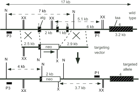
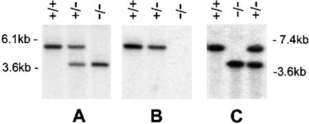
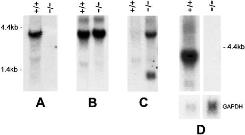
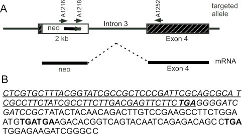
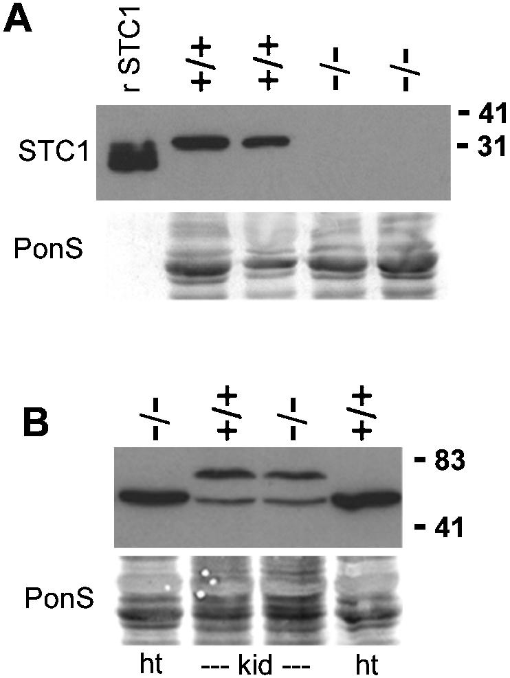
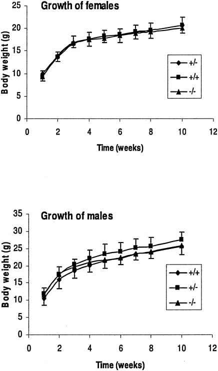
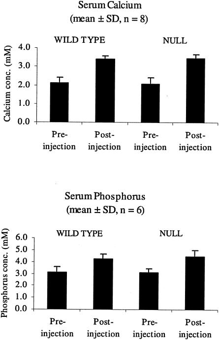
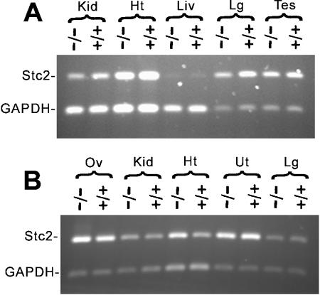
The stanniocalcin 1 (STC1) gene is expressed in a wide variety of tissues, including the kidney, prostate, thyroid, bone, and ovary. STC1 protein is considered to have roles in many physiological processes, including bone development, reproduction, wound healing, angiogenesis, and modulation of inflammatory response. In fish, STC1 is a hormone that is secreted by the corpuscles of Stannius and is involved in calcium and phosphate homeostasis. To determine the role of STC1 in mammals, we generated Stc1-null mice by gene targeting. The number of Stc1-/- mice obtained was in accordance with Mendelian ratios, and both males and females produced offspring normally. No anatomical or histological abnormalities were detected in any tissues. Our results demonstrated that Stc1 function is not essential for growth or reproduction in the mouse.
Figures
Similar articles
-
Prospect of a stanniocalcin endocrine/paracrine system in mammals.Am J Physiol Renal Physiol. 2002 Mar;282(3):F367-75. doi: 10.1152/ajprenal.00364.2000. Am J Physiol Renal Physiol. 2002. PMID: 11832417 Review.
-
The murine stanniocalcin 2 gene is a negative regulator of postnatal growth.Endocrinology. 2008 May;149(5):2403-10. doi: 10.1210/en.2007-1219. Epub 2008 Feb 7. Endocrinology. 2008. PMID: 18258678
-
Stanniocalcin 1 as a pleiotropic factor in mammals.Peptides. 2004 Oct;25(10):1663-9. doi: 10.1016/j.peptides.2004.04.015. Peptides. 2004. PMID: 15476933 Review.
-
Four stanniocalcin genes in teleost fish: structure, phylogenetic analysis, tissue distribution and expression during hypercalcemic challenge.Gen Comp Endocrinol. 2012 Jan 15;175(2):344-56. doi: 10.1016/j.ygcen.2011.11.033. Epub 2011 Nov 29. Gen Comp Endocrinol. 2012. PMID: 22154646
-
Stanniocalcin (STC) in the endometrial glands of the ovine uterus: regulation by progesterone and placental hormones.Biol Reprod. 2006 May;74(5):913-22. doi: 10.1095/biolreprod.106.050807. Epub 2006 Feb 1. Biol Reprod. 2006. PMID: 16452456
Cited by
-
Stanniocalcin-2 inhibits mammalian growth by proteolytic inhibition of the insulin-like growth factor axis.J Biol Chem. 2015 Feb 6;290(6):3430-9. doi: 10.1074/jbc.M114.611665. Epub 2014 Dec 22. J Biol Chem. 2015. PMID: 25533459 Free PMC article.
-
Targeting stanniocalcin-1-expressing tumor cells elicits efficient antitumor effects in a mouse model of human lung cancer.Cancer Med. 2021 May;10(9):3085-3100. doi: 10.1002/cam4.3852. Epub 2021 Apr 7. Cancer Med. 2021. PMID: 33826244 Free PMC article.
-
Combined Effects of Polystyrene Nanosphere and Homosolate Exposures on Estrogenic End Points in MCF-7 Cells and Zebrafish.Environ Health Perspect. 2024 Feb;132(2):27011. doi: 10.1289/EHP13696. Epub 2024 Feb 21. Environ Health Perspect. 2024. PMID: 38381479 Free PMC article.
-
Pharmacological reactivation of inactive X-linked Mecp2 in cerebral cortical neurons of living mice.Proc Natl Acad Sci U S A. 2018 Jul 31;115(31):7991-7996. doi: 10.1073/pnas.1803792115. Epub 2018 Jul 16. Proc Natl Acad Sci U S A. 2018. PMID: 30012595 Free PMC article.
-
Women with polycystic ovary syndrome present with altered endometrial expression of stanniocalcin-1†.Biol Reprod. 2020 Feb 14;102(2):306-315. doi: 10.1093/biolre/ioz180. Biol Reprod. 2020. PMID: 31494675 Free PMC article.
References
-
- Chang, A. C. M., M. A. Dunham, K. J. Jeffrey, and R. R. Reddel. 1996. Molecular cloning and characterization of mouse stanniocalcin cDNA. Mol. Cell. Endocrinol. 124:185-187. - PubMed
-
- Chang, A. C. M., J. Janosi, M. Hulsbeek, D. De Jong, K. J. Jeffrey, J. R. Noble, and R. R. Reddel. 1995. A novel human cDNA highly homologous to the fish hormone stanniocalcin. Mol. Cell. Endocrinol. 112:241-247. - PubMed
-
- Chang, A. C. M., D. A. Jellinek, and R. R. Reddel. 2003. Mammalian stanniocalcins and cancer. Endocr. Relat. Cancer 10:359-373. - PubMed
-
- Chang, A. C. M., and R. R. Reddel. 1998. Identification of a second stanniocalcin cDNA in mouse and human: stanniocalcin 2. Mol. Cell. Endocrinol. 141:95-99. - PubMed
-
- Deol, H. K., R. Varghese, G. F. Wagner, and G. E. DiMattia. 2000. Dynamic regulation of mouse ovarian stanniocalcin expression during gestation and lactation. Endocrinology 141:3412-3421. - PubMed
Publication types
MeSH terms
Substances
LinkOut - more resources
Full Text Sources
Other Literature Sources
Molecular Biology Databases