. 2005 Dec;25(23):10611-27.
doi: 10.1128/MCB.25.23.10611-10627.2005.

The Sho1 adaptor protein links oxidative stress to morphogenesis and cell wall biosynthesis in the fungal pathogen Candida albicans

Affiliations

The Sho1 adaptor protein links oxidative stress to morphogenesis and cell wall biosynthesis in the fungal pathogen Candida albicans

Elvira Román et al. Mol Cell Biol. 2005 Dec.

Abstract

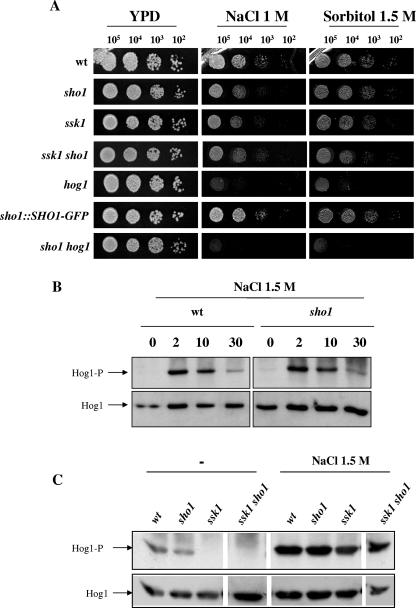
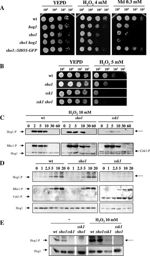
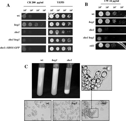
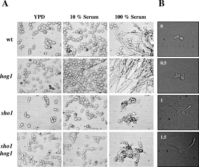
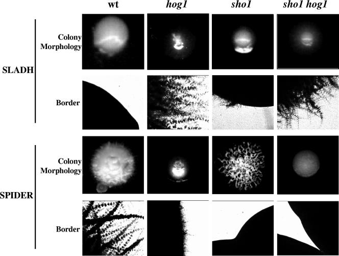
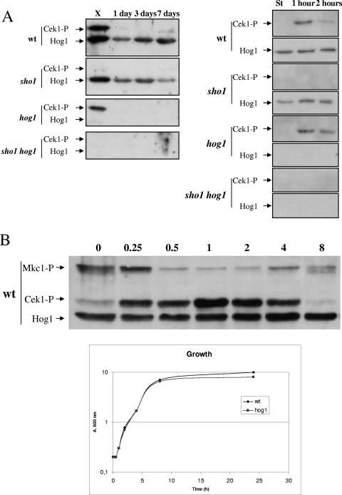
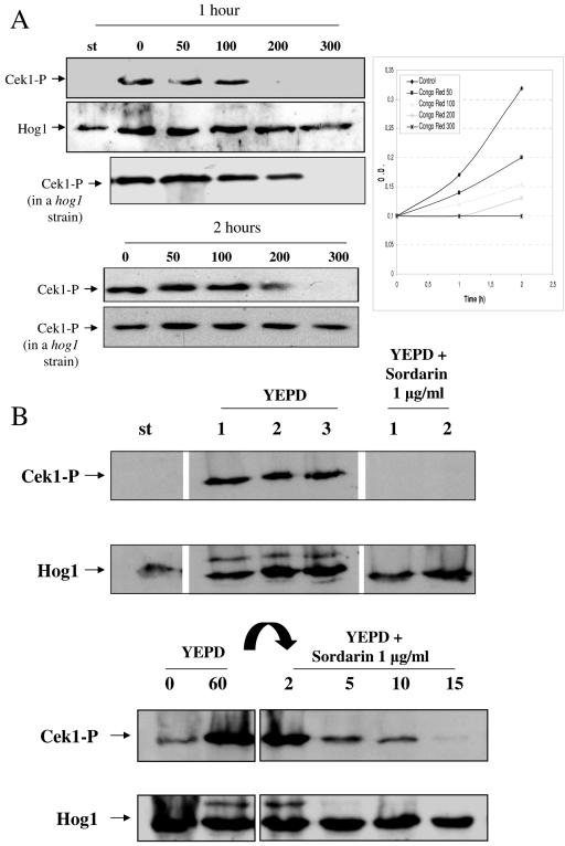
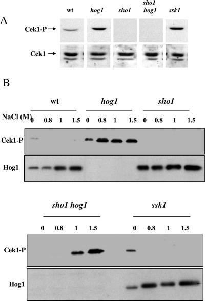
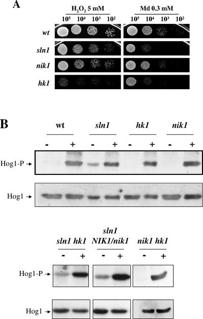
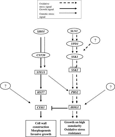
The Sho1 adaptor protein is an important element of one of the two upstream branches of the high-osmolarity glycerol (HOG) mitogen-activated protein (MAP) kinase pathway in Saccharomyces cerevisiae, a signal transduction cascade involved in adaptation to stress. In the present work, we describe its role in the pathogenic yeast Candida albicans by the construction of mutants altered in this gene. We report here that sho1 mutants are sensitive to oxidative stress but that Sho1 has a minor role in the transmission of the phosphorylation signal to the Hog1 MAP kinase in response to oxidative stress, which mainly occurs through a putative Sln1-Ssk1 branch of the HOG pathway. Genetic analysis revealed that double ssk1 sho1 mutants were still able to grow on high-osmolarity media and activate Hog1 in response to this stress, indicating the existence of alternative inputs of the pathway. We also demonstrate that the Cek1 MAP kinase is constitutively active in hog1 and ssk1 mutants, a phenotypic trait that correlates with their resistance to the cell wall inhibitor Congo red, and that Sho1 is essential for the activation of the Cek1 MAP kinase under different conditions that require active cell growth and/or cell wall remodeling, such as the resumption of growth upon exit from the stationary phase. sho1 mutants are also sensitive to certain cell wall interfering compounds (Congo red, calcofluor white), presenting an altered cell wall structure (as shown by the ability to aggregate), and are defective in morphogenesis on different media, such as SLAD and Spider, that stimulate hyphal growth. These results reveal a role for the Sho1 protein in linking oxidative stress, cell wall biogenesis, and morphogenesis in this important human fungal pathogen.


Figures

FIG. 1.
FIG. 1.
Osmotic response of the sho1 mutant. (A) 105, 104, 103, and 102 exponentially growing cells (from a culture at an A600 of 1) of strains wt (RM100), hog1 (CNC13), sho1 (REP3), sho1 hog1 (REP8), sho1::SHO1-GFP (REP5), ssk1 (CSSK21), and ssk1 sho1 (REP12) were spotted on solid agar plates of YEPD rich medium supplemented with sorbitol and sodium chloride at the concentrations indicated. Plates were incubated for 24 h at 37°C and scanned. (B) Sho1-dependent activation of Hog1 under osmotic stress was studied. Overnight cultures of the strains wt (RM100) and sho1 (REP3) were inoculated in fresh YEPD medium to an A600 of 0.05. At an A600 of 1, the same volume of 3 M NaCl (in YEPD) was added to the cultures to reach the final concentration of 1.5 M NaCl. Samples were taken at the times indicated and processed for the preparation of a total protein extract (see Material and Methods). The presence of phosphorylated Hog1 (Hog1-P) was visualized by probing a blot with an anti-phospho p38 antibody, and Hog1 protein (Hog1) was detected by using anti-ScHog1 polyclonal antibody. The labels indicate the number of minutes after the addition of the osmostress agent. (C) Hog1 phosphorylation was analyzed in wt, sho1, ssk1, and ssk1 sho1 exponentially growing cells (A600 = 1) in the absence (left) or presence (right) of 1.5 M NaCl (10 min of exposure to this agent). In parallel, another portion of the culture was subjected to 10 mM H2O2 instead of osmotic stress and processed in a similar way (shown in the next figure). The autoradiography was intentionally overexposed. Lanes are arranged in this figure from a scanned Western blot (as shown by blank intermediate lines) to avoid incorporation of samples not related to this particular experiment. They correspond to the same experiment and were processed simultaneously in the same gel.
FIG. 2.
FIG. 2.
Oxidative stress response in HOG pathway mutants. (A) Serial dilutions of exponentially growing cells of the strains wt (RM100), hog1 (CNC13), sho1 (REP3), sho1 hog1 (REP8), and sho1::SHO1-GFP (REP5) (from a culture at an A600 of 1) were spotted onto plates supplemented with hydrogen peroxide and menadione at the concentrations indicated. (B) The hydrogen peroxide susceptibility of ssk1 (CSSK21) and ssk1 sho1 (REP12) mutants was analyzed by solid medium studies in the presence of a 5 mM concentration of the mentioned oxidant. (C) The role of Sho1 and Ssk1 in the transmission of H2O2 signal to Hog1 was analyzed by Western blotting. H2O2 (10 mM) was added to exponentially growing cultures (at an A600 of 1), and samples were collected at 2, 5, 10, 30, and 60 min after treatment with the oxidant. phospho-Mkc1 (Mkc1-P) and phospho-Cek1 (Cek1-P) were detected by blotting the membranes with the anti-phospho-p44/p42 antibody. (D) Exponentially growing cultures of the mentioned strains were challenged with 0, 1, 2.5, 5, 10, and 20 mM H2O2 (indicated at the top of each lane), and the pattern of MAP kinase activation was detected after 10 min. Cells were collected and processed for immunodetection analysis to detect the activated (-P) forms of the MAP kinases. (E) Hog1 phosphorylation was analyzed in a ssk1 sho1 mutant under oxidative stress. Exponentially growing cultures of the indicated strains (A600 of 1) (−) were treated with 10 mM hydrogen peroxide for 10 min, and samples were processed as described above. Control lanes of this experiment are exactly the same as those from Fig. 1C, as this experiment was performed simultaneously, and are included here to facilitate comparison.
FIG. 3.
FIG. 3.
Alterations in cell wall biogenesis in sho1 mutants. Serial dilutions of exponentially growing cells of the strains indicated (from a culture at an A600 of 1) were spotted onto plates supplemented with Congo red (CR) (A) or calcofluor white (CW) (B) at the concentrations indicated. (C) Stationary-phase cells from the strains indicated were inoculated in YEPD fresh medium, grown at 37°C until they reached an A600 of 1, and photographed 2 to 4 min after swirling of the cultures in a glass tube was stopped. Phase-contrast photographs were also taken under these conditions and are shown; a portion of the sho1 photograph is further amplified to appreciate its altered morphology.
FIG. 4.
FIG. 4.
Effect of Sho1 in the serum-induced morphological transition. (A) Stationary-phase cells were inoculated at 106 cells/ml in prewarmed YEPD medium (YPD), YEPD supplemented with 10% fetal bovine serum (10% Serum), or 100% serum (100% Serum). Phase-contrast microphotographs were taken after 3 h of incubation at 30°C. (B) The dimorphic transition of individual sho1 cells was studied on solid serum medium (1% agar serum) at 37°C. Hypha formation was monitored by phase-contrast microscopy, taking photographs at the times indicated (in hours).
FIG. 5.
FIG. 5.
Effect of Sho1 on colonial morphology. Exponentially growing cells (from a culture at an A600 of 1) from wt (RM100), hog1 (CNC13), sho1 (REP3), and sho1 hog1 (REP8) were obtained, washed in phosphate-buffered saline, and counted. Approximately 50 CFU were spread onto YEPD, SLADH, or Spider medium plates and incubated for 7 days at 37°C before photographs were taken. Colony morphologies and borders are shown for each medium and strain.
FIG. 6.
FIG. 6.
Growth phase-dependent activation of the Cek1 MAP kinase. (A) Left panel: cells from 1-, 3-, and 7-day stationary-phase cultures (indicated at the top of the lane) were analyzed for MAP kinase activation and compared to exponentially growing cells at an A600 of 1 (labeled with X, for exponentially). Cells were collected and prepared for Western blot analysis. Right panel: 1-day stationary-phase cultures of the mentioned strains (wt [RM100], sho1 [REP3], hog1 [CNC13], and sho1 hog1 [REP8]) (labeled with St, for stationary) were diluted at an A600 of 0.2 and allowed to grow. Samples were taken after 1 and 2 h (indicated in each lane) and processed for the analysis of Cek1 activation. In both panels, Cek1-P refers to phospho-Cek1 (detected with anti-phospho-p44/p42 antibody), while Hog1 refers to CaHog1 protein (detected with anti- S. cerevisiae Hog1 antibody). (B) A stationary-phase culture of the wild-type strain was diluted in YEPD medium at an A600 of 0.2, and samples were taken at the times indicated (in hours) and processed for MAP kinase activation. The symbols are similar to those in panel A. The growth in mass, as determined using the absorbance of the cultures, is shown below.
FIG. 7.
FIG. 7.
Growth-dependent activation of the Cek1 MAP kinase. (A) Stationary-phase cells of the wt strain (RM100) were diluted at an A600 of 0.1 in either YEPD or YEPD in the presence of Congo red at 50, 100, 200, and 300 μg/ml. Samples were collected after 1 and 2 h of growing at 37°C, and cells were processed for MAP kinase phosphorylation by Western blotting analysis. The increase in A600 is shown in the diagram at the right. Symbols are the same as described in the legend to Fig. 6. Results for the hog1 mutant under the same conditions are also shown. (B) An overnight culture of a wt strain (labeled st) was diluted in fresh YEPD to an A600 of 0.2 and incubated at 37°C, taking samples after 1, 2, and 3 h of growth in these conditions (shown in each lane; YEPD lanes 1, 2, and 3). A portion of the overnight culture was also transferred to a prewarmed flask with YEPD medium, where 1 μg/ml sordarin was added, and samples were taken after 1 and 2 h of incubation at 37°C and processed (labeled YEPD + sordarin, lanes 1 and 2). Lanes are arranged in this figure from a scanned Western blot (as shown by blank intermediate lines) to avoid incorporation of samples not related to this particular experiment. They correspond to the same experiment and were processed simultaneously in the same gel. (C) Similarly, stationary growing cells of a wt strain were diluted in fresh YEPD to an A600 of 0.2 and incubated at 37°C. After 1 h, 1 μg/ml sordarin was added. Samples were collected at the times indicated in each lane (in minutes). Symbols are same as described in the legend to Fig. 6.
FIG. 8.
FIG. 8.
Cek1 can be activated in a Sho1-independent manner under high osmolarity. (A) Pattern of activation of Cek1 in the strains wt (RM100), hog1 (CNC13), sho1 (REP3), sho1 hog1 (REP8), and ssk1 (CSSK21) under exponential growth in YEPD medium. Arrows indicate the phosphorylated Cek1 protein (Cek1-P) and Cek1 (Cek1) protein as revealed by Western blotting using either anti-p44/p42 antibodies (upper panel) or a rabbit polyclonal serum against a GST-Cek1 fusion (lower panel). Lanes are arranged in this figure from a scanned Western blot (as shown by blank intermediate lines) to avoid incorporation of samples not related to this particular experiment. They correspond to the same experiment and were processed simultaneously in the same gel. (B) Stationary growing cells of the strains wt (RM100), hog1 (CNC13), sho1 (REP3), sho1 hog1 (REP8), and ssk1 (CSSK21) were diluted in fresh medium to an A600 of 0.05 and incubated at 37°C until they reached an A600 of 1. A twofold-concentrated NaCl-YEPD (0, 1.6, 2, and 3 M) was then added to the cultures to reach the final concentration indicated at the top of each lane (0, 0.8, 1, and 1.5 M). After 10 min, cells were collected, processed as indicated (see Materials and Methods), and analyzed for MAP kinase activation.
FIG. 9.
FIG. 9.
sln1 mutants constitutively activate the HOG pathway. (A) Exponentially growing cells (A600 of 1) (105, 104, 103, and 102 cells) were spotted onto YEPD plates supplemented with 5 mM hydrogen peroxide (H2O2) and 0.3 mM menadione (Md). The plates were then incubated at 37°C for 24 h and scanned. (B) Hog1 phosphorylation in histidine kinase mutants upon H2O2 treatment. Overnight cultures of the strains indicated were reinoculated into fresh YEPD medium to an A600 of 0.05 and incubated at 37°C. At an A600 of 1, samples were taken as a control (−) before adding H2O2 to a final concentration of 10 mM. After 10 min, cells were collected (+) and processed for MAP kinase activation.
FIG. 10.
FIG. 10.
Proposed model. Schematic diagram showing the activating (→) or inhibitory (⊥) stimuli leading to activation of the MAP kinases Cek1, Mkc1, and Hog1 in C. albicans in response to different stimuli (see box at top of figure). The putative hypothetical elements that could lead to the activation of the corresponding MAP kinase in response to osmotic or oxidative stress are shown as question marks.

Similar articles

Cited by

References

    1. Alex, L. A., C. Korch, C. P. Selitrennikoff, and M. I. Simon. 1998. COS1, a two-component histidine kinase that is involved in hyphal development in the opportunistic pathogen Candida albicans. Proc. Natl. Acad. Sci. USA 95:7069-7073. - PMC - PubMed
    1. Alonso-Monge, R., F. Navarro-García, G. Molero, R. Diez-Orejas, M. Gustin, J. Pla, M. Sánchez, and C. Nombela. 1999. Role of the mitogen-activated protein kinase Hog1p in morphogenesis and virulence of Candida albicans. J. Bacteriol. 181:3058-3068. - PMC - PubMed
    1. Alonso-Monge, R., F. Navarro-García, E. Roman, A. I. Negredo, B. Eisman, C. Nombela, and J. Pla. 2003. The Hog1 mitogen-activated protein kinase is essential in the oxidative stress response and chlamydospore formation in Candida albicans. Eukaryot. Cell 2:351-361. - PMC - PubMed
    1. Arana, D. M., C. Nombela, R. Alonso-Monge, and J. Pla. 2005. The Pbs2 MAP kinase kinase is essential for the oxidative-stress response in the fungal pathogen Candida albicans. Microbiology 151:1033-1049. - PubMed
    1. Ausubel, F. M., et al. (ed.). 1993. Current protocols in molecular biology. Greene Publishing Associates, New York, N.Y.

Publication types

MeSH terms

LinkOut - more resources