. 2005 Nov 22;102(47):16961-6.
doi: 10.1073/pnas.0506482102. Epub 2005 Nov 15.

MicroRNAs control translation initiation by inhibiting eukaryotic initiation factor 4E/cap and poly(A) tail function

Affiliations

MicroRNAs control translation initiation by inhibiting eukaryotic initiation factor 4E/cap and poly(A) tail function

David T Humphreys et al. Proc Natl Acad Sci U S A. .

Abstract

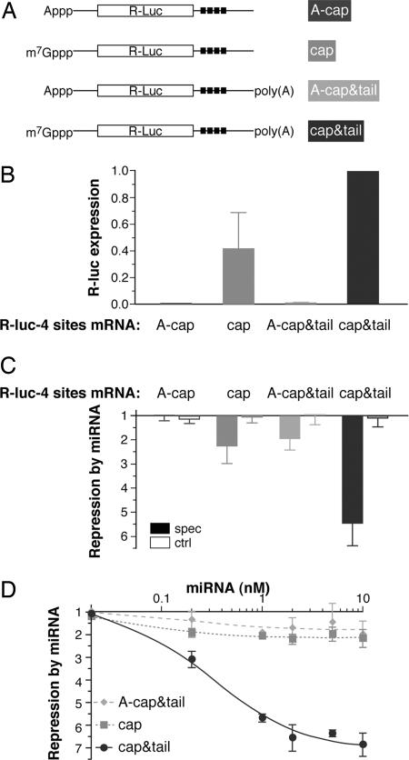
MicroRNAs (miRNAs) repress translation of target mRNAs by interaction with partially mismatched sequences in their 3' UTR. The mechanism by which they act on translation has remained largely obscure. We examined the translation of mRNAs containing four partially mismatched miRNA-binding sites in the 3' UTR in HeLa cells cotransfected with a cognate miRNA. The mRNAs were prepared by in vitro transcription and were engineered to employ different modes of translation initiation. We find that the 5' cap structure and the 3' poly(A) tail are each necessary but not sufficient for full miRNA-mediated repression of mRNA translation. Replacing the cap structure with an internal ribosome entry site from either the cricket paralysis virus or the encephalomyocarditis virus impairs miRNA-mediated repression. Collectively, these results demonstrate that miRNAs interfere with the initiation step of translation and implicate the cap-binding protein eukaryotic initiation factor 4E as a molecular target.


Figures

Fig. 1.
Fig. 1.
miRNAs target the initiation step of translation. HeLa cells were cotransfected with an R-luc mRNA, the F-luc control mRNA, and (where indicated) a synthetic miRNA. For each R-luc mRNA, transfections were performed with specific CXCR4 miRNA, with let-7 control miRNA, and without miRNA. R-luc activity from each transfection was normalized to the corresponding F-luc measurement. (A) The four R-luc test mRNAs. R-luc-4 sites and -0 sites are m7G(5′)ppp(5′)G-capped and polyadenylated and either contain or lack four target sites for miRNA CXCR4 (black squares). Two further variants of the R-luc-4 sites mRNA carried the wild-type CrPV intergenic IRES or an inactive mutant (indicated by the asterisks, ref. 37). The CrPV IRES mRNAs carry an A-cap and are not polyadenylated. (B) Repression by CXCR4 miRNA. Repression was calculated by dividing the normalized R-luc activity without miRNA by the normalized R-luc activity in the presence of miRNA. Bars represent averaged results mostly from three to five independent triplicate experiments (filled bars, CXCR4; open bars, let-7). Error bars indicate standard deviation where appropriate. (C) Activity of the CrPV IRES. Bars represent normalized R-luc activity with mutant or wild-type CrPV IRES mRNA (no miRNA added; expression from the wild-type IRES is expressed as 1.0). R-luc expression driven by the wild-type CrPV IRES was inefficient but still ≈3 orders of magnitude above background readings from mock-transfected controls. Averaged results from two independent triplicate experiments are shown.
Fig. 2.
Fig. 2.
miRNA-mediated repression requires the 5′ cap structure and poly(A) tail. HeLa cells were cotransfected with mRNA and miRNAs as in Fig. 1. (A) Schematic of the R-luc-4 sites mRNAs. (B) Normalized R-luc activity from the four R-luc-4 sites mRNAs (no miRNA added; expression from the cap&tail mRNA is set to 1.0). Averaged results from two (A-cap) or five experiments are shown with standard deviation where appropriate. (C) Repression by a miRNA (calculated as in Fig. 1B). Averaged results from three to five experiments are shown with standard deviation (filled bars, CXCR4; open bars, let-7). (D) Repression of different R-luc-4 site mRNAs by CXCR4 miRNA is saturated at similar miRNA levels. The effect was measured as a function of miRNA concentration in the transfection mix and calculated as in Fig. 1B. Most data points represent averaged results from three experiments with standard deviation where appropriate (squares, cap; diamonds, tail; circles, cap&tail).
Fig. 3.
Fig. 3.
The miRNA/target interaction does not trigger accelerated mRNA decay. (A) Multiple aliquots of HeLa cells were cotransfected with R-luc-4 sites and F-luc control mRNAs, either with (▪) or without CXCR4 miRNA (•), and harvested at different times. R-luc expression was not normalized to F-luc. Five time series of this kind were each scaled to the total level of recovered R-luc protein and then averaged. Error bars represent standard deviation. The functional half-life of an mRNA in cells is defined as the time required for half-maximal accumulation of R-luc activity (43). (B) HeLa cells were cotransfected with an R-luc-4 sites mRNA, CXCR4 miRNA (as specified above the lanes), and the F-luc control mRNA (lanes 4-8). Total RNA was isolated 7 h after transfection, and 1-μg aliquots were subjected to dual-probe ribonuclease protection assays. Undigested probe mix (lane 1), a model reaction with input R-luc and F-luc mRNAs (marker, lane 2), and total RNA from untransfected HeLa cells (lane 3) are shown for comparison. Positions of probes and protected fragments are indicated on the right. Different “exposures” of the same gel are shown in the top and bottom half of the insert. The box below lists the R-luc/F-luc band intensity ratios relative to the value in lane 7. Each assay was also performed with 5 μg of total RNA to ascertain conditions of probe excess (data not shown). (C) Cells were cotransfected and total RNA was isolated as in B, followed by RT-PCR analyses (see Materials and Methods) with primer pairs specific for either R-luc-4 sites mRNA (across the miRNA target sites) or the F-luc control mRNA. Because the PCR primers anneal to sites on either side of the miRNA targets, cleavage induced by the miRNA should reduce or abolish the R-luc PCR product. PCR products were analyzed after 15, 18, 21, and 24 cycles to demonstrate subsaturation of the PCR amplification (the panel only shows PCR products after 18 cycles). miRNA-mediated R-luc repression in this experiment was ≈4.5-fold (data not shown). Equivalent results were obtained with RNA isolated 16 h after transfection (data not shown) and in three independent transfection experiments.
Fig. 4.
Fig. 4.
The EMCV IRES element impairs miRNA repression. HeLa cells were cotransfected with mRNA and miRNAs as in Figs. 1 and 2. (A) Schematic of the R-luc-4 sites mRNAs used in this experiment: They carry either the standard 5′ UTR or the EMCV IRES, either with or without a poly(A) tail; all are A-capped. (B) Normalized R-luc activity from the four versions of R-luc-4 sites mRNA (no miRNA added; expression from the EMCV&tail mRNA is set to 1.0). Averaged results from two (A-cap) to four experiments are shown with standard deviation where appropriate. (C) Repression by a miRNA (calculated as in Figs. 1B and 2C). Averaged results from three to five experiments are shown with standard deviation (filled bars, CXCR4; open bars, let-7).

References

    1. Bartel, D. P. (2004) Cell 116, 281-297. - PubMed
    1. He, L. & Hannon, G. J. (2004) Nat. Rev. Genet. 5, 522-531. - PubMed
    1. Meister, G. & Tuschl, T. (2004) Nature 431, 343-349. - PubMed
    1. Ambros, V. (2004) Nature 431, 350-355. - PubMed
    1. Cullen, B. R. (2004) Mol. Cell 16, 861-865. - PubMed

Publication types

MeSH terms

LinkOut - more resources