. 2006 Feb 15;107(4):1703-11.
doi: 10.1182/blood-2005-08-3445. Epub 2005 Nov 15.

Absence of beta7 integrin results in less graft-versus-host disease because of decreased homing of alloreactive T cells to intestine

Affiliations

Absence of beta7 integrin results in less graft-versus-host disease because of decreased homing of alloreactive T cells to intestine

Elisha Waldman et al. Blood. .

Abstract

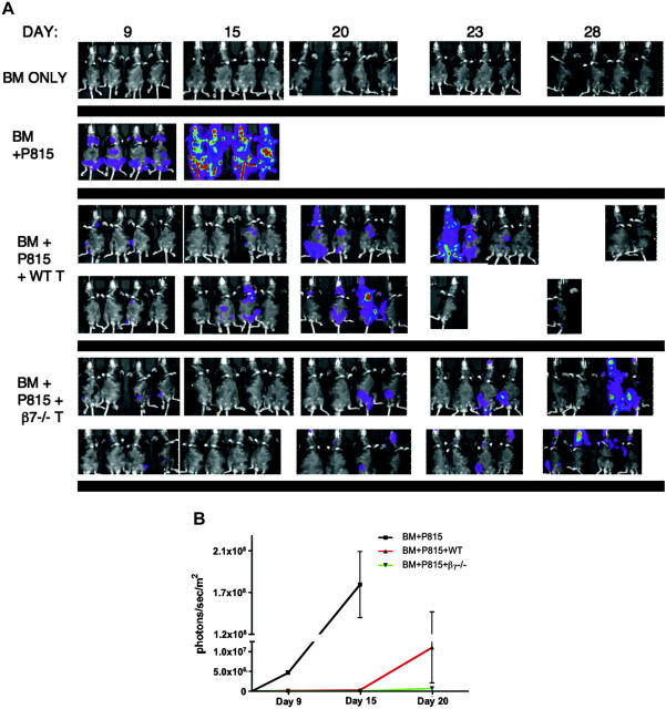
The alpha4beta7 integrin plays a central role in the homing of T cells to the gut. We hypothesized that absence of the beta7 subunit would result in a reduction of intestinal graft-versus-host disease (GVHD) and an improvement in overall GVHD morbidity and mortality in recipients of hematopoietic stem cell transplantation (HSCT). Analysis of alloreactive beta7-/- T cells showed intact activation, proliferation, cytokine production, and cytotoxicity. However, recipients of beta7-/- donor T cells in murine HSCT models experienced less GVHD morbidity and mortality than recipients of wild-type (WT) T cells, associated with a decrease in donor T-cell infiltration of the liver and intestine and with an overall significant decrease in hepatic and intestinal GVHD. In graft-versus-tumor (GVT) experiments, we demonstrated intact or even enhanced GVT activity of beta7-/- donor T cells. In conclusion, beta7-/- donor T cells caused less GVHD morbidity and mortality than WT donor T cells because of selectively decreased T-cell infiltration of the liver and intestines. Our data suggest that strategies to target the beta7 integrin have the clinical potential to alleviate or prevent GVHD while sparing or potentiating GVT activity.


Figures

Figure 1.
Figure 1.
Recipients of β7-/- donor T cells have less GVHD mortality and morbidity. (A) Lethally irradiated (1300 cGy) B10.BR recipients underwent transplantation with 5 × 10⁶ WT TCD BM and either 1 × 10⁶ WT (□) or β7-/- (▴) splenic T cells. Survival from 3 combined experiments (n = 30) is depicted as a Kaplan-Meier curve. Statistical analysis: ▴ versus □ (P = < .04). (B) Clinical GVHD score curve is shown for mice (A), again representing 3 combined experiments (n = 30), where □ versus ▴ (P = .023). (C) Lethally irradiated (1300 cGy) B10.BR recipients underwent transplantation with 5 × 10⁶ WT TCD BM only and 2 × 10⁶ WT (□) or β7-/- (▴) splenic T cells or 3 × 10⁶ WT (♦) or β7-/- (+) splenic T cells. Statistical analysis: ▴ versus □ (P = NS), ♦ versus + (P = NS). TCD BM only group (n = 4), 2 × 10⁶ WT or β7-/- T cells group (n = 8), 3 × 10⁶ WT or β7-/- T cells group (n = 5).
Figure 2.
Figure 2.
WT and β7-/- T cells do not differ in activation or alloreactive proliferation and have intact cytotoxicity. WT and β7-/- donor T cells were labeled with CFSE and were injected intravenously into sublethally irradiated (750 cGy) B10.BR recipients. Donor splenic T cells from recipient mice were analyzed 72 hours after infusion. (A) Histogram overlay of dividing donor CD4+ and CD8+ CFSE-labeled T cells (shaded area, β7-/-; outline, WT) show nearly identical proliferation kinetics. (B) CD44, CD62L, and CD25 expression on CD4+ and CD8+ donor T cells, with percentage positive T cells expressed as a percentage of fast proliferating T cells (outline = β7-/-; shaded area = WT). (C) Lethally irradiated (1300 cGy) B10.BR recipients underwent transplantation with 5 × 10⁶ WT TCD BM and either 1 × 10⁶ WT or β7-/- splenic T cells, and spleens were analyzed at day 11 after HSCT by flow cytometry. Graph represents the percentage of donor-derived CD4+CD25+FoxP3+ T cells (P = NS). (D) Mice underwent transplantation and harvest as in panel C. Percentage of donor CD4+CD44hiCD62Llo (effector) and CD4+CD44hiCD62Lhi (central memory) populations. (E) Percentage of donor CD8+CD62Llo population. (F) Percentage of donor CD8+CD44hi population. (G) Splenocytes from B6D2F1 HSCT recipients of WT versus β7-/- T cells were analyzed at day 14 after HSCT against syngeneic (EL4) and allogeneic (P815) target cells (n = 5).
Figure 3.
Figure 3.
Recipients of β7-/- T cells have significantly lower numbers of T cells in their intestinal mucosa but significantly higher numbers of circulating T cells. (A) Lethally irradiated (1300 cGy) B10.BR mice underwent transplantation such that each mouse received WT TCD-BM (5 × 10⁶) and WT (Thy1.1) T cells in combination with β7-/- (Thy1.2) T cells (2 × 10⁶ of each type). T cells were analyzed before transfer into recipient mice to ascertain that equivalent percentages of CD4+/CD8+ cells were being given (not shown). Mice were killed at day 8, and T cells were isolated and analyzed, as described in “Materials and methods.” Donor origin of the isolated T cells was determined by multicolor flow cytometry. Statistical analysis is as follows: for mesenteric lymph nodes and spleen, P < .001; for gut, P = .004 (n = 5); experiment repeated 3 times. (B) Lethally irradiated (1300 cGy) B10.BR recipients underwent transplantation with 5 × 10⁶ WT TCD-BM and either 1 × 10⁶ WT or β7-/- splenic T cells. Cardiac puncture to obtain blood for complete blood counts was performed on day 7 and day 14. *P ≤ .05 (n = 8).
Figure 4.
Figure 4.
Recipients of β7-/- donor T cells have significantly less gastrointestinal target organ abnormality and no significant difference in skin GVHD. Lethally irradiated (1300 cGy) B10.BR recipients underwent transplantation with 5 × 10⁶ WT TCD BM and either 1 × 10⁶ WT or β7-/- splenic T cells. On day 7 (A) and day 14 (B), mice were killed and gastrointestinal target organs were harvested for evaluation of histopathology, as described in “Materials and methods.” Shown are combined results of pathology scores for 10 mice in each group (*P < .001). (C) Ear and tongue were harvested on days 7 and 14 for evaluation of skin histopathology. (D) Lymphocytes per high-power field (600 ×) from organs on day 7. Shown are results of 5 fields per organ. *P < .001 (n = 10). (E) Representative photomicrographs from day-7 organs. Green arrows show typical apoptotic cells, and black arrows show typical lymphocytes. Original magnification × 600. Images were captured with an Olympus BX 40 microscope (Olympus, Melville, NY) equipped with a 10 ×/0.40 numerical aperture objective lens. Image acquisition was performed with a JVC GC-Qx 5HDU digital camera (JVC, Wayne, NJ).
Figure 5.
Figure 5.
Recipients of β7-/- donor T cells and of WT T cells generate similar levels of serum IFNγ. Lethally irradiated (1300 cGy) B10.BR recipients underwent transplantation with 5 × 10⁶ WT TCD-BM and either 1 × 10⁶ WT or β7-/- splenic T cells. Serum levels of IFNγ were determined by ELISA at day 7 and 14 after transplantation. Shown are combined results of 2 experiments (n = 9). P = .08 at day 7, and P = .751 at day 14.
Figure 6.
Figure 6.
GVT activity is preserved in recipients of β7-/- T cells. Lethally irradiated (1100 cGy split dose) B6D2F1 mice underwent transplantation on day 0 with 5 × 10⁶ WT TCD-BM cells with or without the addition of splenic T cells. Recipients were given 1 × 10⁵ P815 murine mastocytoma cells as a separate intravenous injection at the time of transplantation. Survival is depicted as a Kaplan-Meier curve representing mice that received TCD-BM + P815 (•), TCD-BM +β7-/- T cells + P815 (▴), TCD-BM + WT T cells + P815 (□). Causes of death (GVHD vs tumor) for all recipients that died during the course of the experiment are shown in Table 1. Statistical analysis: ▴ versus □ (P = .005). Shown are combined results of 3 experiments (n = 30).
Figure 7.
Figure 7.
Spatiotemporal analysis of alloreactive T cells and tumor cells by bioluminescence imaging after allogeneic BMT. Lethally irradiated (1300cGy) B6D2F1 recipients underwent transplantation with 5 × 10⁶ WT TCD BM and either 0.5 × 10⁶ WT or β7-/- splenic T cells. P815 tumor cells (0.5 × 10⁶) that had been transduced with an LTR-HSV1 TK-EGFP-Luc retroviral vector were infused into each mouse at the time of transplantation. (A) Mice were tracked for in vivo luminescence 10 minutes after intraperitoneal injection of firefly luciferin. (B) Average luminescence, quantified as photons per sec/m2 at time points with all mice still alive, demonstrates delay in tumor growth in recipients of β7-/- T cells (P = .08).

References

    1. Ferrara JL, Deeg HJ. Graft-versus-host disease. N Engl J Med. 1991;324: 667-674. - PubMed
    1. Sackstein R. Lymphocyte migration following bone marrow transplantation. Ann N Y Acad Sci. 1995;770: 177-188. - PubMed
    1. Murai M, Yoneyama H, Ezaki T, et al. Peyer's patch is the essential site in initiating murine acute and lethal graft-versus-host reaction. Nat Immunol. 2003;4: 154-160. - PubMed
    1. Murai M, Yoneyama H, Harada A, et al. Active participation of CCR5(+)CD8(+) T lymphocytes in the pathogenesis of liver injury in graft-versus-host disease. J Clin Invest. 1999;104: 49-57. - PMC - PubMed
    1. D'Ambrosio D, Panina-Bordignon P, Sinigaglia F. Chemokine receptors in inflammation: an overview. J Immunol Methods. 2003;273: 3-13. - PubMed

Publication types