. 2005 Nov 15;19(22):2727-43.
doi: 10.1101/gad.1338505.

HTP-1-dependent constraints coordinate homolog pairing and synapsis and promote chiasma formation during C. elegans meiosis

Affiliations

HTP-1-dependent constraints coordinate homolog pairing and synapsis and promote chiasma formation during C. elegans meiosis

Enrique Martinez-Perez et al. Genes Dev. .

Abstract

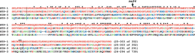
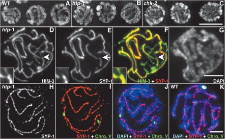
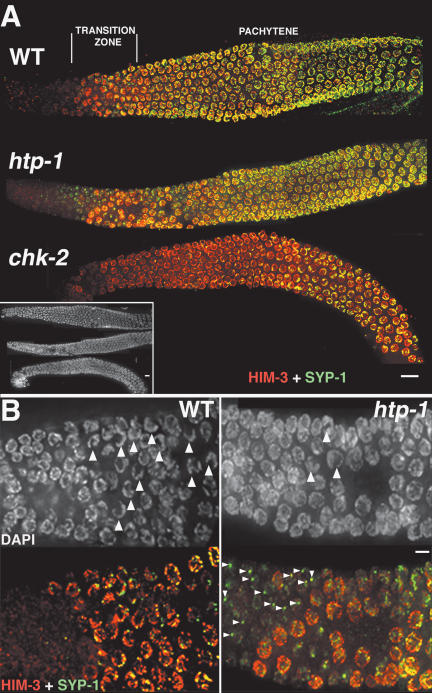
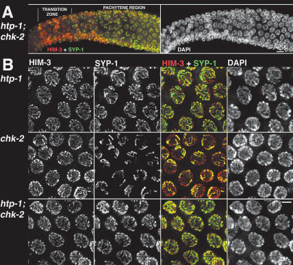
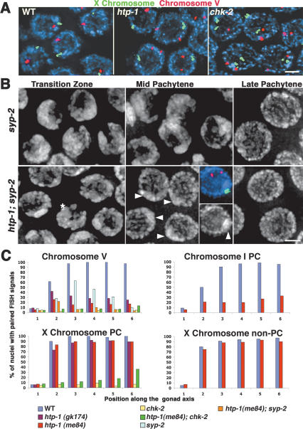
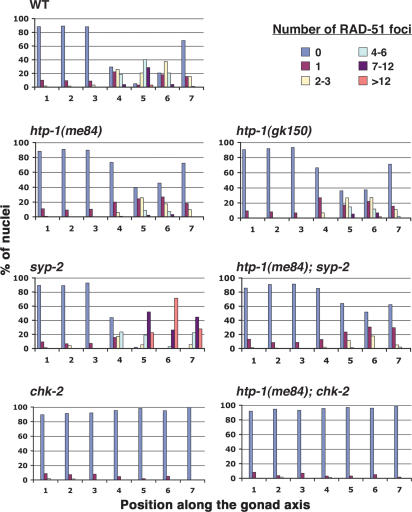
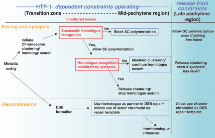
Synaptonemal complex (SC) assembly must occur between correctly paired homologous chromosomes to promote formation of chiasmata. Here, we identify the Caenorhabditis elegans HORMA-domain protein HTP-1 as a key player in coordinating establishment of homolog pairing and synapsis in C. elegans and provide evidence that checkpoint-like mechanisms couple these early meiotic prophase events. htp-1 mutants are defective in the establishment of pairing, but in contrast with the pairing-defective chk-2 mutant, SC assembly is not inhibited and generalized nonhomologous synapsis occurs. Extensive nonhomologous synapsis in htp-1; chk-2 double mutants indicates that HTP-1 is required for the inhibition of SC assembly observed in chk-2 gonads. htp-1 mutants show a decreased abundance of nuclei exhibiting a polarized organization that normally accompanies establishment of pairing; analysis of htp-1; syp-2 double mutants suggests that HTP-1 is needed to prevent premature exit from this polarized nuclear organization and that this exit stops homology search. Further, based on experiments monitoring the formation of recombination intermediates and crossover products, we suggest that htp-1 mutants are defective in preventing the use of sister chromatids as recombination partners. We propose a model in which HTP-1 functions to establish or maintain multiple constraints that operate to ensure coordination of events leading to chiasma formation.


Figures

Figure 1.
Figure 1.
Alignment of the C. elegans HIM-3 homologs: For each protein, the residues included and the total length are indicated in brackets. Characters above the HTP-1 sequence indicate residues that match (*) or are similar to (•) the HORMA domain model from the Conserved Domain Database (CDD) (Marchler-Bauer et al. 2005). Residues are colored using HTP-1 as the reference sequence. Red indicates identity with HTP-1, blue indicates conservative substitutions, and green indicates semiconservative substitutions. The arrow marks the position of the substitution (G to E) caused by the me84 mutation. Vertical black lines delimit the HORMA domain.
Figure 2.
Figure 2.
htp-1 mutants exhibit nonhomologous synapsis. (AC) DAPI-stained nuclei from the pachytene region of whole-mount gonads. Wild-type and htp-1 mutant nuclei show the presence of parallel DAPI-stained chromatin tracks, which are not observed in chk-2 nuclei. (DG) Squash preparation of htp-1(me84) pachytene nucleus labeled with α-HIM-3 antibodies, α-SYP-1 antibodies, and DAPI. Thick HIM-3 lines in D correspond to regions stained with SYP-1 in E and F, demonstrating synapsis of chromosome axes; DAPI shows that regions of overlap of HIM-3 and SYP-1 are flanked by parallel tracks of DAPI (G). The overlay of HIM-3 and SYP-1 also shows that some axis segments are unsynapsed. The region indicated by the arrow is magnified in the inset and shows a pairing partner switch. (HJ) Squash preparation of htp-1(me84) pachytene nucleus stained with α-SYP-1 antibodies, hybridized with a FISH probe for the 5S rDNA locus on chromosome V, and counterstained with DAPI. FISH signals are separated but are associated with two different α-SYP-1 signals that are located between parallel tracks of DAPI-stained chromatin, demonstrating nonhomologous synapsis. (K) Squash preparation of wild-type pachytene nucleus; both FISH signals are colocalized and associated with a single α-SYP-1 track. Bars, 5 μm.
Figure 3.
Figure 3.
Loading of SC components in htp-1, chk-2, and wild-type (WT) gonads. (A) Whole gonads stained with α-HIM-3 and α-SYP-1 antibodies. The three gonads are aligned according to the DAPI image (inset); the positions of the transition zone and the pachytene region are indicated in reference to wild type. HIM-3 loads in the normal position in both htp-1 and chk-2 mutant gonads. SYP-1 appears to be loaded at roughly wild-type levels in the pachytene region of htp-1(me84) mutant gonads, but loading of SYP-1 is substantially delayed and restricted in chk-2 mutants. (B) High-magnification image of the transition zone region of the gonad. In wild-type (WT) gonads, DAPI staining reveals the presence of numerous nuclei with clustered chromosomes (arrowheads), while in htp-1 mutant gonads, nuclei with clustered chromosomes are very infrequent. In wild-type gonads, SYP-1 signals are only detected in nuclei that have accumulated substantial amounts of HIM-3, while in htp-1 mutant gonads, SYP-1 is detected as bright aggregates (arrowheads) before HIM-3 is present on the chromosomes. All images are projections from 3D data stacks designed to include the whole depth of the nuclei shown. Bars: A, 10 μm; B, 5 μm.
Figure 4.
Figure 4.
HTP-1 prevents SYP-1 loading when homolog pairing fails. (A) htp-1; chk-2 double-mutant gonad showing nuclei from early to mid prophase stained with α-HIM-3 and α-SYP-1 antibodies. As in the htp-1 mutant, and in contrast with the chk-2 single mutant, bright SYP-1 foci are present before HIM-3 is detected on chromosomes, and extensive loading of SYP-1 is seen in the early–mid pachytene region. DAPI staining reveals the absence of nuclei displaying chromosome clustering in htp-1; chk-2 double-mutant gonads. (B) High-magnification images of late-pachytene region nuclei. Both the htp-1 mutant and the htp-1; chk-2 double mutant display almost complete colocalization of HIM-3 and SYP-1, showing that most chromosomal regions are synapsed; in contrast, only a small subset of chromosome axes are associated with SYP-1 in the chk-2 mutant. All images are projections from 3D data stacks designed to include the whole depth of the nuclei shown. Bars: A, 10 μm; B, 5 μm.
Figure 5.
Figure 5.
htp-1 mutants are defective in the establishment of homolog pairing. (A) Pachytene nuclei from whole-mount gonads hybridized with probes for the 5S rDNA locus on chromosome V (red) and for the X-chromosome PC end (green). FISH signals for both probes are paired in all wild-type nuclei; in the htp-1 mutant, signals for the X-chromosome probe are paired, while signals for chromosome V are unpaired in most nuclei; in the chk-2 mutant, lack of pairing for both probes is seen in all nuclei. (B) High-magnification images of DAPI-stained nuclei. The top panels show maintenance of clustered chromosomes in the mid-pachytene region of the syp-2 mutant, followed by eventual dispersal of chromosomes at the very end of the pachytene region. The bottom panels show the corresponding regions of an htp-1; syp-2 gonad. The transition zone region contains very few nuclei with chromosome clustering (asterisk), and by the early-pachytene region, all nuclei show dispersed chromosomes. A DAPI-bright region is evident in midpachytene region nuclei (arrowheads); the inset shows a pachytene nucleus hybridized with probes for chromosomes V and X, demonstrating that the X chromosomes are associated and coincide with the DAPI-bright region visible at this stage. Bars, 2 μm. (C) Graphs displaying quantitation of pairing levels for different chromosome regions in wild-type and mutant gonads. Numbers along the X-axis correspond to different zones along the length of the gonad; zone 1 contains premeiotic nuclei, zone 2 is composed mostly of nuclei in the transition zone, and zones 3–6 are early to late pachytene. The Y-axis indicates the percent of nuclei in each zone that displayed paired FISH signals.
Figure 6.
Figure 6.
Quantitative time-course analysis of RAD-51 foci. Graphs depicting quantitation of RAD-51 foci in gonads of the indicated genotypes. Numbers along the X-axes indicate position along the length of the gonad (zones 1 and 2 contain premeiotic nuclei, zone 3 contains mostly nuclei in the transition zone, and zones 4–7 represent early- to late-pachytene nuclei). Bars represent the percentages of nuclei in a given zone with the numbers of RAD-51 foci indicated by the color code.
Figure 7.
Figure 7.
An htp-1 mutation suppresses the accumulation of RAD-51 foci associated with translocation heterozygosity. Late-pachytene region nuclei stained with α-SYP-1 and α-RAD-51 antibodies. In the wild type and htp-1 mutant, most nuclei in this region contain few or no RAD-51 foci; in contrast, in otherwise wild-type worms heterozygous for the reciprocal translocation eT1 (eT1/+), multiple RAD-51 foci are present in most nuclei in this region of the gonad. In contrast, most nuclei in the corresponding region of an htp-1; eT1/+ mutant gonad contain few or no RAD-51 foci. Images are projections from a 3D data stack designed to include the whole depth of the nuclei shown. Bar, 5 μm.
Figure 8.
Figure 8.
γ-Irradiation of htp-1 mutants does not fully rescue chiasma formation between the X chromosomes. (A) α-RAD-51 immunostaining of mid-pachytene region nuclei from an untreated htp-1 mutant worm (left) and an htp-1 worm treated with 5000 rads of γ-irradiation and fixed 1 h later (right). (B) Each panel shows the DAPI-stained chromosomes (white or blue) from a single diakinesis-stage oocyte nucleus of the indicated genotype. Crooked arrows indicate nuclei from worms that had been exposed to 5000 rads of γ-irradiation and then fixed and stained 18 h later. Orange in the right panels corresponds to FISH signals used to identify the X chromosomes. Wild-type (WT) nuclei show six bivalents, while htp-1 and spo-11 mutants typically show 12 unattached chromosomes (univalents). Following γ-irradiation, chiasma formation is efficiently restored in spo-11 mutants (six bivalents present) but not in htp-1 mutants. Bars, 5 μm. (C) The table shows quantitation of diakinesis nuclei showing chiasmata between the X chromosomes in htp-1 mutants.
Figure 9.
Figure 9.
Model in which HTP-1-dependent constraints govern key aspects of the meiotic program. We propose that HTP-1 functions to establish or maintain constraints that operate during early prophase to (1) couple SC polymerization with successful homolog recognition, (2) couple release from chromosome clustering and termination of homology search with stabilization of pairing through synapsis, and (3) inhibit the use of sister chromatids as recombination partners. Whereas during wild-type meiosis the conditions for proceeding with synapsis and chromosome dispersal are usually met early within the domain in which the proposed constraints operate, analysis of mutants provides evidence for continued operation of these constraints through the mid-pachytene region of the gonad. We further propose that these constraints are lifted in the late-pachytene region of the gonad. Red boxes indicate monitored events; blue boxes indicate functions that require HTP-1. See text for further discussion.

References

    1. Alpi A., Pasierbek, P., Gartner, A., and Loidl, J. 2003. Genetic and cytological characterization of the recombination protein RAD-51 in Caenorhabditis elegans. Chromosoma 112: 6–16. - PubMed
    1. Aravind L. and Koonin, E.V. 1998. The HORMA domain: A common structural denominator in mitotic checkpoints, chromosome synapsis and DNA repair. Trends Biochem. Sci. 23: 284–286. - PubMed
    1. Armstrong S.J., Caryl, A.P., Jones, G.H., and Franklin, F.C. 2002. Asy1, a protein required for meiotic chromosome synapsis, localizes to axis-associated chromatin in Arabidopsis and Brassica. J. Cell Sci. 115: 3645–3655. - PubMed
    1. Bailis J.M., Smith, A.V., and Roeder, G.S. 2000. Bypass of a meiotic checkpoint by overproduction of meiotic chromosomal proteins. Mol. Cell Biol. 20: 4838–4848. - PMC - PubMed
    1. Brenner S. 1974. The genetics of Caenorhabditis elegans. Genetics 77: 71–94. - PMC - PubMed

Publication types

MeSH terms

Substances