. 2005 Nov 21;171(4):641-50.
doi: 10.1083/jcb.200505072. Epub 2005 Nov 15.

The cell cycle-apoptosis connection revisited in the adult brain

Affiliations

The cell cycle-apoptosis connection revisited in the adult brain

Sylvian Bauer et al. J Cell Biol. .

Abstract

Adult neurogenesis is studied in vivo using thymidine analogues such as bromodeoxyuridine (BrdU) to label DNA synthesis during the S phase of the cell cycle. However, BrdU may also label DNA synthesis events not directly related to cell proliferation, such as DNA repair and/or abortive reentry into the cell cycle, which can occur as part of an apoptotic process in postmitotic neurons. In this study, we used three well-characterized models of injury-induced neuronal apoptosis and the combined visualization of cell birth (BrdU labeling) and death (Tdt-mediated dUTP-biotin nick end labeling) to investigate the specificity of BrdU incorporation in the adult mouse brain in vivo. We present evidence that BrdU is not significantly incorporated during DNA repair and that labeling is not detected in vulnerable or dying postmitotic neurons, even when a high dose of BrdU is directly infused into the brain. These findings have important implications for a controversy surrounding adult neurogenesis: the connection between cell cycle reactivation and apoptosis of terminally differentiated neurons.


Figures

Figure 1.
Figure 1.
Validation of the BrdU/TUNEL double staining in the adult mouse OE 36–48 h after unilateral right OBX. (A) Unilateral OBX leads to massive apoptosis of olfactory sensory neurons in the ipsilateral OE only, visualized by TUNEL staining (red). The basal lamina (dashed line) separates the OE proper (arrow) from underlying lamina propria (arrowhead). Bar, 200 μm. (B) The lamina propria contains axon bundles of olfactory sensory neurons, expressing high levels of casp-3 (green). Bar, 200 μm. (C) After a single BrdU injection, the OE contains only a few scattered BrdU+ nuclei next to the basal lamina (green). Bar, 200 μm. (D) Repeated injections of BrdU over a 24-h period leads to many more BrdU+ nuclei, extending toward the surface of the OE. Bar, 200 μm. (E and F) Combination of BrdU staining (green) and TUNEL labeling (red) in the OE of the latter animal visualized under epifluorescence indicates that colocalization is very rare. Bars, 50 μm.
Figure 2.
Figure 2.
2 d after unilateral OBX, the OE contains many dying neurons but rare BrdU/TUNEL colocalization, detected only when BrdU is given repeatedly. (A) TUNEL staining (red) colocalizes with BrdU (green) in various patterns with considerable overlap. (B) BrdU staining is shown inside a TUNEL+ nucleus. (C) TUNEL staining is shown inside a BrdU+ nucleus. (A and B) Vertical images show orthogonal projections along the z axis. (C–E) Colocalization is most often detected in the basal OE, where progenitor cells proliferate and immature neurons (TuJ1 in blue) are generated. Bars: (A) 5 μm; (B) 10 μm; (C) 50 μm; (D and E) 20 μm.
Figure 3.
Figure 3.
Detection of BrdU and TUNEL staining 8.5 h after brain irradiation shows a large increase in neuronal death but a decrease in BrdU incorporation. Brain irradiation (IR) induces a >44-fold increase in TUNEL+ nuclei in the SVZ of the lateral ventricle (A, dotted line) and the SGZ of the hippocampus (C, dotted line). In the control brain (CC), sporadic cell death is detected (arrows). After brain irradiation, BrdU incorporation is reduced more than twofold in both germinal zones (compare B and D with A and C, respectively) and virtually all BrdU+ nuclei are simultaneously TUNEL+ (A'–A''' and insets in C, corresponding to areas marked by asterisks in A and C, respectively). (B' and B”) BrdU/TUNEL colocalization is also found in the normal adult brain but to a considerably lesser extent and only in the SVZ. (B”) Vertical image shows orthogonal projection along the z axis. (E and F) After brain irradiation, some of the BrdU/TUNEL double-stained nuclei are also positive for Ki67 (blue), indicating cellular proliferation. Bars: (A–D) 50 μm; (A'–B”) 20 μm; (C, insets) 30 μm; (E and F) 10 μm.
Figure 4.
Figure 4.
Detection of IdU and CldU staining 8 h after brain irradiation indicates a reduction in both labels. IdU (green) was injected 1.5 h before irradiation, whereas CldU (red) was injected 6 h after irradiation. (A and B) Brain irradiation (IR) decreases IdU+ and CldU+ nuclei by 40 and 75%, respectively, in the SVZ of the lateral ventricle (dotted lines) compared with control (CC). Insets in B correspond to high-power magnification pictures showing single IdU+ or CldU+ cells, as well as double-stained cells in the control SVZ (B' and B”, arrows). After brain irradiation, most of the CldU+ nuclei are also labeled with IdU (A'–A''', corresponding to areas marked by asterisks in A). (C) Brain irradiation induces strong expression of casp-3 (green) in the SVZ, as well as massive TUNEL staining (red). Interestingly, when BrdU is injected repeatedly after brain irradiation, most of the BrdU staining (blue) is found in TUNEL+ cells and only in a few caspase 3+ cells (arrows). Bars: (A and B) 50 μm; (A'–B” and C) 10 μm.
Figure 5.
Figure 5.
KA-induced seizure in adult FVB/N mice leads to massive neuronal cell death. Postmitotic neurons (NeuN, green) are extremely TUNEL+ (red) in the CA region of the hippocampus (A and B) and in the somatosensory cortex (C) by 3 d after lesion. Bars: (A) 200 μm; (B) 50 μm; (C) 100 μm.
Figure 6.
Figure 6.
BrdU and TUNEL staining 3–4 d after KA injection shows a major increase in neuronal death but rare double labeling. (A and B) BrdU was either injected i.p. (100 mg/kg) 2 h before killing (Inj) or directly infused in the lateral ventricle for 24 h (Inf). Massive cell death is detected in both the hippocampus (A, B, and CA1) and the somatosensory cortex (CX). (A) However, BrdU/TUNEL colocalization is absent in animals receiving a single BrdU injection. (B) Similarly, after infusion of BrdU, which leads to a much higher uptake of the marker into brain cells, the majority of dying cells is not positive for BrdU. Rare colocalization is nonetheless detectable in this case, specifically in areas close to the lateral ventricle, such as the SVZ (C) or the corpus callosum (D). A total of three BrdU+/TUNEL+ nuclei are detected in the CA3 area in six sections containing >24,000 TUNEL+ nuclei (E, arrow and insets). In the hippocampal formation, most of the double-stained nuclei are found in the stratum radiatum and colocalize with Mac-1, a marker of microglia (blue in F, arrow and insets) but not with NeuN (blue in G, arrow). Vertical images represent orthogonal projections along the z axis. Insets in E and F are 18 and 34 μm wide, respectively. BrdU is in green, and TUNEL is in red. Bars: (A and B) 200 μm; (C and D) 10 μm; (E) 30 μm; (CA1, CX, F, and G) 50 μm.
Figure 7.
Figure 7.
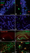
Markers of DNA repair (p53 protein) and cell cycle (cyclin D1) do not label BrdU + cells. After KA injection, a few NeuN+ postmitotic neurons (blue) express a high level of nuclear p53 protein (A–D, red) but are never positive for BrdU (green), either in the CA1/CA3 areas (A and B) or the cortex (C and D). Surprisingly, the intact adult mouse brain (E–G) contains populations of postmitotic neurons (NeuN+, green) showing a high level of nuclear cyclin D1 (E–H, red) immunoreactivity, including pyramidal neurons of the CA1 area (E and F), where cyclin D1 immunoreactivity abruptly stops at the junction with CA2/CA3 (E and F, arrows), and neurons of the piriform cortex (G). After KA injection, cyclin D1 is highly expressed in hypertrophic reactive glial fibrillary acidic protein (GFAP+) astrocytes (green) in the cortex (H). Bars: (A–D and G) 50 μm; (E and F) 500 μm; (H) 30 μm.

References

    1. Alvarez-Dolado, M., R. Pardal, J.M. Garcia-Verdugo, J.R. Fike, H.O. Lee, K. Pfeffer, C. Lois, S.J. Morrison, and A. Alvarez-Buylla. 2003. Fusion of bone-marrow-derived cells with Purkinje neurons, cardiomyocytes and hepatocytes. Nature. 425:968–973. - PubMed
    1. Bauer, S., S. Rasika, J. Han, C. Mauduit, M. Raccurt, G. Morel, F. Jourdan, M. Benahmed, E. Moyse, and P.H. Patterson. 2003. Leukemia inhibitory factor is a key signal for injury-induced neurogenesis in the adult mouse olfactory epithelium. J. Neurosci. 23:1792–1803. - PMC - PubMed
    1. Bauer, S., M. Hay, B. Amilhon, A. Jean, and E. Moyse. 2005. In vivo neurogenesis in the dorsal vagal complex of the adult rat brainstem. Neuroscience. 130:75–90. - PubMed
    1. Becker, E.B., and A. Bonni. 2004. Cell cycle regulation of neuronal apoptosis in development and disease. Prog. Neurobiol. 72:1–25. - PubMed
    1. Bernier, P.J., A. Bedard, J. Vinet, M. Levesque, and A. Parent. 2002. Newly generated neurons in the amygdala and adjoining cortex of adult primates. Proc. Natl. Acad. Sci. USA. 99:11464–11469. - PMC - PubMed

Publication types

MeSH terms