. 2005 Dec;132(24):5425-36.
doi: 10.1242/dev.02149. Epub 2005 Nov 16.

Wnt3a links left-right determination with segmentation and anteroposterior axis elongation

Affiliations

Wnt3a links left-right determination with segmentation and anteroposterior axis elongation

Masa-aki Nakaya et al. Development. 2005 Dec.

Abstract

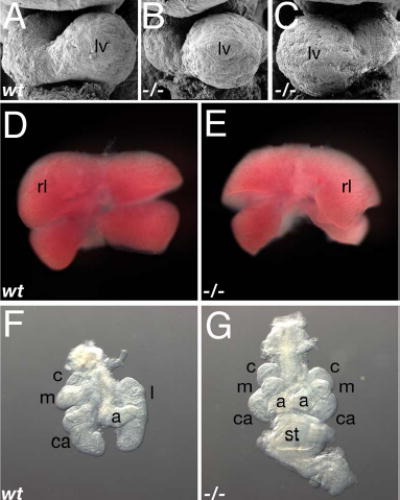
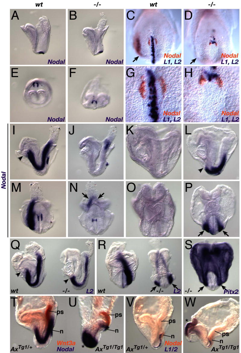
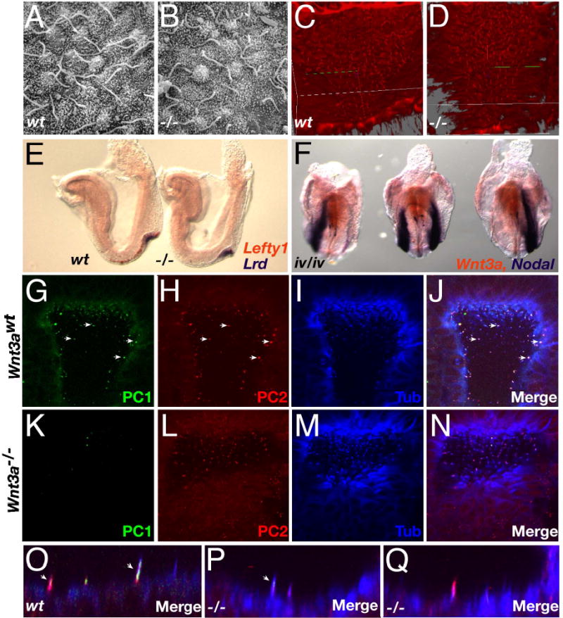
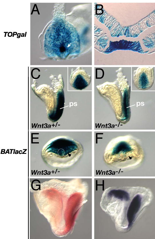
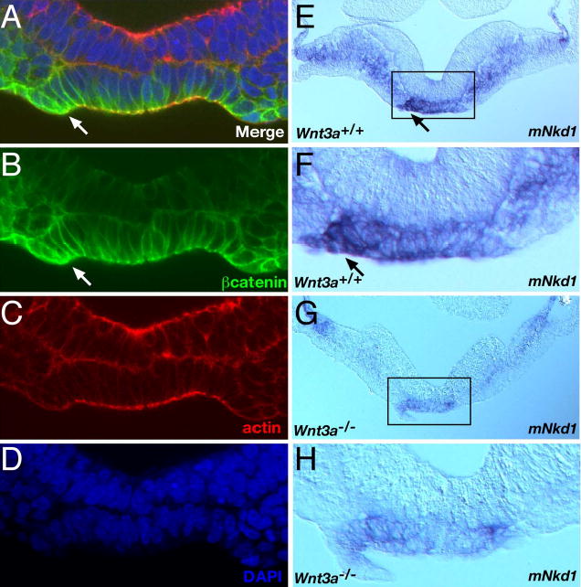
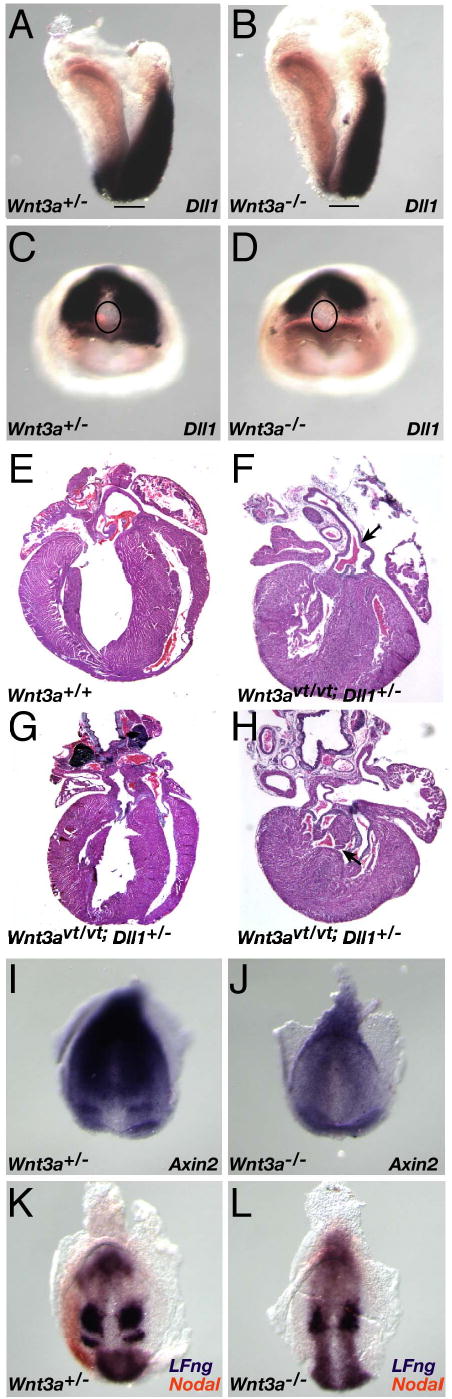
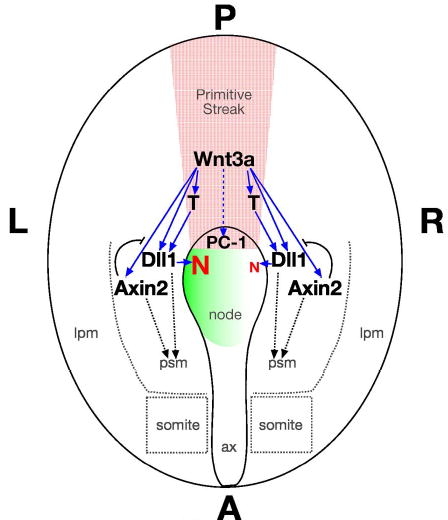
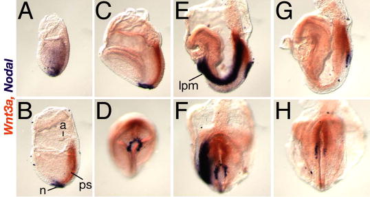
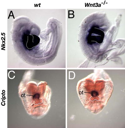
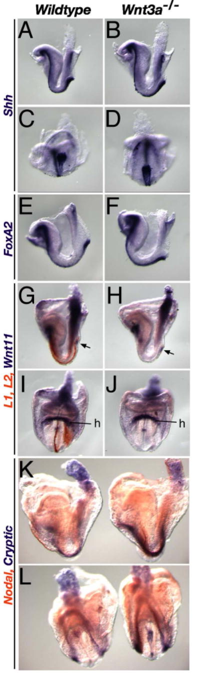
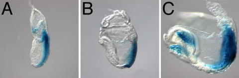
The alignment of the left-right (LR) body axis relative to the anteroposterior (AP) and dorsoventral (DV) axes is central to the organization of the vertebrate body plan and is controlled by the node/organizer. Somitogenesis plays a key role in embryo morphogenesis as a principal component of AP elongation. How morphogenesis is coupled to axis specification is not well understood. We demonstrate that Wnt3a is required for LR asymmetry. Wnt3a activates the Delta/Notch pathway to regulate perinodal expression of the left determinant Nodal, while simultaneously controlling the segmentation clock and the molecular oscillations of the Wnt/beta-catenin and Notch pathways. We provide evidence that Wnt3a, expressed in the primitive streak and dorsal posterior node, acts as a long-range signaling molecule, directly regulating target gene expression throughout the node and presomitic mesoderm. Wnt3a may also modulate the symmetry-breaking activity of mechanosensory cilia in the node. Thus, Wnt3a links the segmentation clock and AP axis elongation with key left-determining events, suggesting that Wnt3a is an integral component of the trunk organizer.


Figures

Fig. 1
Fig. 1. Loss of Wnt3a leads to laterality defects.
(A–C) SEM micrographs of E9.5 wildtype (A), and Wnt3a/ (B, C) hearts displayed normal (B) and inverted (situs inversus) looping (C). (D, E) E11.5 livers. Situs inversus was observed in the asymmetric arrangement of the Wnt3a/ liver (E), compared to the control (D). (F, G) E11.5 lungs and stomach. A midline stomach and right pulmonary isomerism was often observed in mutants (G), in contrast to the wildtype lungs (F, stomach not shown). Abbr: lv, left ventricle; rl, right lateral lobe; c, cranial lobe; m, medial lobe; ca, caudal lobe; a, accessory lobe; l, left lobe; st, stomach.
Fig. 2
Fig. 2. Wnt signaling controls LR asymmetric gene expression.
(A–P) WISH analysis of Nodal expression (see text for details). Headfold-stage wildtype (A, E), and Wnt3a/ (B, F) embryos express Nodal (purple) in the node. Two color WISH showing Nodal (orange), and Lefty1 and Lefty2 expression (purple), in wildtype (C, G) and mutant (D, H) 3 somite stage embryos. (G, H) High power ventral views of the nodes of the wildtype and mutant embryos depicted in (C) and (D). Nodal expression in wildtype 4 somite (I, M) and similarly-staged Wnt3a/ (J, N) embryos. Arrows in N indicate bilateral Nodal expression in the mutant posterior LPM. Nodal was not expressed in the wildtype LPM after the 6–7 somite stage (K, O), but was bilaterally expressed in the mutant LPM (L, P). (Q–S) Similar abnormal expression patterns were observed for Lefty2 in 7-somite stage mutants (right embryos in Q and R, cf. to 4-somite wildtype embryos on the left side), and for Pitx2 expression in 6-somite mutants (S). (T, U) Two color WISH showing Nodal (purple) and Wnt3a (orange) expression in 4- somite wildtype (T) and AxinTg1/Tg1 (U) littermates. (V, W) Lefty1/2 (purple) and Nodal (orange) expression in 2-somite wildtype (V) and AxinTg1/Tg1 (W) littermates. Asterisks indicate ectopic, bilateral expression of Nodal (U) and Lefty1/2 (W) in anterior domains. A–D, I–L, Q, T–W, lateral views; E–H, M, N, S, ventral posterior views; O, P, R, anterior views; arrows indicate the LPM; arrowheads, the anterior limit of LPM expression. Abbr.: ps, primitive streak; n, node.
Fig. 3
Fig. 3. Examination of cilia in the Wnt3a−/− mutants.
(A, B) High-power SEM micrographs of cilia in wildtype (A) and Wnt3a/ (B) nodes of headfold stage embryos (~E7.75–8). (C, D) Projection images of node cilia visualized using anti-acetylated tubulin antibodies. (E) Two color WISH analysis of Lefty1 (orange) and Lrd (purple) expression in 3 somite wildtype (left) and Wnt3a/ (right) embryos. (F) Wnt3a (orange) and Nodal (purple) expression in three iv/iv embryos. (G–Q) Confocal microscopy images of PC1 (G, K), PC2 (H, L), acetylated tubulin (I, M) and the merged images (J, N, O, P, Q) in E7.75 wildtype (G–J, O) and Wnt3a/ (K–N, P, Q) nodes. The abundance of white spots in global views of the node indicated co-expression of all three markers in individual wildtype cilium (arrows, G–J). In contrast, Wnt3a/ cilia predominantly appeared as purple spots (N) indicating coexpression of only PC2 and tubulin. Views were selected to present sufficient numbers of labeled cilia and therefore depict slightly different regions of the wildtype and mutant node, however, images are representative of the entire node. Profiles of labeled cilia illustrate the coexpression of PC1 and PC2 in distinct spatial domains in wildtype cilia (arrows, O). Rare mutant cilia coexpressed PC1 and PC2 but these domains were unusually small and not easily detected (arrow, P). Cilia co-expressing PC2 and tubulin were easily detected in the mutant node (Q).
Fig. 4
Fig. 4. Expression of Wnt/βcatenin reporter transgenes in vivo is Wnt3a-dependent.
(A) A 5 somite TOPgal embryo showed strong expression in the node (arrow), primitive streak (ps), and posterior mesoderm. (B) Cross-section through the node of embryo shown in (A). (C, E) A headfold stage BATlacZ embryo showing βgal expression in the primitive streak (for posterior view, see inset), anterior psm (black arrow) and the node (curved white line). (D, F) BATlacZ expression was reduced in the Wnt3a/ streak, and was not expressed in the node (curved white line) and anterior psm (black arrow). (G) βgal activity in 6 somite BATlacZ embryo visualized with Salmon-gal (red). (H) lacZ mRNA expression in a 5 somite BATlacZ embryo visualized by WISH. (A, and insets in C, D) Ventral-posterior views. (E, F) Ventral views, left side of the embryo is on the left. (C, D, G, H) Lateral views, anterior is to the left.
Fig. 5
Fig. 5. Asymmetric distribution of canonical Wnt/βcatenin signaling pathway components in the node.
All images are posterior views of cross sections, the left side of the embryo is facing left. (A) Merge of confocal microscopy images of E7.75 wildtype embryo labeled with anti-βcatenin antibody (B), rhodamine phalloidin (C), and DAPI (D). (E–H) Expression of the Wnt/βcatenin target gene mNkd1 in 4 somite stage wildtype (E, F) and Wnt3a/ embryos. Arrows indicate sites of asymmetric expression in the wildtype node. Boxed regions in E and G are represented as high-power views of the nodes in panels F and H, respectively. Note that all assessments of mNkd1 distribution were performed on whole embryos by WISH (not shown), and subsequently sectioned for confirmation and clarity.
Fig. 6
Fig. 6. Components of the Notch pathway participate in a Wnt3a-dependent pathway that controls laterality and somitogenesis.
(A–D) WISH analysis of Dll1 expression in E8 wildtype (A, C) and Wnt3a/ (B, D) embryos. Note that Dll1 expression in the psm surrounds the wildtype node (C), but only abuts the posterior-most node in a Wnt3a/ embryo (D). The lines in A and B, and ovals in C and D, indicate the location of the node. (E–H) Histological sections of wildtype neonatal heart (E), and three examples of cardiac laterality defects in compound Wnt3avt; Dll1 mutants, including PTA (arrow, F), TGA (G) and VSD (arrow, H). Axin2 expression in the streak, psm, and in an anterior stripe (presomite 0) in wildtype 1-somite embryos (I), was down-regulated in Wnt3a/ mutants and no psm stripes were observed (J). Two color WISH on 4 somite stage embryos illustrating cycling LFng (purple), and Nodal expression (orange) in the wildtype left LPM. Dynamic Lfng expression was not observed in the absence of Wnt3a, as only a single psm stripe was observed in the Wnt3a mutants (L). Nodal was not expressed in the mutant LPM at these stages.
Fig. 7
Fig. 7. Wnt3a functions as a trunk organizer.
The diagram depicts the ventral view of an E8 embryo. Wnt3a (red stippling) is expressed in the streak and node where it directly activates (solid blue arrows) T (Brachyury), Dll1, and Axin2 via the Wnt/βcatenin pathway. Please see the text for details. Abbr.: red N, Nodal; solid blue arrow, direct gene regulation; dashed blue arrow, indirect regulation; green gradient, left-sided Ca2+ flux; curved black line, negative feedback loop. Ax, axial mesendoderm.
Supplementary Figure 1
Supplementary Figure 1. Wnt3a and Nodal are expressed in adjacent node cells.
Two color whole-mount in situ hybridization showing Wnt3a (orange) and Nodal (purple) expression in E7.5–8.5 embryos; (A, B, C, E, G) lateral views of left side of embryo, anterior is to the left, (D, F, H) ventral views of the embryos depicted above, posterior end points up. (A) E7.5 early alantoic bud stage; Wnt3a transcripts were first detected in primitive streak ectoderm, prior to node formation, as Nodal expression is down-regulated in embryonic mesoderm and distally restricted to the presumptive node. (B) E7.5 alantoic bud stage, (C, D) E7.75 neural plate stage; note that Wnt3a expression in the ectoderm overlaps Nodal expression in the underlying posterior ventral node endoderm. (E, F) E8.2, 4 somite stage; Nodal is expressed asymmetrically in the node and in the left lateral plate mesoderm (LPM), while Wnt3a continues to be symmetrically expressed in the streak. (G, H) E8.5, 8 somite stage; Nodal expression is undetectable in the left LPM but remains asymmetric in the node, while Wnt3a expression persists in the streak. N, node; ps, primitive streak; a, allantois; lpm, lateral plate mesoderm.
Supplementary Figure 2
Supplementary Figure 2. Heart looping defects are not due to abnormal cardiac specification.
Reduced Wnt/βcatenin signaling is associated with heart formation (Lickert et al., 2002; Marvin et al., 2001). To rule out the possibility that the heart looping defects observed in the Wnt3a mutants were secondary to defects in cardiogenesis, we examined the expression of the heart markers Cripto, and Nkx2.5, the latter of which is required for proper looping morphogenesis (Lyons et al., 1995; Tanaka et al., 1999). WISH was performed on wildtype (A, C) and Wnt3a/ littermates (B, D) to examine Nkx2.5 expression in the heart at E8.75 (A, B), and Cripto expression in the outflow tract at the 7 somite stage (C, D). Both genes continued to be strongly expressed in hearts undergoing aberrant looping morphogenesis. Note that the mutant Cripto-positive outflow tract remained in the midline at stages (E8.5) when it would normally have undergone a rightward shift in wildtype embryos. The white dashed line outlines the looping ventricles. ot, outflow tract.
Supplementary Figure 3
Supplementary Figure 3. Node and axial markers are expressed in Wnt3a mutants.
WISH of E8.2–8.5 wildtype (left) and mutant (right) embryos, analyzed for marker gene expression. Markers of the node and axial mesendoderm, such as Shh (A–D) and FoxA2 (E, F), continued to be expressed in 3-4-somite stage Wnt3a/ embryos (B, D, F). (G–J) Despite the loss of Lefty1 and Lefty2 (orange), normally expressed in the midline PFP and left LPM (G, I), Wnt11 (purple) continued to be expressed in the mutant node (arrow), posterior primitive streak and heart (H, J) as it was in wildtype embryos (G, I). (K, L) Two color WISH analysis of Cryptic (purple) and Nodal (orange) expression. Cryptic was expressed in the midline, including the node, and bilaterally in the LPM of wildtype embryos (left embryos). Cryptic expression in the LPM and node was up-regulated in 6–7 somite stage Wnt3a/ embryos, suggesting that Wnt3a, directly or indirectly, represses Cryptic transcription. We have previously shown that Brachyury is also expressed in the Wnt3a/ node and notochord at these stages (Yamaguchi et al., 1999). C, D, L, ventral posterior view; I, J, anterior view; the rest are lateral views; h, heart.
Supplementary Figure 4
Supplementary Figure 4. TOPgal expression from E7.5–8.5.
The TOPgal reporter indicates that the canonical Wnt/βcatenin signaling pathway is active in the primitive streak at E7.5, when Wnt3a is first expressed there (A). Expression is easily detected in the node at E7.75 (B), persisting in the node and streak through E8.5 stages (C). Expression is also detected in the mid-hindbrain and heart tube. Staining was not observed in axial derivatives of the node such as the notochord or PFP.

References

    1. Aulehla A, Herrmann BG. Segmentation in vertebrates: clock and gradient finally joined. Genes Dev. 2004;18:2060–2067. - PubMed
    1. Aulehla A, Wehrle C, Brand-Saberi B, Kemler R, Gossler A, Kanzler B, Herrmann BG. Wnt3a plays a major role in the segmentation clock controlling somitogenesis. Dev Cell. 2003;4:395–406. - PubMed
    1. Barrantes IB, Elia AJ, Wunsch K, Hrabe de Angelis MH, Mak TW, Rossant J, Conlon RA, Gossler A, de la Pompa JL. Interaction between Notch signalling and Lunatic fringe during somite boundary formation in the mouse. Curr Biol. 1999;9:470–480. - PubMed
    1. Beddington RS. Induction of a second neural axis by the mouse node. Development. 1994;120:613–620. - PubMed
    1. Bouillet P, Oulad-Abdelghani M, Ward SJ, Bronner S, Chambon P, Dolle P. A new mouse member of the Wnt gene family, mWnt-8, is expressed during early embryogenesis and is ectopically induced by retinoic acid. Mech Dev. 1996;58:141–152. - PubMed

Publication types