Comparative Study
. 2005 Nov 29;102(48):17507-12.
doi: 10.1073/pnas.0505789102. Epub 2005 Nov 16.

The epithelial Ca2+ channel TRPV5 is essential for proper osteoclastic bone resorption

Affiliations
Comparative Study

The epithelial Ca2+ channel TRPV5 is essential for proper osteoclastic bone resorption

Bram C J van der Eerden et al. Proc Natl Acad Sci U S A. .

Abstract

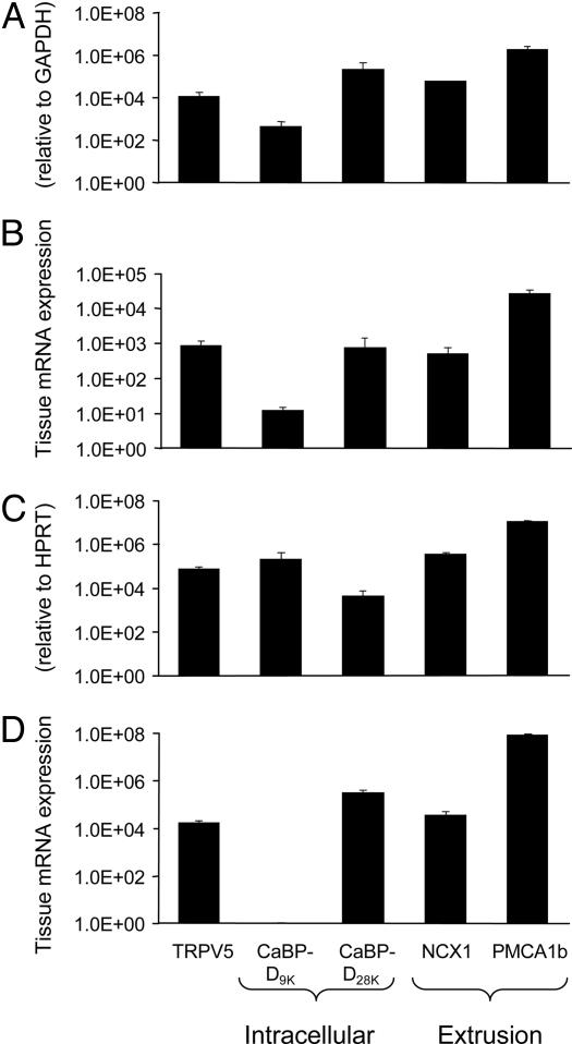
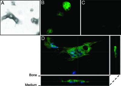
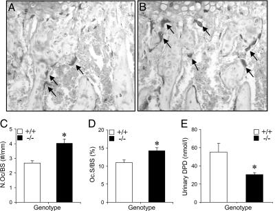
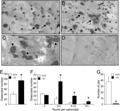
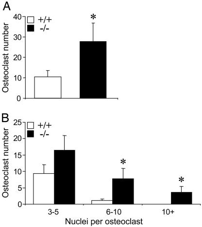
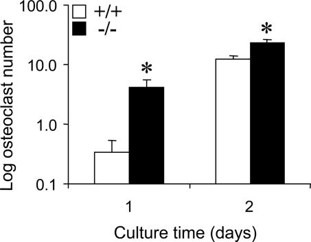
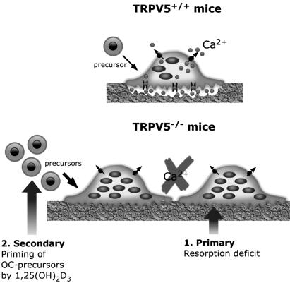
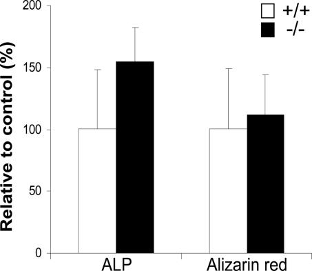
Bone remodeling involves the interplay of bone resorption and formation and is accurately controlled to maintain bone mass. Both processes require transcellular Ca(2+) transport, but the molecular mechanisms engaged remain largely elusive. The epithelial Ca(2+) channel TRPV5 is one of the most Ca(2+)-selective transient receptor potential (TRP) channels. In this study, the functional role of TRPV5 in bone was investigated. TRPV5 mRNA was expressed in human and murine bone samples and in osteoclasts along with other genes involved in transcellular Ca(2+) transport, including calbindin-D(9K) and calbindin-D(28K), Na(+)/Ca(2+) exchanger 1, and plasma membrane Ca(2+)-ATPase 1b. TRPV5 expression in murine osteoclasts was confirmed by immunostaining and showed predominant localization to the ruffled border membrane. However, TRPV5 was absent in osteoblasts. Analyses of femoral bone sections from TRPV5 knockout (TRPV5(-/-)) mice revealed increased osteoclast numbers and osteoclast area, whereas the urinary bone resorption marker deoxypyridinoline was reduced compared with WT (TRPV5(+/+)) mice. In an in vitro bone marrow culture system, the amount of osteoclasts and number of nuclei per osteoclast were significantly elevated in TRPV5(-/-) compared with TRPV5(+/+) mice. However, using a functional resorption pit assay, we found that bone resorption was nearly absent in osteoclast cultures from TRPV5(-/-) mice, supporting the impaired resorption observed in vivo. In conclusion, TRPV5 deficiency leads to an increase in osteoclast size and number, in which Ca(2+) resorption is nonfunctional. This report identifies TRPV5 as an epithelial Ca(2+) channel that is essential for osteoclastic bone resorption and demonstrates the significance of transcellular Ca(2+) transport in osteoclastic function.


Figures

Fig. 1.
Fig. 1.
TRPV5 is expressed in human and murine osteoclasts. Total RNA was isolated from human femoral biopsy samples (A), human PBMC-derived osteoclast-like cells (B), murine femurs (C), and osteoclasts derived from murine tibial bone marrow cultures (D). The mRNA expression of TRPV5, the intracellular Ca2+ binding proteins calbindin-D9K (CaBP-D9K) and calbindin-D28K (CaBP-D28K), and the Ca2+ extrusion proteins NCX1 and PMCA1b was measured. The accolades refer to the supposed function of the different genes: TRPV5 for uptake of Ca2+, the calbindins for intracellular Ca2+ transport, and NCX1 and PMCA1b for extrusion of Ca2+. HPRT, hypoxanthine-guanine phopshoribosyl transferase.
Fig. 2.
Fig. 2.
TRPV5 is predominantly visualized in the ruffled border membrane of murine osteoclasts. Murine osteoclasts derived from bone marrow were cultured on bone slices in the presence of M-CSF and RANKL for 6 days. (A) Osteoclasts were identified by TRACP staining. (B and C) TRPV5 staining (green) was demonstrated in osteoclasts derived from TRPV5+/+ mice (B) but not from TRPV5-/- mice (C). (D) Confocal laser scanning microscopy showed TRPV5 staining predominantly at the ruffled border membrane, where bone resorption occurs. Nuclei are stained with DAPI (blue).
Fig. 3.
Fig. 3.
TRPV5-/- mice exhibit enhanced osteoclastogenesis but reduced resorption. (A and B) TRACP staining was performed on femoral bone sections from TRPV5+/+ (A) and TRPV5-/- (B) mice, and images were taken of the metaphyses. Arrows indicate osteoclasts. (C and D) Osteoclast number (C) and osteoclast surface area (D) per bone surface area was quantified (n = 7-8). (E) Urinary DPD levels were measured in TRPV5+/+ and TRPV5-/- mice (n = 9). Values are presented as mean ± SEM. *, P < 0.05 versus TRPV5+/+ mice.
Fig. 4.
Fig. 4.
Osteoclastogenesis in vitro is enhanced but resorption is diminished. (A and B) Murine bone marrow cells from TRPV5+/+ (A) and TRPV5-/- (B) mice were cultured on bovine cortical bone slices for 6 days and stained for TRACP to visualize multinucleated osteoclasts. Arrows indicate multinuclear osteoclasts. (C and D) Coomassie brilliant blue was used to stain the resorption pits in TRPV5+/+ (C) and TRPV5-/- (D) cultures. Arrowheads indicate resorption pits. (E-G) In these cultures, osteoclast number (E), the number of nuclei per osteoclast (F), and the number of resorption pits per bone slice (G) were quantified (n = 6-9). Values are presented as mean ± SEM. *, P < 0.05 versus TRPV5+/+ cultures.
Fig. 5.
Fig. 5.
Diminished resorption does not enhance osteoclastogenesis in TRPV5-/- mice. Murine bone marrow cells from TRPV5+/+ and TRPV5-/- mice were cultured on plastic instead of cortical bone slices for 6 days and stained for TRACP. The amount of osteoclasts (A) and the number of nuclei per osteoclast (B) was measured for both cultures (n = 6). Values are presented as mean ± SEM. *, P < 0.05 versus TRPV5+/+ cultures.
Fig. 6.
Fig. 6.
Increased number of osteoclast precursors in TRPV5-/- mice. Murine bone marrow cells from TRPV5+/+ and TRPV5-/- mice were cultured on plastic and stained for TRACP at days 1 and 2 of culture. The number of TRACP-positive cells was quantified (n = 6). Values are presented as mean ± SEM. *, P < 0.05 versus TRPV5+/+ cultures.
Fig. 7.
Fig. 7.
Osteoclast phenotype in TRPV5-/- mice. The findings in this study reveal two phenomena, which contribute to the osteoclast phenotype in TRPV5-/- mice. First, an intrinsic defect occurs in TRPV5-/- osteoclasts, leading to a bone resorption deficit, as opposed to TRPV5+/+ mice. Second, priming of bone marrow by high 1,25(OH)2D3 levels may result in more osteoclast precursors in TRPV5-/- compared with TRPV5+/+ mice, leading to an increase in the amount and size of osteoclasts.
Fig. 8.
Fig. 8.
Osteoblast differentiation is not altered in TRPV5-/- mice. Murine bone marrow cells from TRPV5+/+ and TRPV5-/- mice were either cultured for 10 days and stained for ALP or cultured for 20 days and stained for mineralization (alizarin red). The number of ALP- and alizarin red-positive colonies were quantified. Values are presented as mean ± SD.

Similar articles

Cited by

References

    1. Hoenderop, J. G. & Bindels, R. J. (2005) J. Am. Soc. Nephrol. 16, 15-26. - PubMed
    1. Bronner, F. (1998) J. Nutr. 128, 917-920. - PubMed
    1. Hoenderop, J. G., Van Der Kemp, A. W., Hartog, A., van de Graaf, S. F., Van Os, C. H., Willems, P. H. & Bindels, R. J. (1999) J. Biol. Chem. 274, 8375-8378. - PubMed
    1. Peng, J. B., Chen, X. Z., Berger, U. V., Vassilev, P. M., Tsukaguchi, H., Brown, E. M. & Hediger, M. A. (1999) J. Biol. Chem. 274, 22739-22746. - PubMed
    1. Montell, C., Birnbaumer, L., Flockerzi, V., Bindels, R. J., Bruford, E. A., Caterina, M. J., Clapham, D. E., Harteneck, C., Heller, S., Julius, D., et al. (2002) Mol. Cell 9, 229-231. - PubMed

Publication types

MeSH terms