Comparative Study
. 2005 Nov 29;102(48):17436-41.
doi: 10.1073/pnas.0506351102. Epub 2005 Nov 16.

Transformation of breast cells by truncated neurokinin-1 receptor is secondary to activation by preprotachykinin-A peptides

Affiliations
Comparative Study

Transformation of breast cells by truncated neurokinin-1 receptor is secondary to activation by preprotachykinin-A peptides

Hiral J Patel et al. Proc Natl Acad Sci U S A. .

Abstract

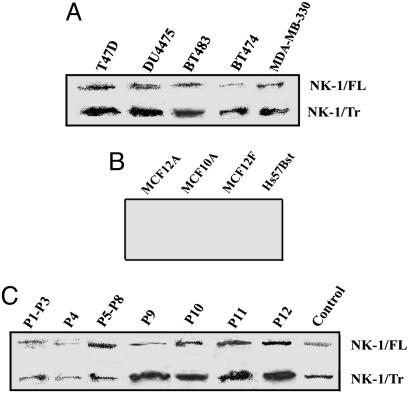
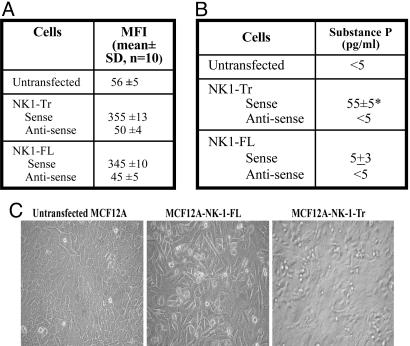
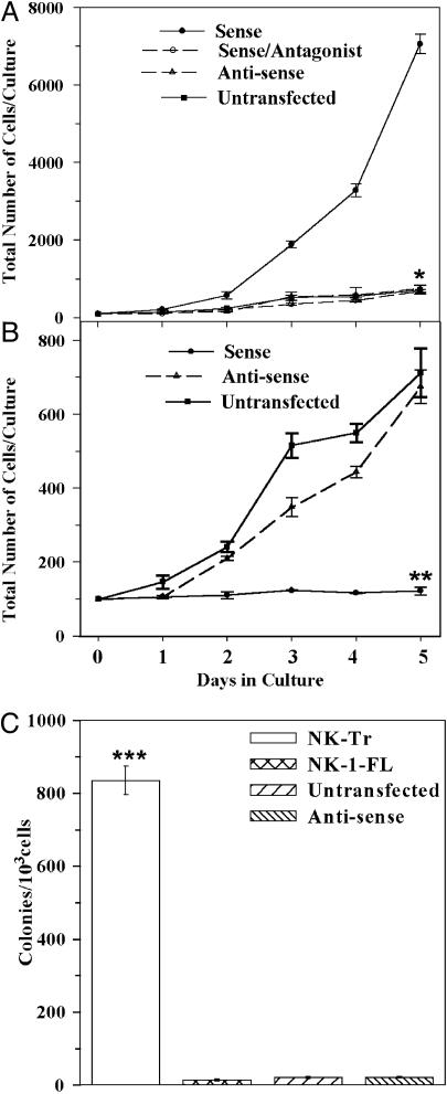
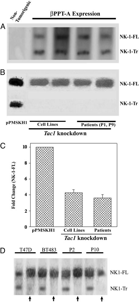
Breast cancer remains the cancer with the highest mortality among women in the United States. Peptides derived from the oncogenic Tac1 gene (full transcript: betaPPT-A) stimulate the proliferation of breast cancer cells (BCCs) via seven-transmembrane G protein-coupled neurokinin 1 (NK1) and NK2 receptors. The NK1 gene could generate full-length (NK1-FL) and truncated (NK1-Tr) transcripts. NK1-Tr lacks 100 residues in their cytoplasmic end, could couple to G proteins, and shows reduced efficiency with respect to internalization and desensitization. This study reports on a role of NK1-Tr in the transformation of nontumorigenic breast cells, and investigates whether Tac1 expression is linked to the generation of NK1-Tr. Western blots and Northern analyses showed coexpressions of NK1-Tr and NK1-FL in BCCs (cell lines and primary cells from patients with different stages of breast cancer). Stable transfections of betaPPT-A or NK1-Tr expression vectors in nontumorigenic cells showed each induces the expression of the other, consequently resulting in a transformed phenotype. Analyses with microarrays indicate similar patterns of cytokine production by NK1-Tr transfectants and BCCs, but not NK1-FL transfectants. These observations indicate tumor-promoting properties by NK1-Tr, but not NK1-FL. Overall, the oncogenic property of Tac1 in breast cells involves concomitant expression of NK1-Tr and vice versa, consequently leading to the production of cytokines with growth promoting functions.


Figures

Fig. 1.
Fig. 1.
Coexpression of NK1-FL and NK1-Tr cDNA in BCCs. Membrane extracts were examined for NK1 by Western blots in BCCs (A); nontumorigenic breast cell lines (B), and primary BCCs, with DU4475 as control (C).
Fig. 2.
Fig. 2.
Stable transfection of NK1-Tr and NK1-FL cDNA in MCF12A and MCF10A. (A) The mean fluorescence intensities (MFI, mean ± SD) are shown for transfectants for five cell passages. (B) Substance P production (mean ± SD, n = 10) in the culture media of NK1-FL and NK1-Tr transfectants. (C) Representative confluent cultures for NK1 transfectants. *, P < 0.05 vs. untransfected and NK1-FL transfectants.
Fig. 3.
Fig. 3.
Growths curves and clonogenic assays for nontumorigenic cell lines. MCF10A and MCF12A, untransfected or stably transfected with NK1-Tr (A) or NK1-FL (B) in the sense and antisense orientations. Cells were seeded at 100 per ml in 2 ml of culture media. Transfectants with NK1-Tr cDNA were cultured in the presence or absence of NK1-specific antagonist (10 nM C-99,994). Beginning at day 1, viable cells were counted daily up to day 5. The mean ± SD, n = 10 represent five cell passages per transfectant. (C) Clonogenic assays were performed with cells from five cell passages at 103 cells per ml. At day 5, colonies with >20 cells were counted, and the results were presented as the mean ± SD, n = 10. *, P < 0.05 vs. sense transfectants; **, P < 0.05 vs. sense transfectants; ***, P < 0.05 vs. NK1-FL transfectants and untransfected/antisense.
Fig. 4.
Fig. 4.
NK1 expression in PPT-A knockdown BCCs and βPPT-A expressing nontumorigenic breast cells (MCF12A, MCF10A, MCF12F, MCF10-2A). Northern analyses for NK1-FL and NK1-Tr mRNA were determined in βPPT-A-expressing cells. (A) Representative untransfected nontumorigenic cell is shown for MCF12A in the left lane. (B) Tac1 knockdown in T47D and BT474 with pPMSKH1-PPT-A. In the left lane, control transfectants with pPMSKH1 alone shown for T47D. (C) The density of NK1-FL is normalized to 10, and fold changes in PPT-A knockdown BCCs are shown for the six breast cancer cell lines described in Materials and Methods. Western blots with anti-NK-1 using membrane extracts from two breast cancer cell lines and two patients are shown. Arrows represent transfectants with pPMSKH1-PPT-A compared to transfectants with pPMSKH1 alone (no arrow).

References

    1. Jemal, A., Murray, T., Samuels, A., Ghafoor, A., Ward, E. & Thun, M. J. (2003) CA Cancer J. Clin. 53, 5-26. - PubMed
    1. Mundy, G. R. (2002) Nat. Rev. Cancer 2, 584-593. - PubMed
    1. Singh, D., Joshi, D. D., Hameed, M., Qian, J., Gascón, P., Maloof, P. B., Mosenthal, A. & Rameshwar, P. (2000) Proc. Natl. Acad. Sci. USA 97, 388-393. - PMC - PubMed
    1. Grotzer, M. A., Janss, A. J., Fung, K. M., Biegel, J. A., Sutton, L. N., Rorke, L. B., Zhao, H., Cnaan, A., Phillips, P. C., Lee, V. M. Y., et al. (2000) J. Clin. Oncol. 18, 1027-1035. - PubMed
    1. Heppeler, A., Froidevaux, S., Eberle, A. N. & Maecke, H. R. (2000) Curr. Med. Chem. 7, 971-994. - PubMed

Publication types

LinkOut - more resources