Comparative Study
. 2006 Feb;17(2):700-10.
doi: 10.1091/mbc.e05-08-0821. Epub 2005 Nov 16.

Full-length dimeric MCAK is a more efficient microtubule depolymerase than minimal domain monomeric MCAK

Affiliations
Comparative Study

Full-length dimeric MCAK is a more efficient microtubule depolymerase than minimal domain monomeric MCAK

Kathleen M Hertzer et al. Mol Biol Cell. 2006 Feb.

Abstract

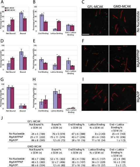
MCAK belongs to the Kinesin-13 family, whose members depolymerize microtubules rather than translocate along them. We defined the minimal functional unit of MCAK as the catalytic domain plus the class specific neck (MD-MCAK), which is consistent with previous reports. We used steady-state ATPase kinetics, microtubule depolymerization assays, and microtubule.MCAK cosedimentation assays to compare the activity of full-length MCAK, which is a dimer, with MD-MCAK, which is a monomer. Full-length MCAK exhibits higher ATPase activity, more efficient microtubule end binding, and reduced affinity for the tubulin heterodimer. Our studies suggest that MCAK dimerization is important for its catalytic cycle by promoting MCAK binding to microtubule ends, enhancing the ability of MCAK to recycle for multiple rounds of microtubule depolymerization, and preventing MCAK from being sequestered by tubulin heterodimers.


Figures

Figure 1.
Figure 1.
FL-MCAK is dimeric and MD-MCAK is monomeric. (A) Schematic representation of the proteins used in this study. NT, N terminus; Neck, class-specific neck region; CD, catalytic domain; CT, C terminus. The dark gray box within the C terminus represents the putative coiled coil region. The numbers correspond to amino acid residues. (B) Equal amounts (0.5 μg) of purified recombinant proteins were run on SDS-PAGE gels and stained with Colloidal Coomassie Blue. Molecular mass markers in kilodaltons are indicated on the left. (C) GMPCPP-stabilized MTs (1.5 μM) were incubated with control buffer (No M) or equal molar amounts (50 nM) of FL-MCAK (FL-M), GFL-MCAK (GFL-M), MD-MCAK (MD-M), GMD-MCAK (GMD-M), or GFP alone for 30 min at 22°C. MTs were separated from soluble tubulin heterodimer by ultracentrifugation. Supernatant (S) and pellet (P) were analyzed by SDS-PAGE followed by Coomassie staining. The position of tubulin (T) is indicated on the right. (D) Purified proteins were analyzed by sucrose gradient sedimentation and gel filtration chromatography to determine estimated molecular weights using Equation 1. All values represent an average ± SEM for at least three experiments.
Figure 2.
Figure 2.
At high ratios of MCAK to MTs, FL-MCAK and MD-MCAK show similar affinities for MTs. (A) FL-MCAK or MD-MCAK (1.24 μM) was incubated in the absence of nucleotide with increasing concentrations of paclitaxel- and GMPCPP-stabilized MTs (0–8 μM) for 15 min at 22°C. FL-MCAK or MD-MCAK bound to MTs was separated from unbound enzyme by ultracentrifugation. Equal volumes of supernatant (S) and pellet (P) were run on SDS-PAGE gels and stained with Coomassie Brilliant Blue. (B) The amount of FL-MCAK or MD-MCAK in the supernatant and the pellet was quantified. The binding affinity curves were derived from Equation 3 and represent averaged data from at least three individual experiments. MT·E is the amount of FL- or MD-MCAK fractionating with MTs. (C) A summary of the binding affinities.
Figure 3.
Figure 3.
At substoichiometric ratios of MCAK to MTs, GFL-MCAK binds more effectively to MT ends than GMD-MCAK. GFL-MCAK or GMD-MCAK (9 nM) was mixed with 400 nM GMPCPP-stabilized MTs for 15 min at room temperature without additional nucleotide (A–C), in the presence of 5 mM MgAMPPNP (D–F), or in the presence 5 mM MgADP (G–I). Reactions were fixed, sedimented onto coverslips, and processed for immunofluorescence. MTs were scored for the percentage of binding events (A, B, D, E, G, and H) without knowledge of the sample identity. Asterisk (*) denotes a statistically significant difference between GFL-MCAK and GMD-MCAK with a p value < 0.05. Representative micrographs of GFL-MCAK (green) or GMD-MCAK (green) binding to MTs (red) without additional nucleotide (C), or in the presence of MgAMPPNP (F) or MgADP (I). Bar, 5 μm. The localization of GFL-MCAK or GMD-MCAK in fluorescence microscopy binding assays was quantified (J). The reported value is the average percentage of MTs with GFL-MCAK or GMD-MCAK localization ± SEM. n is the total number of MTs counted from five independent experiments.
Figure 4.
Figure 4.
FL-MCAK depolymerizes MTs more efficiently than MD-MCAK. (A) GMPCPP-stabilized MTs (1 μM) were incubated with increasing concentrations (0–128 nM) of either FL-MCAK or MD-MCAK in the presence of saturating MgATP for 15 min at 22°C. Soluble tubulin heterodimer was separated from the remaining MTs by centrifugation. Equal volumes of supernatant (S) and pellet (P) were analyzed by SDS-PAGE followed by Coomassie staining, and the amount of depolymerization was then quantified using NIH Image. (B) The combined data derived from at least three separate experiments were fit to the dose-response curve (Equation 2). (C) Summary of the data derived from the graphs in B.
Figure 5.
Figure 5.
MD-MCAK has lower ATPase activity than FL-MCAK. FL-MCAK, and MD-MCAK (50 nM) were assayed for steady-state ATPase activity in the presence of increasing concentrations of (A) paclitaxel- and GMPCPP-stabilized MTs (0–15 μM) or (B) tubulin heterodimer (0–20 μM) at 2 mM MgATP. The data in A and B were fit to Equations 4 (for MTs) and 5 (for tubulin heterodimer). (C) The steady-state kinetics for FL-MCAK or MD-MCAK (50 nM) were determined as a function of MgATP concentration at 8 μM MTs for FL-MCAK and 4 μM MTs for MD-MCAK with the fit of the data to equation 5. (D) Summary of the kinetic parameters derived from A to C.
Figure 6.
Figure 6.
MD-MCAK binds with higher affinity to tubulin heterodimer than FL-MCAK. (A) FL-MCAK (FL-M) or MD-MCAK (MD-M) at 500 nM was incubated in the absence of nucleotide with paclitaxel- and GMPCPP-stabilized MTs (3.25 μM tubulin) as a function of soluble GDP tubulin heterodimer (0–13 μM) for 15 min at 22°C followed by centrifugation. Equal volumes of the supernatant (S) and pellet (P) for each reaction were analyzed by SDS-PAGE followed by Colloidal Coomassie staining. Because of the high ratios of total tubulin to MCAK enzyme in each reaction, the regions of each gel containing FL-MCAK or MD-MCAK were equally contrast-enhanced relative to the tubulin portion of the gel. T, tubulin. B, FL-MCAK and MD-MCAK partitioning to the supernatant and the pellet were quantified. The fraction of MCAK in the pellet in the absence of additional GDP tubulin heterodimer (0:1) was considered 100%. The partitioning of MCAK to the pellet as a function of GDP tubulin heterodimer was normalized to the 0:1 control. The data represent mean ± SEM for three individual experiments.
Figure 7.
Figure 7.
Model for the role of dimerization of MCAK. The catalytic cycle is diagrammed at only one end of the MT, although MCAK depolymerizes MTs from both ends. (A) With FL-MCAK, a critical number of molecules (for clarity, only one is diagrammed) are necessary to induce ATP-promoted depolymerization at the end of the MT. The tubulin heterodimer·MCAK complex is released from the shortening MT. The affinity of FL-MCAK for the tubulin heterodimer is weak, resulting in release of MCAK with rapid rebinding to the MT end. (B) MD-MCAK also can depolymerize MTs, but it requires a greater number of molecules than FL-MCAK because of the lower binding specifically to the ends of the MT. The higher affinity of MD-MCAK for the tubulin heterodimer delays its release to the solution and therefore slows its overall recycling rate for MT end binding and subsequent MT depolymerization.

References

    1. Berliner, E., Young, E. C., Anderson, K., Mahtani, H. K., and Gelles, J. (1995). Failure of a single-headed kinesin to track parallel to microtubule protofilaments. Nature 373, 718-721. - PubMed
    1. Bringmann, H., Skiniotis, G., Spilker, A., Kandels-Lewis, S., Vernos, I., and Surrey, T. (2004). A kinesin-like motor inhibits microtubule dynamic instability. Science 303, 1519-1522. - PubMed
    1. Cassimeris, L., and Spittle, C. (2001). Regulation of microtubule-associated proteins. Int. Rev. Cytol. 210, 163-226. - PubMed
    1. Cochran, J. C., Gatial, J. E., 3rd, Kapoor, T. M., and Gilbert, S. P. (2005). Monastrol inhibition of the mitotic kinesin Eg5. J. Biol. Chem. 280, 12658-12667. - PMC - PubMed
    1. Crevel, I. M., Lockhart, A., and Cross, R. A. (1996). Weak and strong states of kinesin and ncd. J. Mol. Biol. 257, 66-76. - PubMed

Publication types