Comparative Study
. 2005 Nov 16;25(46):10598-606.
doi: 10.1523/JNEUROSCI.2990-05.2005.

Binding of the positron emission tomography tracer Pittsburgh compound-B reflects the amount of amyloid-beta in Alzheimer's disease brain but not in transgenic mouse brain

Affiliations
Comparative Study

Binding of the positron emission tomography tracer Pittsburgh compound-B reflects the amount of amyloid-beta in Alzheimer's disease brain but not in transgenic mouse brain

William E Klunk et al. J Neurosci. .

Abstract

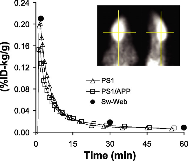
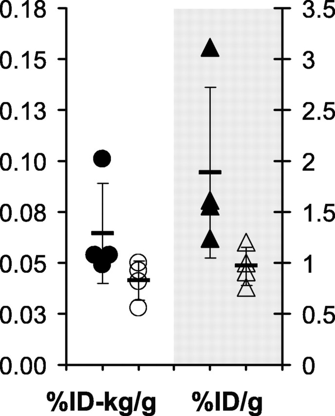
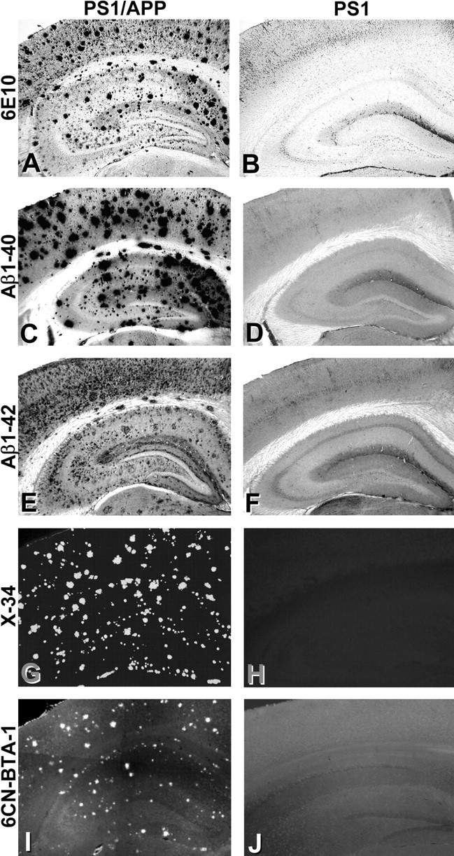
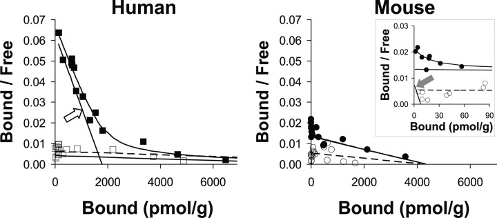
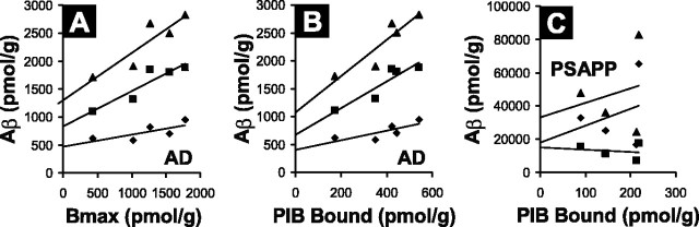
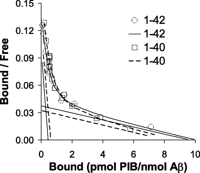
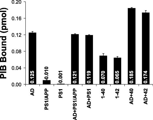
During the development of in vivo amyloid imaging agents, an effort was made to use micro-positron emission tomography (PET) imaging in the presenilin-1 (PS1)/amyloid precursor protein (APP) transgenic mouse model of CNS amyloid deposition to screen new compounds and further study Pittsburgh Compound-B (PIB), a PET tracer that has been shown to be retained well in amyloid-containing areas of Alzheimer's disease (AD) brain. Unexpectedly, we saw no significant retention of PIB in this model even at 12 months of age when amyloid deposition in the PS1/APP mouse typically exceeds that seen in AD. This study describes a series of ex vivo and postmortem in vitro studies designed to explain this low retention. Ex vivo brain pharmacokinetic studies confirmed the low in vivo PIB retention observed in micro-PET experiments. In vitro binding studies showed that PS1/APP brain tissue contained less than one high-affinity (K(d) = 1-2 nm) PIB binding site per 1000 molecules of amyloid-beta (Abeta), whereas AD brain contained >500 PIB binding sites per 1000 molecules of Abeta. Synthetic Abeta closely resembled PS1/APP brain in having less than one high-affinity PIB binding site per 1000 molecules of Abeta, although the characteristics of the few high-affinity PIB binding sites found on synthetic Abeta were very similar to those found in AD brain. We hypothesize that differences in the time course of deposition or tissue factors present during deposition lead to differences in secondary structure between Abeta deposited in AD brain and either synthetic Abeta or Abeta deposited in PS1/APP brain.


Figures

Figure 1.
Figure 1.
Micro-PET time-activity curves show no difference in the retention of [11C]PIB in PS1/APP and PS1 mice. Brain radioactivity was determined with micro-PET in a 12-month-old PS1/APP mouse (□) and a PS1 littermate (▵) imaged simultaneously. [11C]PIB retention is expressed as %ID per gram of brain normalized to the body weight (in kilograms). The [11C]PIB retention from an ex vivo study with Swiss-Webster (Sw-Web) mice (n = 3 at each time point; •) is also shown. Inset, Axial (left) and sagittal (right) image of the PS1/APP mouse.
Figure 2.
Figure 2.
Ex vivo assessment of radioactivity shows no significant difference in the retention of [11C]PIB in PS1/APP and PS1 mice. Fifteen-month-old PS1/APP mice (filled symbols) and PS1 littermate controls (open symbols) were given intravenous injections of [11C]PIB in the tail vein and killed 15 min later. The brains were removed rapidly, and retained radioactivity was assessed in a gamma counter. The same data are expressed as %ID per gram on the right and as the weight-normalized %ID-kilogram/gram on the left. Neither show a significant difference.
Figure 3.
Figure 3.
Histological analysis shows extensive amyloid deposition in the PS1/APP mice but not the PS1 mice. Tissue sections from PS1/APP (A, C, E, G, I) and PS1 (B, D, F, H, J) mice were immunostained with antibodies to total Aβ (A, B; 6E10), Aβ1-40 (C, D), and Aβ1-42 (E, F). Sections also were stained with the fluorescent Congo red derivative X-34 (G, H), which stains fibrillar amyloid, and the fluorescent PIB analog 6-CN-BTA-1 (I, J).
Figure 4.
Figure 4.
Scatchard analyses of [3H]PIB binding shows a significant high-affinity binding component in human AD brain but not in PS1/APP brain. Left, This human AD brain (▪) showed a large high-affinity component (open arrow) with a Kd value of 2.8 nm and a Bmax value of 1780 pmol/g (BP = 636) and a low-affinity component with a Kd value of 264 nm and a Bmax value of 11,000 pmol/g (BP = 42). The human control brain (□) showed only the low-affinity component with a Kd value of 242 nm and a Bmax value of 15,000 pmol/g (BP = 62). Right, This PS1/APP brain (•) showed a very small high-affinity component (filled arrow) with a Kd value of 0.10 nm and a Bmax value of 7.78 pmol/g (BP = 78), along with a much larger low-affinity component with a Kd value of 32 nm and a Bmax value of 4300 pmol/g (BP = 134). The PS1 brain (○) showed the low-affinity component with a Kd value of 79 nm and a Bmax value of 4200 pmol/g (BP = 53).
Figure 5.
Figure 5.
The binding of [3H]PIB reflects the total amount of Aβ in AD brain but not in PS1/APP brain. Correlation of the Bmax for [3H]PIB binding (A) or correlation of the amount of [3H]PIB bound (B) (using a concentration of 1 nm [3H]PIB) to levels of Aβ1-40 (♦), Aβ1-42 (▪), and total insoluble Aβ (▴) in AD brain (n = 5) is shown. C, Correlation analogous to that in B performed in PS1/APP (PSAPP) brain (n = 4).
Figure 6.
Figure 6.
The binding of [3H]PIB to synthetic Aβ1-40 and Aβ1-42 is nearly identical, and the Bmax is very small as in PS1/APP brain (see Table 3).
Figure 7.
Figure 7.
Mixing experiments do not indicate the presence of an easily transferable factor responsible for high-affinity [3H]PIB binding in AD brain. Homogenates of AD, PS1/APP, and PS1 brain (containing 100μg of tissue) and suspensions of Aβ1-40 and Aβ42-1 fibrils (containing 200 pmol of Aβ) or the mixtures indicated were incubated with 1 nm [3H]PIB in a total volume of 1 ml. The picomoles of [3H]PIB bound are indicated inside or above the bars. The amount of [3H]PIB bound to the mixtures is approximately equal to the sum of the binding to the two individual components, indicating no synergy.

References

    1. Bacskai BJ, Hickey GA, Skoch J, Kajdasz ST, Wang Y, Huang GF, Mathis CA, Klunk WE, Hyman BT (2003) Four-dimensional multiphoton imaging of brain entry, amyloid binding, and clearance of an amyloid-beta ligand in transgenic mice. Proc Natl Acad Sci USA 100: 12462-12467. - PMC - PubMed
    1. Cherry SR (2001) Fundamentals of positron emission tomography and applications in preclinical drug development. J Clin Pharmacol 41: 482-491. - PubMed
    1. Duff K, Eckman C, Zehr C, Yu X, Prada CM, Perez-Tur J, Hutton M, Buee L, Harigaya Y, Yager D, Morgan D, Gordon MN, Holcomb L, Refolo L, Zenk B, Hardy J, Younkin S (1996) Increased amyloid-beta42(43) in brains of mice expressing mutant presenilin 1. Nature 96: 710-713. - PubMed
    1. Guidotti A, Baraldi M, Schwartz JP, Costa E (1979) Molecular mechanisms regulating the interactions between the benzodazepines and GABA receptors in the central nervous system. Pharmacol Biochem Behav 10: 803-807. - PubMed
    1. Holcomb L, Gordon MN, McGowan E, Yu X, Benkovic S, Jantzen P, Wright K, Saad I, Mueller R, Morgan D, Sanders S, Zehr C, O'Campo K, Hardy J, Prada CM, Eckman C, Younkin S, Hsiao K, Duff K (1998) Accelerated Alzheimer-type phenotype in transgenic mice carrying both mutant amyloid precursor protein and presenilin 1 transgenes. Nat Med 4: 97-100. - PubMed

Publication types

Substances