Comparative Study
. 2005 Nov 16;25(46):10717-28.
doi: 10.1523/JNEUROSCI.4112-05.2005.

Critical role of calcitonin gene-related peptide 1 receptors in the amygdala in synaptic plasticity and pain behavior

Affiliations
Comparative Study

Critical role of calcitonin gene-related peptide 1 receptors in the amygdala in synaptic plasticity and pain behavior

Jeong S Han et al. J Neurosci. .

Abstract

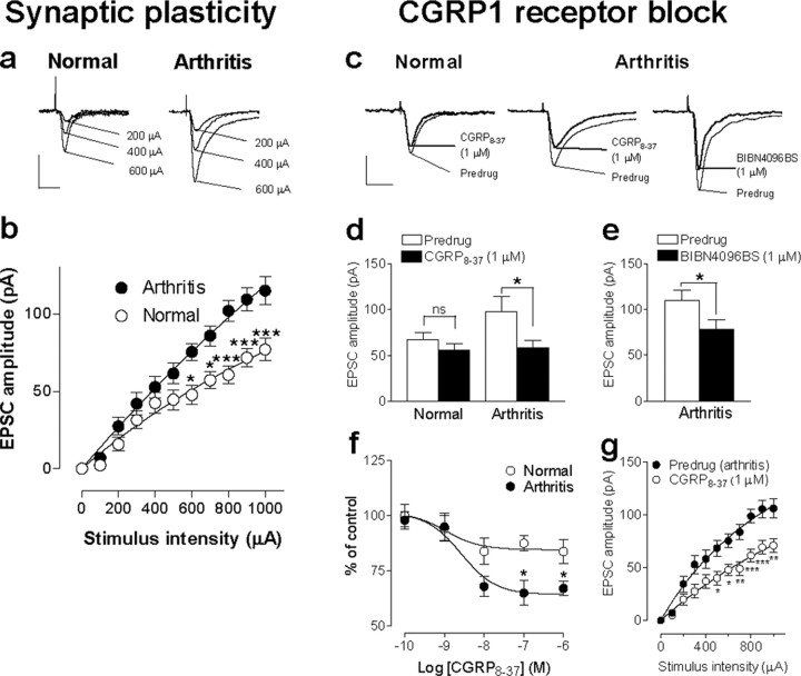
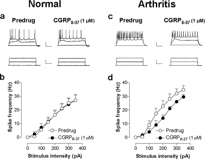
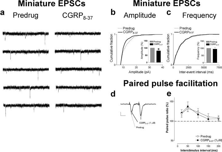
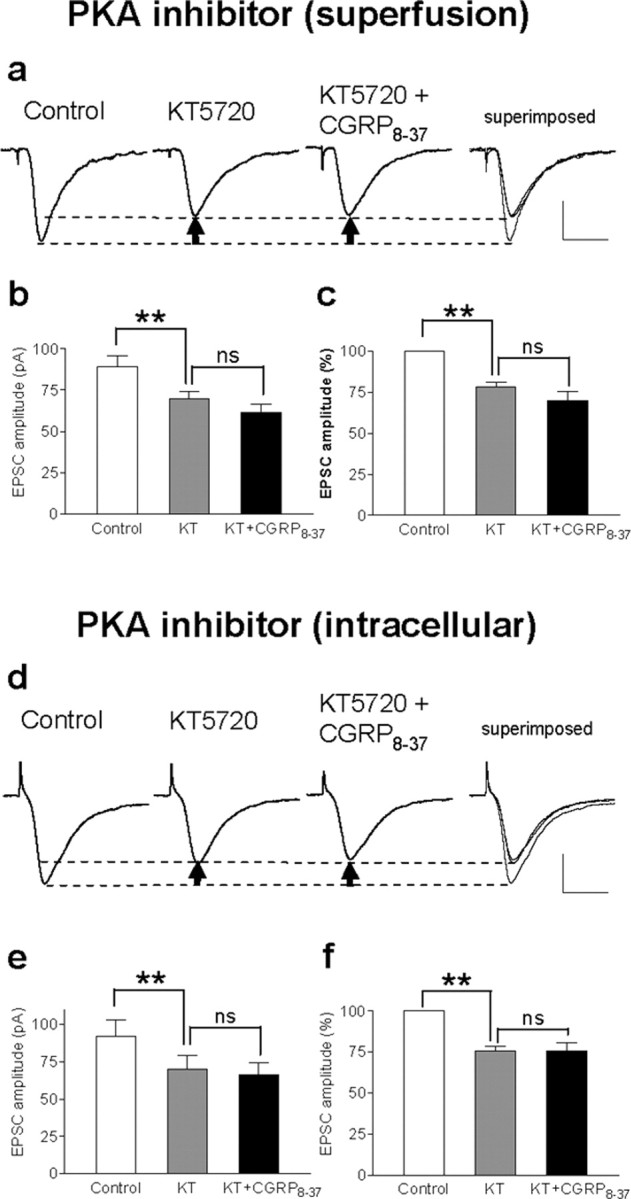
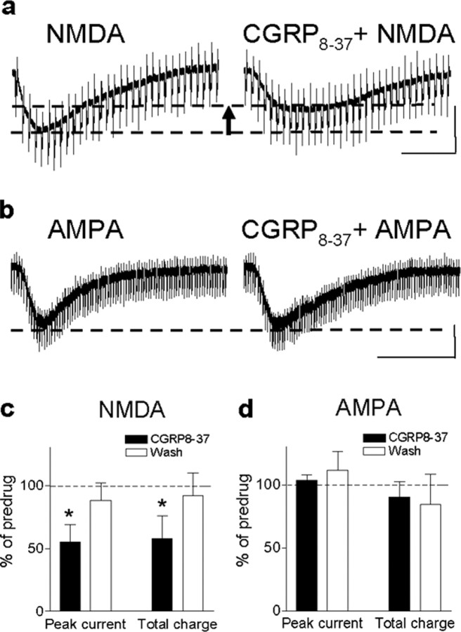
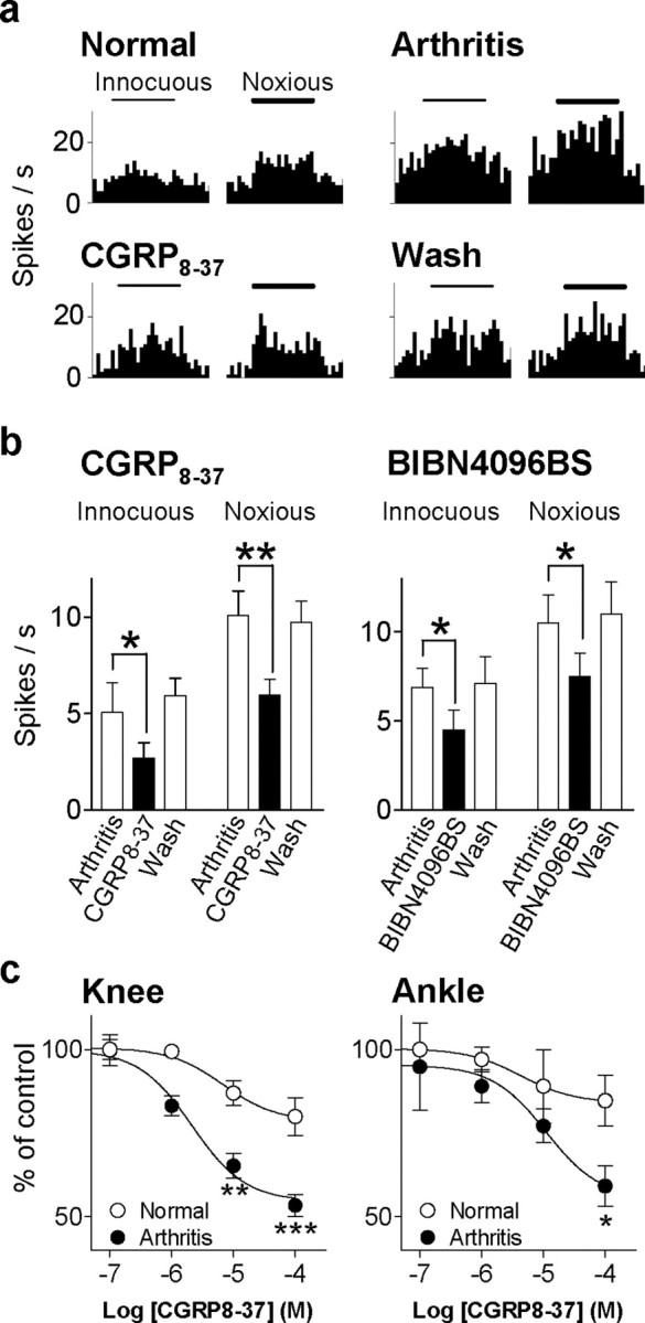
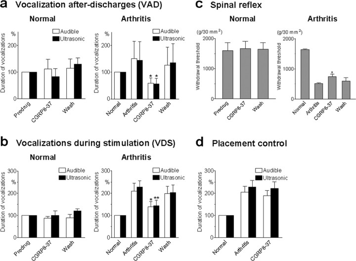
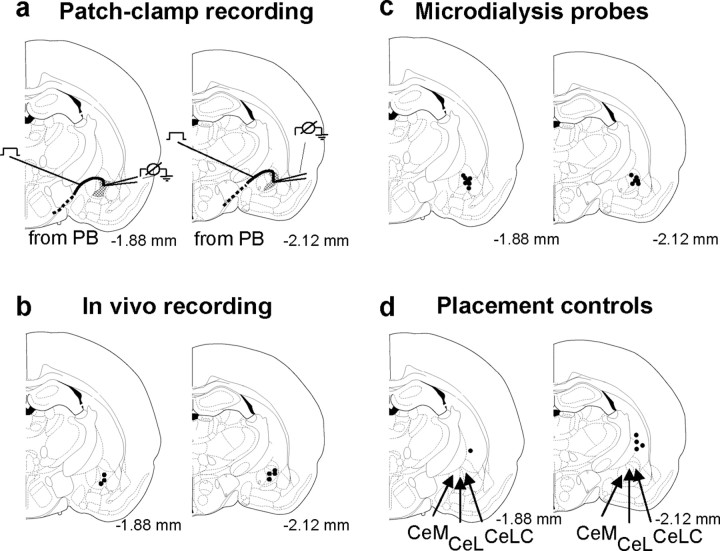
The role of neuropeptides in synaptic plasticity is less well understood than that of classical transmitters such as glutamate. Here we report the importance of the G-protein-coupled calcitonin gene-related peptide (CGRP1) receptor as a critical link between amygdala plasticity and pain behavior. A key player in emotionality and affective disorders, the amygdala has been implicated in the well documented, but mechanistically unexplained, relationship between pain and affect. Our electrophysiological and pharmacological in vitro (patch-clamp recordings) and in vivo (extracellular single-unit recordings) data show that selective CGRP1 receptor antagonists (CGRP(8-37) and BIBN4096BS) in the amygdala reverse arthritis pain-related plasticity through a protein kinase A (PKA)-dependent postsynaptic mechanism that involves NMDA receptors. CGRP1 receptor antagonists inhibited synaptic plasticity in the laterocapsular division of the central nucleus of the amygdala (CeLC) in brain slices from arthritic rats compared with normal controls. The effects were accompanied by decreased neuronal excitability and reduced amplitude, but not frequency, of miniature EPSCs; paired-pulse facilitation was unaffected. The antagonist effects were occluded by a PKA inhibitor. CGRP1 receptor blockade also directly inhibited NMDA-evoked, but not AMPA-evoked, membrane currents. Together, these data suggest a postsynaptic site of action. At the systems level, the antagonists reversed the sensitization of nociceptive CeLC neurons in anesthetized rats in the arthritis pain model. Importantly, CGRP1 receptor blockade in the CeLC inhibited spinal (hindlimb withdrawal reflexes) and supraspinal pain behavior of awake arthritic rats, including affective responses such as ultrasonic vocalizations. This study provides direct evidence for the critical dependence of pain behavior on CGRP1-mediated amygdala plasticity.


Figures

Figure 1.
Figure 1.
Pain-related synaptic plasticity in the amygdala depends in part on the activation of CGRP1 receptors. a, Peak amplitudes of monosynaptic EPSCs, a measure of synaptic strength, were larger in a CeLC neuron recorded in a brain slice from an arthritic rat (right) than in a control neuron from a normal rat (left). Individual traces show monosynaptic EPSCs (average of 8-10 EPSCs) evoked at the PB-CeLC synapse with increasing stimulus intensities. Calibration: 50 ms, 50 pA. b, Input-output functions were measured by increasing the stimulus intensity in 100 μA steps. CeLC neurons from arthritic animals (n = 19) showed significantly enhanced synaptic transmission compared with control neurons (n = 37) (p < 0.0001, F(1,593) = 60.29, two-way ANOVA followed by Bonferroni's post hoc tests). c, A selective CGRP1 receptor antagonist (CGRP8-37, 1 μm) inhibited synaptic plasticity in a CeLC neuron from an arthritic animal (middle trace) but had little effect on basal synaptic transmission in a CeLC neuron from a normal animal (left trace). Likewise, the selective nonpeptide CGRP1 receptor antagonist (BIBN4096BS, 1 μm) inhibited synaptic plasticity in a CeLC neuron from an arthritic animal (right trace). Individual traces show monosynaptic EPSCs (average of 8-10 EPSCs) evoked at the PB-CeLC synapse with the stimulus intensity set to 70-80% of that required for generating maximum EPSC amplitude. Calibration: 50 ms, 50 pA. d, Averaged raw (current) data show that CGRP8-37 (1 μm) inhibited the increased EPSC amplitude in neurons (n=17) from arthritic rats (right; p < 0.05, paired t test) but had no significant effects on the amplitude of EPSCs recorded in control neurons (n = 29) from normal rats (left). e, BIBN4096BS (1 μm) also decreased the EPSC amplitude (averaged current data) significantly (p < 0.05, paired t test; n = 5). f, Concentration-response relationships show that CGRP8-37 was more efficacious in neurons from arthritic rats (n = 17) than in control neurons (n = 29) from normal rats (p < 0.01; F(1,220) = 10.74, two-way ANOVA). Peak amplitudes of monosynaptic EPSCs during each concentration of CGRP8-37 were averaged and expressed as percentage of predrug (baseline) control (100%). CGRP8-37 was applied for at least 15 min, and measurements were made at 12 min. g, Input-output function of the PB-CeA synapse (see b) was significantly reduced by CGRP8-37 (1 μm) in CeLC neurons from arthritic rats (n = 12; p < 0.0001; F(1,242) = 76.32, two-way ANOVA followed by Bonferroni's post hoc tests). Whole-cell voltage-clamp recordings were made from CeLC neurons held at -60 mV. *p < 0.05; **p < 0.01; ***p < 0.001.
Figure 2.
Figure 2.
CGRP8-37 inhibits neuronal excitability of CeLC neurons in the arthritis pain model but not under normal conditions. Action potentials were evoked in CeLC neurons by direct (through the patch electrode) intracellular injections of current pulses (250 ms) of increasing magnitude (50 pA steps) before and during CGRP8-37 administration. a, b, CGRP8-37 did not affect the action potential firing rate in CeLC neurons in slices from normal rats. c, d, However, the action potential firing rate was significantly decreased by CGRP8-37 in CeLC neurons from arthritic rats (n = 10; p < 0.0001; F(1,144) = 14.15, two-way ANOVA), suggesting a functional change of CGRP1 receptor activation that has postsynaptic effects in arthritis but not under normal conditions. For the measurement of action potential firing in current clamp, neurons were recorded at -60 mV. Calibration (in a, c): top traces, 100 ms, 25 mV; bottom traces, 100 ms, 150 pA. Symbols and error bars in b and d represent mean ± SE.
Figure 3.
Figure 3.
mEPSC analysis and PPF suggest postsynaptic rather than presynaptic effects of CGRP8-37. a, Original current traces of mEPSC recordings in an individual CeLC neuron in the presence of TTX (1μm) showed that CGRP8-37 (1μm) reduced amplitude but not frequency of mEPSCs. Calibration: 1 s, 20 mV. The CeLC neuron was recorded in a slice from an arthritic rat. b, c, Normalized cumulative distribution analysis of mEPSC amplitude and frequency showed that CGRP8-37 caused a significant shift toward smaller amplitudes (b) (p < 0.005; maximal difference in cumulative fraction, 0.175; Kolmogorov-Smirnov test) but had no effect on the interevent interval (frequency) distribution (c). CGRP8-37 selectively decreased mean mEPSC amplitude (p < 0.05, paired t test) but not mEPSC frequency (events per second) in the sample of neurons (n = 4; see bar histograms in d, e). PPF, a measure of presynaptic mechanisms, was not changed by CGRP8-37. PPF was calculated as the ratio of the second and the first of two consecutive EPSCs evoked by two electrical stimuli of equal intensity at increasing interstimulus intervals. Peak EPSC amplitudes were measured as the difference between the current level before the stimulus artifact and the peak of the EPSC. d, Current traces (average of 8-10 EPSCs) recorded in an individual CeLC neuron illustrate that PPF evoked at a 50 ms interval was not affected by CGRP8-37. Calibration: 25 ms, 50 pA. e, CGRP8-37 had no significant effect on PPF at various stimulus intervals in the whole sample of neurons (n = 6; p > 0.05, paired t test), further arguing against a presynaptic action. Symbols and error bars represent mean ± SE. Neurons were recorded in voltage clamp at -60 mV.
Figure 4.
Figure 4.
Effects of CGRP8-37 are occluded by a PKA inhibitor. a-c, Superfusion of the slices with a selective membrane-permeable PKA inhibitor (KT5720; 1μm in ACSF) decreased synaptic plasticity and abolished the inhibitory effects of CGRP8-37 (1 μm), suggesting that CGRP1 receptor activation involves PKA activation. a, Individual traces show monosynaptic EPSCs (average of 8-10 EPSCs) in a CeLC neuron in a brain slice from an arthritic rat. Recordings were made before drug application, in the presence of KT5720 alone and during coapplication of CGRP8-37 and KT5720; superimposed traces are shown on the right. Calibration: 20 ms, 40 pA. Arrows indicate difference of peak amplitudes. b, c, Averaged raw (picoamperes; b) and normalized (percentage; c) data show the significant (**p < 0.01, repeated measures ANOVA with Tukey's post hoc test) inhibitory effect of a PKA inhibitor [KT5720 (KT)] but no additional inhibition by CGRP8-37 when coapplied with KT5720. d-f, Direct intracellular application of the PKA inhibitor through the patch pipette filled with internal solution containing KT5720 (1 μm) also occluded the inhibitory effects of CGRP8-37, suggesting a direct postsynaptic mechanism. d, Monosynaptic EPSCs were measured immediately after whole-cell patch configuration was obtained control (left). EPSC amplitude decreased 10 min after the patch formation when the PKA inhibitor had entered the cell. Coapplication of CGRP8-37 (superfusion) did not further reduce EPSC amplitude. e, f, Averaged raw (picoamperes; e) and normalized (percentage; f) data show the significant (**p < 0.01, repeated measures ANOVA with Tukey's post hoc test) inhibitory effect of a PKA inhibitor (KT5720, intracellular application) but no additional inhibition by CGRP8-37. Calibration: 20 ms, 40 pA.
Figure 5.
Figure 5.
CGRP8-37 effects involve inhibition of NMDA, but not AMPA, receptor function. a, CGRP8-37 decreased the inward current evoked by application of exogenous NMDA (1 mm in the chamber) in a neuron from an arthritic rat. Calibration: 20 s, 200 pA. The arrrow indicates difference of peak amplitude of NMDA current before and during CGRP8-37 application. b, CGRP8-37 had no effect on the inward current evoked by application of exogenous AMPA (30 μm in the chamber) in a CeLC neuron from an arthritic rat. Calibration: 100s, 200pA. c, Averaged data show the significant inhibitory effect of CGRP8-37 on NMDA-evoked membrane currents in terms of peak current and area under the curve (n=5; paired t test, p < 0.05). d, AMPA-evoked membrane currents were not affected significantly by CGRP8-37 (n = 5; paired t test, p > 0.05). Symbols and error bars represent mean ± SE. Neurons were recorded in voltage clamp at -60 mV. *p < 0.05.
Figure 6.
Figure 6.
CGRP1 receptor antagonists inhibit nociceptive sensitization of CeLC neurons in anesthetized intact animals. a, Extracellular recordings of the responses of one multireceptive CeLC neuron to innocuous (100 g/30 mm2) and noxious (2000 g/30 mm2) mechanical stimulation of the knee joint before and 6 h after induction of the knee joint arthritis (see Materials and Methods). CGRP8-37 (100μm) was administered into the CeLC by microdialysis for 20 min. Histograms show action potentials (spikes) per second (bin width, 1 s). Horizontal bars indicate duration of stimuli (15 s). b, Averaged raw data (spikes per second) for the sample of neurons tested with CGRP8-37 (100 μm; n = 7) and BIBN4096BS (100 μm; n = 4). Both antagonists significantly reduced the increased responses to normal levels (p < 0.01-0.05, paired t test). The inhibitory effects were reversible after washout for 20-30 min with ACSF in the microdialysis fiber. Bar histograms and error bars represent mean ± SE. *p < 0.05; **p < 0.01. c, Concentration-response relationships show that CGRP8-37 was more efficacious in sensitized neurons 6 h after induction of arthritis (n = 7) than under normal conditions before arthritis (n = 7). The differences of drug effects between arthritis and normal conditions were statistically significant (knee, p < 0.001, F(1,25) = 26.57; ankle, p < 0.05, F(1,25) = 6.36; two-way ANOVA). *p < 0.05; **p < 0.01; ***p < 0.001 (Bonferroni's post hoc tests). Responses to brief (15 s) noxious stimulation of the knee or ankle during each concentration of CGRP8-37 were averaged and expressed as percentage of predrug (baseline) control (100%). CGRP8-37 was administered by microdialysis for 20 min, and measurements were made at 15-20 min. Concentrations shown indicate concentrations in the microdialysis fiber.
Figure 7.
Figure 7.
CGRP8-37 inhibits pain-related behavior (audible and ultrasonic vocalizations and hindlimb withdrawal reflexes) in awake animals with arthritis but not in normal animals. Audible and ultrasonic vocalizations were measured in normal rats (left column) and arthritic rats (right column). Duration of vocalizations was measured as the arithmetic sum of the duration of each individual vocalization event as described previously (Han and Neugebauer, 2005). a, VADs, which continue beyond the actual stimulus and are organized in the limbic forebrain (Borszcz and Leaton, 2003; Han and Neugebauer, 2005), were evoked by noxious (2000 g/30 mm2) stimulation (15 s) of the knee. Application of CGRP8-37 (100 μm, concentration in the microdialysis probe; 15-20 min) into the CeLC of normal rats had no significant effect (left; n = 4; p > 0.05, paired t test). In arthritic animals (6 h after induction), CGRP8-37 (100μm) significantly reduced audible and ultrasonic VADs (right; n = 9; p < 0.05, paired t test). Vocalizations of arthritic animals were expressed as percentage of vocalizations of the same animals before arthritis induction (normal, set to 100%). b, Audible and ultrasonic VDSs, which are organized at the medullary brainstem level (Borszcz and Leaton, 2003; Han and Neugebauer, 2005), were evoked by noxious (2000 g/30 mm2) stimulation (15 s) of the knee. Administration of CGRP8-37 (100μm, concentration in the microdialysis probe; 15-20 min) into the CeLC did not affect the duration of audible and ultrasonic VDSs in normal animals (left; n = 4; p > 0.05, paired t test) but significantly inhibited VDSs of arthritic rats (right; n = 9; p < 0.05, paired t test). c, Hindlimb withdrawal reflexes were evoked by mechanical stimulation (compression) of the knee (15 s) with increasing intensity (steps of 50 g/30 mm2). Withdrawal thresholds, a measure of pain sensitivity, were defined as the minimum stimulus intensity that evoked a withdrawal reflex. Thresholds decreased after arthritis. Application of CGRP8-37 into the CeLC significantly increased the reduced thresholds in arthritic animals (right; n = 6; p < 0.05, paired t test) but had no effect in normal rats (left; n = 4; p > 0.05, paired t test). d, Placement control experiments show that application of CGRP8-37 into the striatum (caudate-putamen dorsolateral to CeLC) did not produce significant changes of audible and ultrasonic vocalizations in arthritic animals (n = 5; p > 0.05, paired t test). Vocalization data in d represent the total duration of VAD plus VDS. Because neither VADs nor VDSs were inhibited by CGRP8-37, the data were pooled for simplification. a-d, CGRP8-37 (100 μm) was administered by microdialysis for 15-20 min. All drug effects were reversible. Bar histograms and error bars represent mean ± SE. *p < 0.05; **p < 0.01.
Figure 8.
Figure 8.
Histologic verification of electrophysiological recording sites and drug application sites. a-d, Standard diagrams (adapted from Paxinos and Watson, 1998) show coronal sections through the right brain hemisphere at different levels posterior to bregma (-1.88 to -2.12 mm). a, In vitro patch-clamp recordings were made in the shaded area (CeLC). Stimulating electrode was positioned on the fiber tract of afferents from the parabrachial area (dashed line). The boundaries of the different amygdaloid nuclei as well as the fibers from the brainstem can be easily visualized under the microscope. b, Recording sites of CeLC neurons in the in vivo electrophysiological studies using extracellular single-unit recordings. c, Sites of drug application into the CeLC by microdialysis in the vocalization experiments. d, Sites of drug application into the striatum by microdialysis as placement controls for any effects attributable to drug diffusion. CeM, Medial division of the central nucleus of the amygdala; CeL, lateral division of the central nucleus of the amygdala.

References

    1. Bernard J-F, Bandler R (1998) Parallel circuits for emotional coping behaviour: new pieces in the puzzle. J Comp Neurol 401: 429-436. - PubMed
    1. Bird GC, Lash LL, Han JS, Zou X, Willis WD, Neugebauer V (2005) Protein kinase A-dependent enhanced NMDA receptor function in pain-related synaptic plasticity in rat amygdala neurones. J Physiol (Lond) 564: 907-921. - PMC - PubMed
    1. Borszcz GS, Leaton RN (2003) The effect of amygdala lesions on conditional and unconditional vocalizations in rats. Neurobiol Learn Mem 79: 212-225. - PubMed
    1. Cabell L, Audesirk G (1993) Effects of selective inhibition of protein kinase C, cyclic AMP-dependent protein kinase, and Ca2+-calmodulin-dependent protein kinase on neurite development in cultured rat hippocampal neurons. Int J Dev Neurosci 11: 357-368. - PubMed
    1. Cartmell J, Schoepp DD (2000) Regulation of neurotransmitter release by metabotropic glutamate receptors. J Neurochem 75: 889-907. - PubMed

Publication types

MeSH terms

Substances