. 2005 Dec 7;24(23):4144-53.
doi: 10.1038/sj.emboj.7600875. Epub 2005 Nov 17.

Structure of unliganded HSV gD reveals a mechanism for receptor-mediated activation of virus entry

Affiliations

Structure of unliganded HSV gD reveals a mechanism for receptor-mediated activation of virus entry

Claude Krummenacher et al. EMBO J. .

Abstract

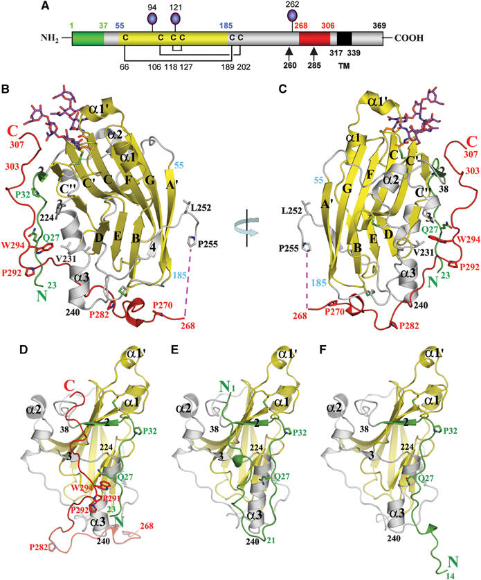
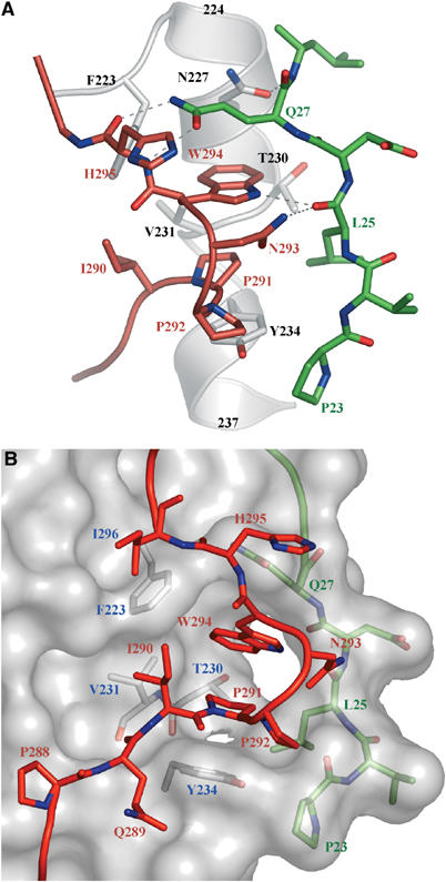
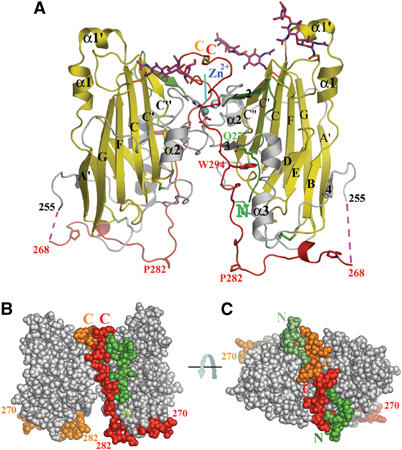
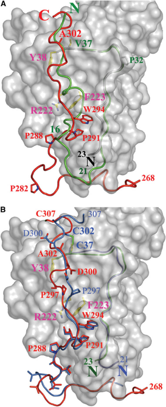
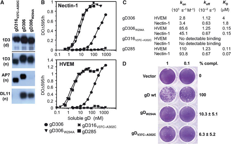
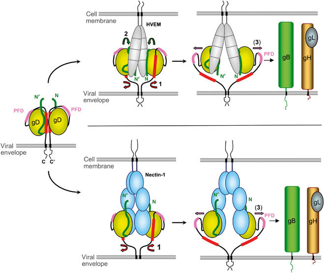
Herpes simplex virus (HSV) entry into cells requires binding of the envelope glycoprotein D (gD) to one of several cell surface receptors. The 50 C-terminal residues of the gD ectodomain are essential for virus entry, but not for receptor binding. We have determined the structure of an unliganded gD molecule that includes these C-terminal residues. The structure reveals that the C-terminus is anchored near the N-terminal region and masks receptor-binding sites. Locking the C-terminus in the position observed in the crystals by an intramolecular disulfide bond abolished receptor binding and virus entry, demonstrating that this region of gD moves upon receptor binding. Similarly, a point mutant that would destabilize the C-terminus structure was nonfunctional for entry, despite increased affinity for receptors. We propose that a controlled displacement of the gD C-terminus upon receptor binding is an essential feature of HSV entry, ensuring the timely activation of membrane fusion.


Figures

Figure 1
Figure 1
gD(23–306)307C structure. (A) Schematic representation of HSV-1 gD. Disulfide bonds are shown as black lines and N-linked oligosaccharides as lollipops. Colors of the N-terminal region, forming the HVEM-binding hairpin in the gD285–HVEM complex, the Ig-like core, and the C-terminal region past residue 255 are represented in green, yellow, and red, respectively. Positions of important amino acids and domain boundaries are numbered according to the mature form of gD. TM: transmembrane region. (B) Ribbon representation of the gD(23–306)307C subunit. The secondary structure elements are labeled as in Carfi et al (2001). The color code used is same as in (A). (C) As in (B) after 180° rotation as indicated. (D) Front view of gD(23–306)307C structure. The model is rotated 90° clockwise with respect to panel B to show the front side of gD, where the N- and C-terminal regions interact. (E) Front view of gD285 from the gD285–HVEM complex, with HVEM removed for clarity. (F) Front view of gD285 in the unbound state.
Figure 2
Figure 2
Interactions of the C-terminal region in the gD(23–306)307C structure. (A) Interactions of the 290–295 region (in red), with residues 23–27 (in green) and the α3 α-helix (in white). Dashed lines represent hydrogen bonds. (B) Surface representation of gD core (in gray) with residues involved in the formation of the Trp294- and Pro291-binding crevice shown underneath in white. The side chains of Pro291, Trp294, and other C-terminal residues are shown in red as ball-and-stick representation, whereas residues from the N-terminal region are shown in green under the surface.
Figure 3
Figure 3
The disulfide-linked dimer of gD(23–306)307C. (A) Ribbon representation of the dimer. A Zn2+ ion, trapped at the dimer interface, and the disulfide bonds are shown. The red dotted lines represent a disordered part of the C-terminal region. N- and C-termini and important amino acids are indicated. (B) Sphere representation of the dimer. The C-termini (268–307) of the two subunits are in red (subunit A, on the right) and orange (subunit B), and the N-terminal residues (23–28) in dark green (A) and pale green (B). The view is as in A. (C) As in panel B after a 90° rotation around an axis parallel to the plane.
Figure 4
Figure 4
Superimposition of gD(23–306)307C and gD285 from the gD285–HVEM complex structure. (A) The gD(23–306)307C core is represented as a white surface. The N-terminal residues of gD285 (1–16, in green) from the gD285–HVEM complex occupy the same space as residues from the C-terminus of gD(23–306)307C (289–307, in red). The side chains of Tyr38, Phe223, and Arg222, three residues involved in nectin-1 binding and buried under C-terminal residues in gD(23–306)307C, are shown. Val37 and Ala302, two residues predicted to form a disulfide if mutated in cysteines, are also shown. (B) The C-terminus of gD(23–306)307C (in red) and of gD316V37C–A302C (in blue) occupy identical positions in the 285–296 region, but adopt different conformations in the 297–307 region. The disulfide bond between Cys302 and Cys37, the side chains of several C-terminal residues as well as those of Tyr38, Phe223, and Arg222 are shown. The N-terminal residues of gD(23–306)307C and gD316V37C–A302C are colored in green and blue, respectively.
Figure 5
Figure 5
Biochemical and functional characterization of gD mutants. (A) Western blots of purified gD ectodomains were probed by various Mabs. Mab 1D3 detects a linear epitope (aa 11–19) under both denaturing/reducing (d) and native conditions (n) electrophoresis. Mabs AP7 and DL11 detect conformation-dependent epitopes under native conditions of electrophoresis. (B) Receptor binding as detected by ELISA. Various concentrations of the indicated forms of purified gD ectodomains were added to immobilized receptor ectodomains (i.e. nectin or HVEM). The bound gD was detected with anti-gD polyclonal serum R7. (C) Kinetic constants (kon and koff) and affinity constant (KD) for gD–HVEM and gD–nectin-1 complexes were measured by surface plasmon resonance and fitted to a 1:1 Langmuir interaction model (see Materials and methods). (D) Complementation of HSV FgDβ with gD mutants and titration of complemented virus on VD60 cells. VD60 cells were stained with crystal violet to visualize plaques. Complemented virus present in the culture medium of transfected–infected cells was undiluted or diluted 10-fold. Titers of complemented viruses are represented as percentage of wt on the right. These numbers are an average of four experiments, including the one presented on the left. Error bars represent ±1 s.d. Vector indicates the use of pcDNA3.1 for transfection as a negative control.
Figure 6
Figure 6
Proposed mechanism for receptor-mediated activation of HSV gD. Envelope gD is shown, as a putative dimer, in its unbound state as well as during interaction with HVEM (top) or nectin-1 (bottom). Conformational changes are chronologically indicated by numbered arrows: (1) displacement of the C-terminus, (2) folding of the gD N-terminus in the case of HVEM binding, and (3) exposure of the PFD. The N-terminus of gD is shown in green, the C-terminus (290–299) is colored red, and the PFD (260–285) is pink.

References

    1. Campadelli-Fiume G, Cocchi F, Menotti L, Lopez M (2000) The novel receptors that mediate the entry of herpes simplex viruses and animal alphaherpesviruses into cells. Rev Med Virol 10: 305–319 - PubMed
    1. Carfi A, Gong H, Lou H, Willis SH, Cohen GH, Eisenberg RJ, Wiley DC (2002) Crystallization and preliminary diffraction studies of the ectodomain of the envelope glycoprotein D from herpes simplex virus 1 alone and in complex with the ectodomain of the human receptor HveA. Acta Crystallogr D 58: 836–838 - PubMed
    1. Carfi A, Willis SH, Whitbeck JC, Krummenacher C, Cohen GH, Eisenberg RJ, Wiley DC (2001) Herpes simplex virus glycoprotein D bound to the human receptor HveA. Mol Cell 8: 169–179 - PubMed
    1. CCP4 (1994) The CCP4 suite: programs for protein crystallography. Acta Crystallogr D 50: 760–763 - PubMed
    1. Chiang H-Y, Cohen GH, Eisenberg RJ (1994) Identification of functional regions of herpes simplex virus glycoprotein gD by using linker-insertion mutagenesis. J Virol 68: 2529–2543 - PMC - PubMed

Publication types