Review
. 2005 Dec 1;392(Pt 2):249-61.
doi: 10.1042/BJ20050908.

Computational modelling of the receptor-tyrosine-kinase-activated MAPK pathway

Affiliations
Review

Computational modelling of the receptor-tyrosine-kinase-activated MAPK pathway

Richard J Orton et al. Biochem J. .

Abstract

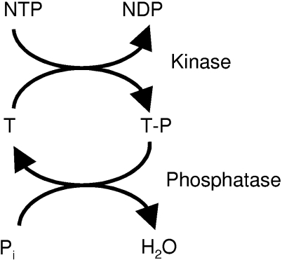
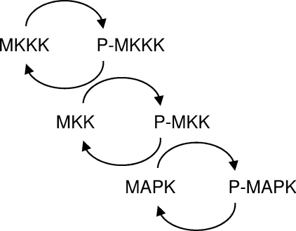
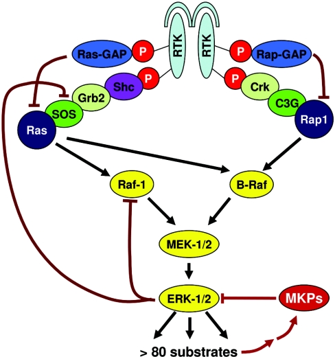
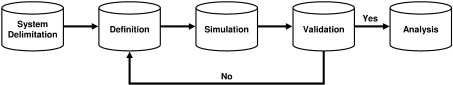
The MAPK (mitogen-activated protein kinase) pathway is one of the most important and intensively studied signalling pathways. It is at the heart of a molecular-signalling network that governs the growth, proliferation, differentiation and survival of many, if not all, cell types. It is de-regulated in various diseases, ranging from cancer to immunological, inflammatory and degenerative syndromes, and thus represents an important drug target. Over recent years, the computational or mathematical modelling of biological systems has become increasingly valuable, and there is now a wide variety of mathematical models of the MAPK pathway which have led to some novel insights and predictions as to how this system functions. In the present review we give an overview of the processes involved in modelling a biological system using the popular approach of ordinary differential equations. Focusing on the MAPK pathway, we introduce the features and functions of the pathway itself before comparing the available models and describing what new biological insights they have led to.


Figures

Figure 1
Figure 1. Kinase/phosphatase signalling reaction
A phospho group is transferred from a nucleotide to a serine, threonine or tyrosine residue of a target protein (T). The reaction is catalysed by a kinase. The phosphorylated target protein (T–P) is in turn dephosphorylated by a phosphatase to complete the cycle. Phosphorylation of a protein often entails conformational changes that modulate the function of the protein. Phosphorylation of MEK, for example, activates the kinase domain of the enzyme. MEK then catalyses the phosphorylation and, as a consequence thereof, activation of MAPK.
Figure 2
Figure 2. General structure of the three-tiered cascade of the MAPK pathway
A signal is propagated along this cascade by sequential phosphorylation as described in the text.
Figure 3
Figure 3. Structure of the ERK pathway
Upon ligand binding, RTK autophosphorylates (phosphates are shown as red circles) on tyrosine residues, which serve as docking sites for adaptor and signalling molecules. Ras and Rap1 are activated by the recruitment of guanosine-nucleotide exchange factors (SOS, C3G) via adaptor proteins (Shc and Grb2; Crk). Ras can activate Raf-1 and B-Raf; Rap1 presumably can activate B-Raf. Raf proteins phosphorylate and activate MEK-1/2, which in turn activate ERK-1/2 (indicated by black arrows). Negative-feedback loops (indicated by red lines) include the induction of MKPs by ERK as well as the inhibitory phosphorylation of Raf-1 and SOS.
Figure 4
Figure 4. The five steps of modelling
This diagram depicts the five steps involved in modelling a biological system. The first step is identifying the biological system to model, followed by actually defining the model to represent the system, simulating the model and validating the simulation results. If the model is valid, it can be analysed further; if it is not, the definition step is revisited, where the model is checked for various types of errors. For more information on each of the steps, see the text.
Figure 5
Figure 5. Timeline of ERK models
This diagram is a timeline of mathematical models that, in some way, incorporate the ERK cascade. Models are represented as ovals labelled with the name of the first author and located above the year in which they were published. White ovals represent models of the core ERK cascade, whereas grey ovals represent larger models generally, including growth-factor receptors, adaptor proteins as well as the ERK cascade itself. Models highlighted in black are the models we have selected for discussion in detail below (for brevity, only the first author is named). 1996: Huang [29]; 1997: Burack [30], Ferrell [31]; 1998: Ferrell [32]; 1999: Bhalla [24], Kholodenko [60]; 2000: Brightman [35], Kholodenko [34], Levchenko: [38]; 2001: Asthagiri [40], González [88]; 2002: Bhalla [33], Moehren [41], Schoeberl [42], Shvartsman [36], Somsen [39], Swain [43]; 2003: Aksan [44], Hatakeyama [46], Hendriks [47], Resat [48], Bluthgen [45], Cho [89], Xiong [49]; 2004: Maly [37], Markevich [51], Oliveira [78], Qiu [52], Yamada [53], Chapman [90], Markevich [50]; 2005: Aksan [91], Perez-Jimenez [92], Oney [93], Sasagawa [54].
Figure 6
Figure 6. Kholodenko et al. [60] model diagram
This schematic representation of a model of EGFR signalling mediated by adaptor and target proteins is taken from Figure 1 of [60] and is reproduced with the permission of the American Society for Biochemistry and Molecular Biology. © 1999.
Figure 7
Figure 7. Brightman and Fell [35] model diagram
This schematic representation of the Brightman and Fell model of EGF signal transduction is taken from Figure 1 of [35]. Reprinted by permission of the Federation of European Biochemical Societies. © 2000. In this schema, GS represents the Grb2–SOS complex.
Figure 8
Figure 8. Schoeberl et al. [42] model diagram
This schematic representation of the Schoeberl et al. model of the EGF-receptor-induced ERK kinase cascade is taken from Figure 1 of [42] and is reproduced with the permission of Nature Biotechnology (http://www.nature.com/). © 2002.

References

    1. Cobb M. H. MAP kinase pathways. Prog. Biophys. Mol. Biol. 1999;71:479–500. - PubMed
    1. Widmann C., Gibson S., Jarpe M. B., Johnson G. L. Mitogen-activated protein kinase: conservation of a three-kinase module from yeast to human. Physiol. Rev. 1999;79:143–180. - PubMed
    1. Chang L., Karin M. Mammalian MAP kinase signalling cascades. Nature (London) 2001;410:37–40. - PubMed
    1. Farooq A., Zhou M. M. Structure and regulation of MAPK phosphatases. Cell. Signalling. 2004;16:769–779. - PubMed
    1. Murphy L. O., MacKeigan J. P., Blenis J. A network of immediate early gene products propagates subtle differences in mitogen-activated protein kinase signal amplitude and duration. Mol. Cell. Biol. 2004;24:144–153. - PMC - PubMed

MeSH terms

Substances